

The Spine care is essential for anyone experiencing neck, back, or disc problems, especially when those issues start interfering with daily life.
Maintaining a strong and flexible spine is essential for mobility and overall well-being. A well-supported spine helps facilitate movement and protects the spinal cord. Structured spinal care, modern rehabilitation methods, and proper posture can help support long-term spinal health.
The spine, or vertebral column, is a key component of the skeletal system, providing stability and flexibility. It consists of five primary regions: cervical, thoracic, lumbar, sacral, and coccygeal. Understanding spinal structure and function helps promote better movement and prevent injuries.
At Chiropractic Specialty Center®, we provide non-invasive chiropractic, physiotherapy, and rehabilitation through gentle, targeted methods that avoid twisting or forceful movements. Our team uses thorough assessments and evidence-informed procedures to address spinal imbalances, disc-related issues, and nerve involvement focusing on long-term structural improvement. Whether you’re facing stiffness, disc bulges, or posture-related concerns, our personalized plans are designed to improve function and prevent worsening. If your spine isn’t functioning as it should, the sooner you act, the better your results.
What You Need to Know About Spine Care Now
Spine care must be non-invasive, precise, and customized – look for providers combining chiropractic, physiotherapy, and rehabilitation in one place.
Not all back or disc problems require surgery – early, structured care helps restore spinal health without surgical risks.
Results depend on accurate assessment – choose a center that uses tech-based diagnostics and gentle methods backed by clinical evidence.
Contact Us Now for Personalized Spine Care
Take the first step toward better spine care; reach out via our contact page to schedule your assessment and learn how our non‑invasive methods can help you.
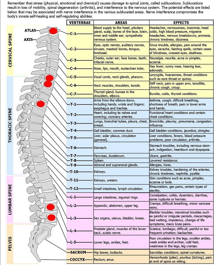
Cervical Spine: The Neck
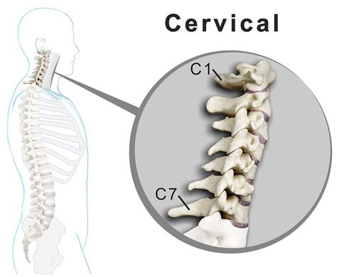
The cervical spine comprises seven vertebrae (C1 to C7) forming the upper section of the spine. It extends from the base of the skull to the upper back and has a natural inward curve (lordosis).
- C1 (Atlas) – Connects the spine to the skull and enables head movement.
- C2 (Axis) – Allows rotational movement of the head.
- Atlanto-Occipital Joints – Facilitate nodding motions.
- Atlantoaxial Joints – Support side-to-side rotation.
Thoracic Spine: Upper and Mid-Back
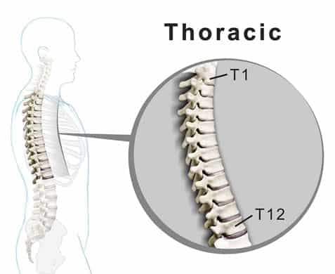
The thoracic spine consists of 12 vertebrae (T1 to T12), forming the middle section of the spine.
- Structure – Connects to the rib cage, forming a protective framework for internal organs.
- Natural Curve – Has a gentle outward curve (kyphosis).
- Function – Supports spinal stability and shields the heart and lungs.
Lumbar Spine and Lumbosacral Region: Lower Back
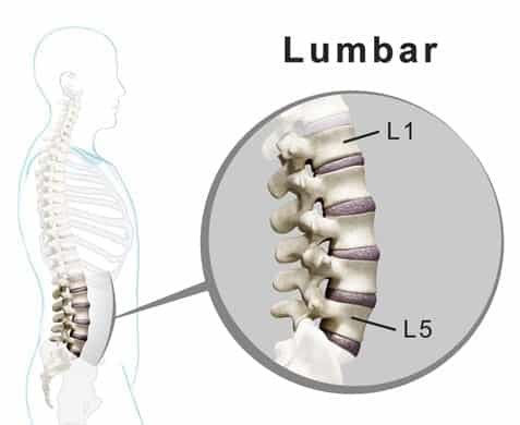
The lumbar spine consists of five vertebrae (L1 to L5) followed by the sacrum (five fused vertebrae) and coccyx (tailbone).
- Lumbar Vertebrae – Support body weight and allow flexibility.
- Sacrum – Links the spine to the pelvis via sacroiliac joints.
- Coccyx – Serves as an attachment point for ligaments and muscles.
The lumbar spine naturally curves inward (lordosis), while the sacrum has an outward curve (kyphosis).
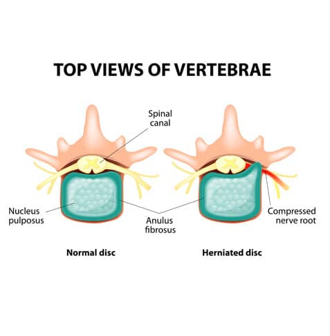
Intervertebral Discs and Their Function
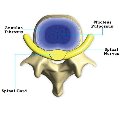
Intervertebral discs act as cushions between vertebrae (except between C1 and C2).
- Annulus Fibrosus – Tough outer layer providing durability.
- Nucleus Pulposus – Gel-like center that absorbs impact.
These discs allow spinal flexibility while helping absorb shock during movement.
Articular Facets: Zygapophyseal Joints
These joints, located at the back of the spine, allow controlled movement.
- Fibrous Capsule – Provides joint stability.
- Ligamentum Flavum – Reinforces joint flexibility.
- Synovial Membrane – Supports smooth joint motion.
These structures help maintain spinal alignment and controlled mobility.
The Spinal Canal and Its Role in Protection
The spinal canal houses and protects the spinal cord.
- Structure – Formed by vertebral bones, extending from the skull down the spine.
- Foramen Magnum – The large opening at the skull base where the spinal cord begins.
- Function – Provides a protective pathway for the spinal cord.
The Spinal Cord: A Vital Communication Pathway
The spinal cord is the primary channel for transmitting nerve signals between the brain and body.
- Length – Approximately 45 cm in males and 43 cm in females.
- Width – Thickest in the neck (~13 mm), narrowing to ~6.4 mm in the lower back.
- Function – Coordinates motor, sensory, and autonomic signals for body movement and function.
Spinal cord compression can affect mobility and nerve function, depending on severity and location.
Spinal Nerves and Their Role in Movement and Sensation
Spinal nerves extend from the spinal cord, transmitting signals between the central nervous system and the body.
- 31 Pairs of Spinal Nerves:
- Cervical Spine – 8 pairs
- Thoracic Spine – 12 pairs
- Lumbar Spine – 5 pairs
- Sacral & Coccygeal Regions – 5 pairs
- Dorsal Root – Transmits sensory signals to the brain.
- Ventral Root – Sends motor commands from the brain to muscles.
- Exit Points – Most spinal nerves pass through foramina, except C1, which exits near the skull base.
Spinal Nerves and Their Influence on Body Function
Spinal nerves play a critical role in regulating movement, sensation, and involuntary functions.
- Motor Signals – Control muscle activity.
- Sensory Signals – Transmit information such as touch, pressure, and temperature.
- Autonomic Signals – Regulate functions like digestion and heart rate.
A well-supported spine is essential for maintaining mobility, flexibility, and overall well-being. Understanding spinal structure and function helps individuals make informed decisions about spinal health.
Nerve Impingement and Spinal Health
Nerve root irritation, often linked to spinal conditions, can contribute to spinal discomfort and mobility challenges.
Common Contributing Factors:
- Slipped discs
- Structural changes such as bone spurs
- Spinal canal narrowing (stenosis)
- Vertebral misalignment (spondylolisthesis)
Effects of Nerve Compression:
Pressure on spinal nerves may lead to changes in function or mobility in areas controlled by the affected nerve. Understanding the structure and role of the spinal column highlights the importance of maintaining spinal health through structured care approaches.
Spinal Health and Common Structural Concerns
The spinal column plays a critical role in supporting movement and protecting the nervous system. Like other parts of the body, it can be affected by structural changes, wear, or degenerative processes.
Severe Spinal Conditions:
Certain conditions affecting the spine may require immediate attention:
- Infections impacting spinal structures
- Growths affecting spinal function
- Post-surgical complications
- Severe spinal cord compression
Cervical Spinal Canal Narrowing (Stenosis):
- This condition requires attention due to its proximity to the brain.
- In some cases, significant narrowing can impact spinal cord function.
Common Spinal Conditions
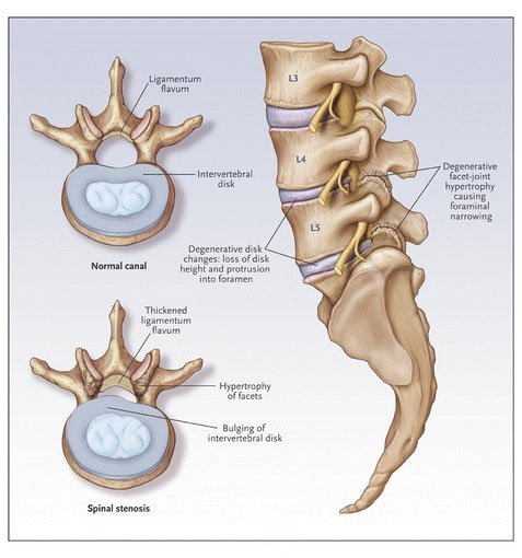
Spinal Stenosis: A Common Concern
- Definition: Spinal stenosis refers to narrowing of the spinal canal, commonly affecting the neck (cervical) or lower back (lumbar) regions.
- Possible Contributing Factors:
- Natural structural changes over time
- Slipped discs
- Disc-related wear
- Joint inflammation
- Thickened spinal ligaments
- Who It Affects:
- Most common in adults over 50, though it can occur at any stage of life.
- Management Strategies:
- Non-surgical approaches, when tailored effectively, may provide support for spinal stenosis-related concerns.
Slipped Discs and Their Impact
A slipped disc occurs when part of an intervertebral disc extends beyond its normal position, affecting nearby nerves or spinal structures.
Types of Disc Issues:
- Protruded Disc – Disc material extends slightly beyond its space.
- Bulging Disc – A broader portion of the disc extends outward.
- Prolapsed Disc – The inner disc material moves further toward the outer layer.
- Extruded Disc – Disc material extends further outward, potentially affecting nearby nerves.
Effects on Daily Life:
Changes in spinal disc structure can impact mobility, flexibility, and overall function.
Facet Joint Concerns and Their Role in Spinal Movement
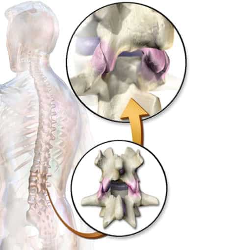
Facet joints, also known as zygapophyseal joints, connect vertebrae and allow controlled movement.
Facet Joint Function:
- Present in each spinal segment except between C1 and the skull.
- Provide weight-bearing support and stability for the spine.
Possible Contributing Factors:
- Excessive twisting or rotational movements
- Stress from repetitive forward bending or extension
Why Facet Joint Health Matters:
- These joints help regulate spinal movement, preventing excessive strain on surrounding structures.
Maintaining spinal health and addressing concerns early can support overall function and mobility. Structured, non-surgical methods using modern rehabilitation technologies may help individuals manage spinal conditions effectively.
Contributing Factors to Degenerative and Arthritic Changes in the Spine
Over time, repeated stress on the spine may contribute to gradual wear, leading to degenerative changes in spinal structures and surrounding joints.
Key Factors That May Contribute to Structural Changes:
- Daily Spinal Stress: Repetitive movements and weight-bearing activities can impact spinal function.
- Injuries: Past trauma may accelerate structural changes.
- Age-Related Changes: Natural processes may influence spinal wear.
- Lifestyle Factors: Limited movement, improper posture, or excessive weight may contribute to spinal stress.
- Underlying Conditions: Conditions affecting bone or joint health may also play a role in spinal changes.
Why Early Assessment Matters:
Structural changes in the spine can sometimes resemble other conditions, such as slipped discs. Early assessment and structured management approaches are key to maintaining spinal function and mobility.
Understanding Spinal Disc Changes and Structural Wear
Types of Spinal Disc Changes:
- Annular Fiber Changes:
- The outer disc structure (annular ligament) may weaken over time, leading to outward bulging.
- Disc Hydration Loss:
- Over time, spinal discs may lose water content, leading to reduced flexibility.
- This process, often referred to as disc degeneration, can contribute to structural changes.
How Disc Degeneration May Affect Spinal Function:
When spinal discs lose hydration, they may become more prone to structural changes, increasing susceptibility to further wear. Understanding these processes allows for proactive spinal care strategies to maintain mobility and flexibility.
Understanding the Differences Between Bulging and Protruding Discs
Spinal disc changes are commonly described using terms such as bulging discs, protruding discs, slipped discs, extruded discs, or fragmented discs. While these terms are often grouped together, there are important distinctions between them.
Bulging Disc
A bulging disc refers to a spinal disc that has expanded beyond its normal shape due to structural stress or wear. This condition does not necessarily involve contact with the spinal cord or spinal nerves. The term “bulge” describes a rounded extension of the disc, which alters its usual shape without immediate nerve compression.
Protruding Disc
A protruding disc represents a more later stage, where the disc extends further outward and may come into contact with nearby spinal structures. While some use the terms bulging and protruding discs interchangeably, the primary difference is the degree of the disc’s extension and its interaction with surrounding nerves.
Distinguishing Herniated and Prolapsed Discs
The terms herniated disc and prolapsed disc describe progressive changes in spinal disc structure but have distinct characteristics.
Herniated Disc
A herniated disc occurs when the outer layer (annular fibers) weakens or sustains small tears, allowing the inner disc material (nucleus pulposus) to shift outward. This movement can lead to increased pressure on surrounding nerve structures.
Prolapsed Disc
A prolapsed disc represents a more later stage of herniation, where the displaced disc material extends further, often shifting downward within the spinal canal. This can result in increased structural changes affecting spinal mobility.
Key Differences Between Disc Changes
- Bulging Disc vs. Protruded Disc:
- Bulging: The disc extends outward without immediate nerve contact.
- Protruded: The disc extension reaches or compresses nearby nerves.
- Herniated Disc vs. Prolapsed Disc:
- Herniated: The inner disc material moves outward due to structural weakening.
- Prolapsed: The disc material shifts further, often pressing into the spinal canal.
Can Bulging or Protruded Discs Affect Spinal Function?
Spinal discs do not have a direct blood supply but contain nerve fibers capable of detecting structural changes.
Bulged Discs
- Bulging discs may not always result in noticeable discomfort.
- As the disc expands further, it can affect nerve fibers in and around the disc, potentially leading to localized or radiating discomfort.
Protruded Discs
- Protruded discs have a greater likelihood of affecting nerve structures.
- Increased pressure from the disc may contribute to nerve-related symptoms.
How Structural Changes Affect Nerve Sensitivity
As discs expand or shift, the mechanical pressure placed on nerve fibers may increase. This process can lead to:
- Increased irritation of nerve fibers in the affected area.
- Changes in the disc’s ability to manage structural stress.
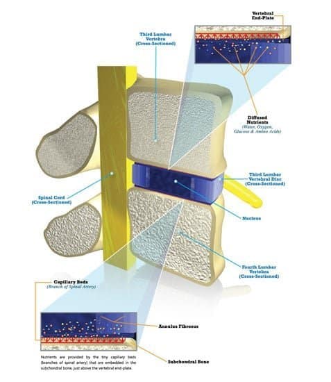
Spinal Disc Blood Supply and Nutrient Flow
Spinal discs rely on diffusion from surrounding capillaries for nutrient absorption, as they do not have a direct blood supply. Small capillaries along the outer disc region provide nutrients to surrounding ligaments and nerve structures.
Effects of Age and Structural Changes
- Spinal changes over time, including bulging, protruding, herniated, or prolapsed discs, can contribute to reduced blood flow in surrounding tissues.
- Extended pressure on small blood vessels may lead to lower oxygen levels in affected areas.
- Prolonged nutrient loss may impact the ability of the disc to maintain normal function.
The Role of the Sinuvertebral Nerve in Spinal Health
The sinuvertebral nerve is responsible for transmitting sensory signals from spinal discs, joints, and ligaments.
Function and Importance
- The sinuvertebral nerve detects structural changes and irritation in the spine.
- Long-term compression of small blood vessels supplying this nerve may affect its function.
Maintaining spinal health through structured, non-invasive strategies can help support long-term mobility and function.
Understanding Nerve Health and Spinal Function
Nerve function is essential for mobility and overall well-being. When spinal nerves experience sustained pressure or compression, changes in nerve sensitivity may occur, affecting spinal structures and movement patterns.
Potential Effects of Prolonged Nerve Compression:
- Initial Changes:
- Nerve irritation in spinal joints, surrounding ligaments, or discs may contribute to discomfort.
- Progression:
- Reduced blood flow to affected nerves may influence their function over time.
- In some cases, nerve signal transmission may be altered, leading to changes in spinal sensation.
These changes may lead some individuals to believe their spinal condition has improved, even if underlying structural concerns remain present.
Recognizing Nerve-Related Symptoms
Spinal changes affecting nerve structures may lead to symptoms extending beyond the spine. These symptoms, referred to as radicular symptoms, may be observed in the arms or legs.
Common Symptoms Associated with Nerve Compression:
- Tingling sensations
- Burning sensations
- Weakness in affected areas
- Numbness
Key Considerations:
- The presence of limb symptoms without noticeable spinal discomfort may indicate progressive nerve involvement.
- Comprehensive assessments can help identify contributing factors to ensure structured spinal care.
Nerve Function and Sensory Changes Over Time
In some cases, nerve adaptation may temporarily reduce spinal symptoms. However, as structural concerns progress:
- Increased pressure on spinal nerves may activate different sensory fibers, leading to changes in limb sensation.
- Individuals may experience altered movement, numbness, or weakness in the arms or legs.
The Importance of Early Spinal Assessment
Minor spinal concerns, if left unaddressed, may contribute to long-term changes in mobility and function. Early evaluation can help identify contributing factors before they lead to progressive spinal challenges. Addressing spinal health proactively can help maintain long-term mobility and function.
Understanding Modic Changes in the Spine

Modic changes refer to variations in the bone marrow near spinal discs. These changes are classified into three types:
- Modic Type I: Associated with inflammatory responses in spinal structures.
- Modic Type II: Characterized by fatty deposits in the bone marrow.
- Modic Type III: Linked to structural changes, including increased bone density.
A structured spinal care approach may help manage Modic changes while addressing overall spinal function.
Approaches to Spinal Health
Spinal health concerns may involve multiple factors, requiring a well-rounded approach to care. Combining chiropractic and physiotherapy-based methods can provide structured support for spinal function and movement.
Key Considerations in Spinal Health:
- Comprehensive assessments help identify co-existing factors.
- A structured care approach supports long-term mobility.
Common Causes of Spinal Strain and Injuries
Repetitive movements, improper posture, and incorrect lifting techniques can place stress on spinal structures.
Potential Contributors to Spinal Strain:
- Lifting Techniques: Using improper lifting mechanics may increase spinal strain.
- Ignoring Discomfort: Unaddressed spinal strain may lead to progressive concerns.
Preventive Strategies for Spinal Health:
- Practicing proper movement techniques during exercise or lifting activities.
- Addressing spinal discomfort early before it leads to more complex issues.
- Maintaining balanced posture and spinal support during daily activities.
A Structured Approach to Scoliosis Management
Managing scoliosis requires a structured, non-surgical approach. Effective care strategies often include:
- Comprehensive spine assessments to evaluate spinal alignment.
- Personalized movement-based strategies for postural support.
- Individualized scoliosis care plans based on specific spinal findings.
Bringing spinal assessments such as X-rays to consultations can help ensure a well-structured evaluation.
Integrative Methods for Spinal Care

Comprehensive spinal care emphasizes a structured, multi-faceted approach, including:
- Accurate spinal assessments to identify underlying factors.
- Chiropractic and physiotherapy-based methods for postural support.
- Rehabilitation strategies to enhance spinal mobility.
By combining structured methods with detailed assessments, spinal health can be supported effectively.
Collaborative Chiropractic and Physiotherapy for Spinal Care
Chiropractic care in Malaysia has expanded as a recognized approach to managing spinal concerns. When combined with physiotherapy, it offers a well-rounded, structured method for supporting spinal mobility and function.
Common areas supported through collaborative chiropractic and physiotherapy approaches include:
- Jaw-related concerns (TMJ issues).
- Slipped discs and postural conditions.
A combined approach allows individuals to receive multi-disciplinary support in a single session.
Non-Surgical Spinal Decompression Methods
Non-surgical spinal decompression focuses on relieving pressure on spinal structures without invasive procedures. modern technology such as RxDecom® supports spinal decompression strategies by providing targeted spinal traction methods. This structured approach helps alleviate pressure on affected spinal areas while maintaining a non-invasive care model.
The Role of Collaborative Spinal Care
Integrative spinal care combines the expertise of chiropractors and physiotherapists to develop structured support strategies.
This dual-care model provides:
- A comprehensive approach to spinal mobility and function.
- Evidence-supported methods tailored to individual needs.
- A focus on structured, research-based techniques.
By emphasizing a multi-disciplinary approach, individuals can benefit from holistic spinal care tailored to their specific needs.