
Modic Type II Changes & Non-Invasive Spine Care

Modic Type II changes are a common, usually settled stage of disc wear in which fat has replaced the normal marrow in the bone beside a disc, and on their own they are not a sign of danger, so a Type II finding on an MRI report is read together with how the back moves and feels. The pattern shows as fatty marrow change in the vertebral bone beside a spinal endplate. It may follow a Type I pattern, remain stable for years, or appear without a clearly documented earlier stage. Like other age- and load-related spinal findings, Modic Type II can be present in people who do not have back pain, so the wording on the scan should not be treated as a complete explanation for symptoms.

Movement tolerance, neurological findings, daily function, other disc or joint changes and the way symptoms respond to load all help determine whether the finding is relevant.

Care for Modic Type II changes is usually conservative: gentle, non-rotatory chiropractic, spinal decompression, physiotherapy and exercise, chosen after an assessment, work on how the segment moves and carries load, and the guide to spine surgery risks and non-invasive alternatives covers the surgical route.

This guide to Modic type II changes, their link to degenerative disc disease and non-invasive spine care, is published by Chiropractic Specialty Center, Bukit Damansara (KL) main center: WhatsApp+60 17 269 1873 Call+603 2093 1000

Modic Type II Changes: Key Takeaways

PointWhat to know

How it looks on MRI

Bright on T1-weighted images and the same or slightly bright on T2, because fat has replaced the normal marrow beside an endplate (Modic 1988).

How common it is

In Modic’s study of 474 people referred for a lower-back MRI, Type II was found in 16%, about four times as often as Type I (Modic 1988).

What it means

Fatty marrow beside a worn disc that, on its own, is not a sign of cancer or infection (de Roos 1987).

Over time

Type II often stays the same for years (Modic 1988), though in one three-year study 14% of discs with Modic changes showed a different type at follow-up (Kuisma 2006).

Link with pain

A Modic change is a fairly specific but insensitive sign of a painful disc: many painful discs have none (Braithwaite 1998).

Antibiotics

People with Type II changes did no better on three months of amoxicillin than on placebo in a Norwegian trial (Bråten 2019).

Care at CSC

Usually conservative: gentle, non-rotatory chiropractic, spinal decompression, physiotherapy and exercise, chosen after an assessment.

Modic Type II Changes: Page Guide & Key Sections

Understanding Modic Changes and Their Role in Spinal Health
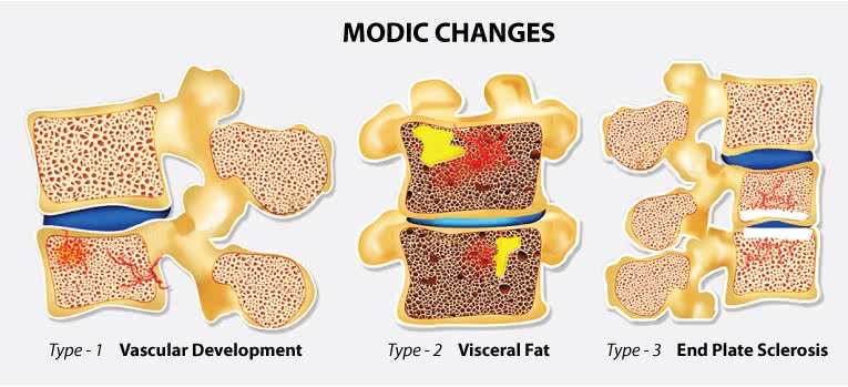
Illustration of Modic changes: type 1 with new blood vessels in the bone, type 2 with fatty marrow and type 3 with endplate sclerosis

Modic changes are changes in the vertebral endplates and the bone marrow beside them, seen on MRI. They are grouped into three types by how they look on T1- and T2-weighted images (Modic 1988):

  1. Modic Type I: dark on T1 and bright on T2, where the marrow beside the endplate holds fibrous tissue with new blood vessels, a change linked with inflammation.
  2. Modic Type II: bright on T1 and the same or slightly bright on T2, where fat has replaced the normal marrow, usually beside a worn disc.
  3. Modic Type III: dark on both, matching hardened, thickened bone (sclerosis).

Modic changes are a common MRI finding in people with low back pain or sciatica, reported in a median of 43% of such patients against 6% of people from non-clinical groups, and they become more common with age (Jensen 2008).

Modic Type II and Degenerative Disc Disease
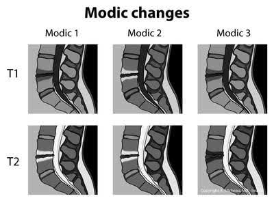
Diagram of how Modic types 1, 2 and 3 look on T1 and T2 MRI images of the lower spine

Modic Type II changes usually follow degenerative disc disease and are often linked to issues such as:

The intervertebral discs receive their nutrients by diffusion from blood vessels at their edges, and that supply can fail when the bone beneath the endplate hardens or the endplate calcifies, which can contribute to disc degeneration (Urban 2004).

Arranging an Assessment for Modic Type II Changes in KL

At a first visit, the chiropractor reviews the MRI report and images, asks how the pain behaves, and checks how the spine moves along with strength, reflexes and sensation, so the Modic finding is read alongside the other disc and joint findings before any care is agreed. Directions and hours for both centers are on the contact page, or call or message either center directly:

Targeted Non-Surgical Care for Modic Changes

CSC plans care for Modic Type II changes after an assessment, using chiropractic and physiotherapy methods matched to how the segment moves and carries load.

Understanding the Causes of Modic Endplate Changes
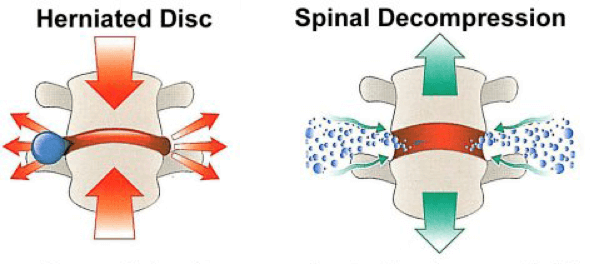
Diagram comparing a compressed, herniated disc with the same disc under spinal decompression

Reduced disc height, often caused by degenerative spinal discs, is a commonly proposed theory behind Modic endplate changes and their association with moderate to severe back pain. A reduction in disc height leads to the loss of the spinal disc’s cushioning effect, exposing vertebral endplates to excessive stress. These stresses are thought to contribute to Modic changes, slipped discs, and pain in the spine.

The redistribution of axial compression loads to adjacent vertebrae is thought to contribute to Modic endplate changes. It remains unclear whether reduced disc height causes these changes or results from them.

Managing the Load Behind Modic Endplate Changes

Modic Type I changes often turn into Type II (Modic 1988), and Type III is thought to be a later stage. Care aimed at the mechanical load on the segment, together with the factors that keep it irritated, is the reasonable approach, and the guide to back pain care by spinal segment explains what that involves.

Research links all three types closely with disc degeneration: in Modic’s original study, every change sat at a level with a worn disc (Modic 1988).

The Link Between Disc Degeneration and Modic Changes

Degenerative disc disease in the lumbar spine is a common source of pain in the back or legs. Lumbar spinal discs, responsible for weight-bearing, shock absorption, and mobility, are especially vulnerable to degeneration and Modic changes.

Modic changes in the spine’s bones or endplates are a response to degenerative disc changes or, less often, to infection or other disease. Degenerative disc changes remain the most common cause of Modic changes, as diagnosed through MRI. Type I Modic changes often involve inflammatory swelling in spinal bones and endplates, while Type II changes reflect fatty replacement of the normal red bone marrow.

NSD Therapy®: How CSC Cares for Modic Changes and Degenerative Disc Disease

At CSC, Modic changes are cared for within NSD Therapy®, a non-invasive method of care for the spine. NSD Therapy® combines targeted chiropractic care, physiotherapy, and progressive therapy devices such as the RxDecom®, so that hands-on care, decompression and graded loading form one plan rather than three. CSC collaborative care works on the mechanical load behind Modic changes and disc degeneration.

Chiropractic Specialty Center: A Non-Invasive Approach to Modic Type II Changes
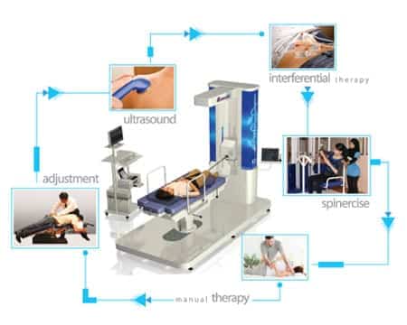
Diagram of NSD Therapy components around a decompression table: adjustment, ultrasound, interferential therapy, spinercise and manual therapy

Chiropractic Specialty Center offers non-invasive care for Modic Type II changes, which are usually found beside a worn disc. Care combines chiropractic techniques, physiotherapy and progressive spinal methods, planned after an assessment of how the segment moves and carries load.

Endplate Sclerosis and Modic Type 2: Same or Different?

Two phrases often show up together on low back reports: “Modic Type 2” and “endplate sclerosis.” They are related, but they are not the same thing.

Modic Type 2 is an MRI finding. It means the marrow in the bone next to a worn disc has turned to fat, so it looks bright on T1 pictures. Endplate sclerosis means the bone right at the endplate has become harder and thicker. It shows up as a white band on X-ray or CT, and as a dark area on MRI, which matches what is called Modic Type 3.

Both belong to the same slow wear-and-tear process, and they often sit side by side at the same level. A 2024 review that looked at 1,510 endplates found that sclerosis became more common as Modic changes moved from Type 1 to Type 2 to Type 3, and mixed types sat between Type 2 and Type 3 (Duan 2024).

In simple terms, sclerosis is the bone reinforcing itself where it carries more load. On its own, it does not mean a bone is weak or breaking. What matters more is how the whole segment moves and whether a nerve is involved, and that is what the exam at CSC checks before any plan is made.

Is Modic Type 2 Dangerous?

No. Modic Type 2 is a common finding on low back MRI in adults, and it is usually a settled stage of disc wear. In Modic’s original study it was about four times more common than Type 1 (Modic 1988). It does not spread like an illness, and it is not a sign of cancer or infection; bright marrow beside a worn disc was described early on as fatty marrow that should not be confused with tumors or infection (de Roos 1987).

Research also points to Type 2 being quieter than the active Type 1 stage. In an MRI study of 74 people, 73% of those with the Type 1 pattern had low back pain, against 11% of those with the Type 2 pattern, which was more common where the disc wear was stable (Toyone 1994). In a Norwegian trial of antibiotics for back pain with Modic changes, people with Type 2 changes did no better on the antibiotic than on placebo (Bråten 2019), which fits with Type 2 being a fatty, settled change rather than an irritated one.

Where a Type 2 change sits next to a disc that is still painful, care focuses on how that segment moves and carries load. At CSC this means gentle non-rotatory chiropractic, spinal decompression to ease pressure on the disc, focused physiotherapy for the deep trunk muscles, and daily habits that spread load more evenly.

A Type 2 change will usually still be there on future scans. That is normal, and it does not mean care has failed. Progress is judged by movement, strength and daily activities, not by the picture alone.

Lifestyle Tips for Spinal Health With Modic Type II Changes

Consider the following strategies:

  • Maintain Proper Posture: Ensuring a neutral spinal position while sitting, standing, and lifting helps reduce unnecessary stress on the vertebrae.
  • Engage in Gentle Movement: Low-impact activities like walking, swimming, or stretching can enhance flexibility and circulation.
  • Support Spinal Alignment During Sleep: Using a supportive mattress and proper pillow positioning reduces strain on the spine.
  • Mindful Daily Activities: Avoid prolonged sitting and repetitive movements that contribute to spinal strain.
  • No Yoga: with Modic changes beside a worn disc, yoga must be avoided, as must any routine built on twisting or deep bending.

Understand Spinal Changes and What They Mean

Modic changes are part of how discs respond to stress over time. CSC also holds spinal disc health talks in Kuala Lumpur from time to time; they are paused at the moment, so for the next date, send a WhatsApp message to the Bukit Damansara main center at +60 17 269 1873.


Serving Nearby Communities for Modic Type II Spine Support

The Bukit Damansara center also serves the nearby communities of Taman Tun Dr Ismail (TTDI), Bangsar & Bangsar South, Mont Kiara, Sri Hartamas, Mid Valley & KL Eco City and Solaris Mont Kiara & Publika, with the same assessment-first care for Modic Type II changes.

For current appointment information, use the two Kuala Lumpur centers below.

Chiropractic Specialty Center: Bukit Damansara

Plaza Damansara, No. 71, Jalan Medan Setia 1, Bukit Damansara, 50490 Kuala Lumpur

  • Telephone: +603 2093 1000
  • WhatsApp / SMS: +60 17 269 1873

Hours: Monday to Friday, 8:00 AM to 8:00 PM; Saturday to Sunday, 8:00 AM to 6:00 PM. Public-holiday hours may differ.

Chiropractic Specialty Center: Bandar Sri Damansara, Kuala Lumpur

8-G, Jalan Damar SD 15/1, Bandar Sri Damansara, 52200 Kuala Lumpur

  • Telephone: +603 6262 5777
  • WhatsApp: +60 12 455 6939

Hours: Monday to Friday, 8:00 AM to 8:00 PM; Saturday to Sunday, 8:00 AM to 6:00 PM. Public-holiday hours may differ.

Bandar Sri Damansara is physically located in Kuala Lumpur and may serve nearby Petaling Jaya communities. Petaling Jaya is a service area, not a separate CSC branch.

What a Modic Finding Does and Does Not Tell You

  • Modic changes are findings on a scan, not a diagnosis of your pain. Degenerative changes of the spine appear on imaging in large numbers of people with no symptoms at all, and the proportion rises steadily with age (Brinjikji W, Luetmer PH, Comstock B, et al. AJNR Am J Neuroradiol. 2015;36(4):811-816).
  • Type I is the active one. It reflects edema and inflammation at the endplate and is the type most often associated with symptoms.
  • Type II reflects fatty change and is generally the more settled state. A report moving from Type I to Type II is not automatically bad news.
  • They can change over time in both directions. A single scan is a snapshot of a moving picture: in a three-year study of 60 people with sciatica, 14% of discs with Modic changes showed a different type at follow-up, and the authors found evidence that Type II changes may be less stable than previously assumed (Kuisma 2006).

What none of this tells you is how much you can do. That is decided by testing what you tolerate, not by reading a report.

What Is Worth Working On

Since the finding sits at the junction between the disc and the vertebral endplate, the practical targets are the things that load that junction repeatedly.

  • Sustained flexion. Long periods of sitting bent forward, and lifting with a rounded back under load.
  • Repeated compression without recovery. Heavy work with no variation in position across the day.
  • Trunk endurance rather than trunk crunches. Endurance of the muscles that control the segment is the useful quality here.
  • Sleep and general health. Both change how inflamed tissue behaves, and both are modifiable.
  • Time. Endplate changes are slow. A four-week program is not a fair test of anything on this page.

When a Back With Modic Changes Needs a Doctor

  • Fever with back pain, or back pain after a recent infection, injection or procedure. Endplate changes and infection can look alike on imaging, and that distinction is a medical one.
  • Unexplained weight loss, night pain that wakes you, or a history of cancer.
  • Loss of bladder and bowel control. Emergency, the same day.
  • Weakness that is getting worse, or a foot that drags; this should be checked promptly by a competent provider, and conservative care may still be a reasonable route for as long as it produces results.

Readers who want to know costs first can check the chiropractic price in Kuala Lumpur and physiotherapy price list in Kuala Lumpur pages.

Modic Type II changes are also read from an MRI report. Parents of a child or teenager with back pain can read the guide to chiropractic care for children, which explains what a first visit for a young person checks.

Modic Type II Changes: Frequently Asked Questions

The questions below are common searches about Modic Type II changes, answered with reference to the studies listed on this page.

Is Modic type 2 dangerous?

No. Modic Type II is usually a settled stage of disc wear in which fat has replaced the normal marrow in the bone beside a disc, and it is a common finding on low back MRI scans in adults. In Modic’s original study of 474 people referred for a lower-back MRI, it was seen in 16%, about four times as often as Type I. It does not spread like an illness, and bright marrow beside a worn disc was described early on as fatty marrow that should not be confused with tumors or infection. What matters more is how the segment moves and whether a nerve is involved, which an examination checks.

What does a Modic type 2 endplate change mean?

A Modic Type II endplate change means the MRI shows the bone marrow beside a spinal endplate as bright on T1-weighted images and the same or slightly bright on T2-weighted images, because fat has replaced the normal marrow there. It is usually found beside a worn disc, and it may follow a Type I change or appear without a known earlier stage. Modic changes can also be present in people without back pain, so the finding is read together with how the back moves, feels and responds to load.

What are the symptoms of Modic type 2 changes?

A Modic Type II change has no symptom that is unique to it, and Modic changes can also be found in people without back pain. When symptoms are present, they usually come from the worn disc or the joints at that level: low back pain, stiffness, and sometimes pain spreading into the buttock or leg. In a discography study of 58 people, 21 of 23 discs beside a Modic change reproduced pain, but pain also came from many discs without one, so a Modic change is a fairly specific but insensitive sign of a painful disc.

What causes Modic type 2 endplate changes?

Modic Type II changes are closely linked with disc degeneration: in Modic’s original study, every change sat at a level with a worn disc. Loss of disc height is a commonly proposed reason, because a thinner disc cushions less and passes more load to the endplates, although it remains unclear whether the lower disc height causes the changes or follows them. A Type II change can also develop from an earlier Type I change, which in Modic’s study often turned into the Type II pattern within 14 months to three years.

What is the treatment for Modic type 2 endplate changes?

Care for Modic Type II endplate changes is usually conservative and is chosen after an assessment of how the segment moves, how the disc and joints respond to load and whether a nerve is involved. At Chiropractic Specialty Center it is given within NSD Therapy®, which combines gentle, non-rotatory chiropractic, spinal decompression, physiotherapy for the deep trunk muscles and exercise that builds up gradually. The evidence does not support antibiotics for Type II changes: in a Norwegian trial, people with Type II changes did no better on three months of amoxicillin than on placebo.

What is endplate sclerosis, and is it serious?

Endplate sclerosis means the bone right at the endplate has become harder and thicker. It shows as a white band on X-ray or CT and as a dark area on MRI, matching the Modic Type III pattern. It belongs to the same slow wear-and-tear process as Modic Type II, and a 2024 review of 1,510 endplates found that sclerosis became more common from Type I to Type II to Type III. On its own it does not mean a bone is weak or breaking; how the segment moves and whether a nerve is involved matter more.

Peer-Reviewed Medical References

  1. Modic MT, Masaryk TJ, Ross JS, Carter JR. Imaging of degenerative disk disease. Radiology. 1988;168(1):177-186.
  2. de Roos A, Kressel H, Spritzer C, Dalinka M. MR imaging of marrow changes adjacent to end plates in degenerative lumbar disk disease. AJR Am J Roentgenol. 1987;149(3):531-534.
  3. Braithwaite I, White J, Saifuddin A, et al. Vertebral end-plate (Modic) changes on lumbar spine MRI: correlation with pain reproduction at lumbar discography. Eur Spine J. 1998;7(5):363-368.
  4. Toyone T, Takahashi K, Kitahara H, et al. Vertebral bone-marrow changes in degenerative lumbar disc disease. An MRI study of 74 patients with low back pain. J Bone Joint Surg Br. 1994;76(5):757-764.
  5. Brinjikji W, Luetmer PH, Comstock B, et al. Systematic literature review of imaging features of spinal degeneration in asymptomatic populations. AJNR Am J Neuroradiol. 2015;36(4):811-816.
  6. Duan Y, Feng D, Zou K, et al. The relationship between Modic changes and endplate sclerosis in patients with lumbar degenerative disease: a systematic review and network meta-analysis. World Neurosurg. 2024;186:7-16.
  7. Modic MT, Steinberg PM, Ross JS, Masaryk TJ, Carter JR. Degenerative disk disease: assessment of changes in vertebral body marrow with MR imaging. Radiology. 1988;166(1 Pt 1):193-199.
  8. Bråten LCH, Rolfsen MP, Espeland A, et al. Efficacy of antibiotic treatment in patients with chronic low back pain and Modic changes (the AIM study): double blind, randomised, placebo controlled, multicentre trial. BMJ. 2019;367:l5654.
  9. Jensen TS, Karppinen J, Sorensen JS, Niinimäki J, Leboeuf-Yde C. Vertebral endplate signal changes (Modic change): a systematic literature review of prevalence and association with non-specific low back pain. Eur Spine J. 2008;17(11):1407-1422.
  10. Kuisma M, Karppinen J, Niinimäki J, et al. A three-year follow-up of lumbar spine endplate (Modic) changes. Spine (Phila Pa 1976). 2006;31(15):1714-1718.
  11. Urban JP, Smith S, Fairbank JC. Nutrition of the intervertebral disc. Spine (Phila Pa 1976). 2004;29(23):2700-2709.

These references describe Modic vertebral endplate (bone-marrow) changes in degenerative disc disease on MRI in general. None of them evaluates a care program at Chiropractic Specialty Center.

The Author of Modic Type II Changes & Non-Invasive Spine Care

Modic Type II Changes & Non-Invasive Spine Care was written by Yama Zafer, D.C., who has 30+ years in chiropractic and physiotherapy and founded Chiropractic Specialty Center in Kuala Lumpur in 2006.

Last Updated: Modic Type II Changes & Non-Invasive Spine Care

Modic Type II Changes & Non-Invasive Spine Care was last reviewed and updated on October 3, 2026.

Share with others: