


Facet Hypertrophy and Non-Invasive Care
Facet hypertrophy means enlargement and degenerative change of the facet joints at the back of the spine. It is common with aging and can occur alongside disc-height loss, osteoarthritis and ligament thickening. The finding may contribute to local stiffness, narrowing around a nerve root or spinal stenosis, but it can also appear on imaging without causing symptoms.
The clinical picture matters more than the word on the scan. Extension, rotation, standing or walking may aggravate some facet-related presentations, while other conditions can behave in a similar way. An assessment therefore considers movement, neurological findings, hip and sacroiliac function, and how the symptoms respond to different positions. Assessment and context also include what facet hypertrophy means, how imaging is interpreted, which symptom patterns may make it relevant, and how conservative care or referral may be considered.
This article explores the causes, symptoms, and biomechanics of facet hypertrophy, while outlining conservative care methods that avoid injections or surgical procedures. Understanding the joint structures involved helps guide proper care decisions aimed at long-term spinal function and stability.
This guide to facet hypertrophy, and how facet joint changes can affect nearby nerves, is published by Chiropractic Specialty Center, Bukit Damansara (KL) main center: WhatsApp+60 17 269 1873 Call+603 2093 1000
Key Takeaways: Facet Hypertrophy Explained Simply
- Facet hypertrophy refers to thickening of small joints at the back of the spine that guide movement
- It often develops alongside disc changes and ligament thickening, not as a single condition
- Reduced disc height may increase stress on these joints over time
- Nearby nerves may be affected when space around them becomes limited
- Signs can include stiffness, reduced movement, or sensations that travel into the arms or legs
- Assessment usually looks at joints, discs, ligaments, and nerve involvement together
- Non-invasive care often focuses on improving joint function, stability, and daily movement patterns
At Chiropractic Specialty Center in Kuala Lumpur, the assessment looks at the severity and likely source of facet-related symptoms, and care combines chiropractic, physiotherapy and rehabilitation using non-invasive methods.
How Facet Joint Changes Can Affect Nearby Nerves
Facet hypertrophy can reduce the space around spinal nerves, especially in the neck and lower back. When facet joints enlarge, they may narrow the openings where nerves exit the spine, leading to nerve impingement. This change in space may affect how nerves respond during daily activities such as sitting, standing, or bending.
What may happen when nerves are affected:
- Tingling or altered sensation: May be felt in the arms or legs depending on the level involved
- Numbness in specific areas: Often follows a pattern linked to the affected nerve
- Reduced strength in certain movements: Can affect grip, lifting, or walking in some cases
- Symptoms that travel away from the spine: For example, from the lower back into the buttock or leg
Facet joint enlargement often occurs together with disc changes. When both are present, the available space for nerves may reduce further, especially during certain positions or movements; causing nerve pain.
What Commonly Leads to Facet Joint Enlargement
When spinal discs lose height or change structure, more load can shift toward the facet joints. Over time, this added stress may lead to thickening of the joint and the formation of bony edges.
Common contributing factors include:
- Changes in spinal discs: Disc height loss or structural changes may increase stress on nearby joints
- Prolonged sitting and posture habits: Long hours of sitting, common in KL work routines, can increase pressure on certain spinal segments
Repetitive loading or physical strain: Daily activities that place repeated stress on the spine may affect joint surfaces - Reduced joint movement over time: Limited motion may affect how joints receive and distribute load
- Natural age-related changes: Joint surfaces and cartilage may change gradually over time
Facet joint changes are often seen together with disc changes and ligament thickening. When these occur together, the way the spine handles movement and load may shift further.
Facet Hypertrophy Assessment at Two KL Centers
Facet joint changes are assessed at both Kuala Lumpur centers of Chiropractic Specialty Center, where movement, posture and any imaging are reviewed together. Readers in TTDI, Bangsar and Bangsar South, Mont Kiara, Sri Hartamas, Mid Valley and KL Eco City and Solaris Mont Kiara and Publika are within reach of one of the two, and opening hours for each are listed on the contact page.
- Bukit Damansara (KL, main center): Call +603 2093 1000 or WhatsApp +60 17 269 1873 to see the Bukit Damansara / KL center.
- Bandar Sri Damansara (KL): Call +603 6262 5777 or WhatsApp +60 12 455 6939 to see the Bandar Sri Damansara center.
How Facet Joints, Discs, and Ligaments Work Together
Facet hypertrophy rarely develops on its own. In most cases, it is linked to degenerative changes in the spinal discs and nearby ligaments (Ligamentum Flavum) that help stabilize each segment of the spine.
Spinal discs act as cushions between the bones of the spine. When discs lose height or change structure, the space between the bones may reduce. As this space changes, more load can shift toward the joints at the back of the spine.
Over time, this shift in load may lead to:
- Increased contact between joint surfaces
- Changes in how joints move and share stress
- Thickening or enlargement of the joint
At the same time, nearby ligaments may also respond to these changes.
One important structure is the ligamentum flavum, a connective tissue that runs along the back of the spinal canal. This ligament works closely with the joints and helps maintain stability.
When disc height reduces and joint loading increases, the ligamentum flavum may:
- Thicken over time
- Fold or “buckle” inward with reduced space
- Contribute to reduced space around nearby nerves
Because these structures are connected and function together, it is common to see facet joint changes, disc changes, and ligament thickening occurring at the same time.
Why a Multi-Structure Assessment Matters
Facet joint changes can involve more than one structure at the same spinal level. Discs, joints, and ligaments often influence each other, especially when changes develop over time.
Because of this, focusing on a single structure may not fully explain what is happening in the spine.
A structured assessment is often used to:
- identify which structures are involved
- understand how load is shared across the spine
- determine how different positions affect symptoms
- guide a plan based on how the spine is functioning as a whole
This type of approach helps explain why symptoms may persist, change, or return if only one part of the spine is considered
Video on How Spinal Disc Changes & Nerve Symptoms Relate to Facet Hypertrophy
Facet hypertrophy commonly develops alongside spinal disc degeneration, disc bulges, protrusions, or spinal canal narrowing. This in-depth educational video explains how spinal discs affect nearby nerves, why symptoms may travel into the buttock or leg, and how disc changes may contribute to stiffness, nerve irritation, and joint overload often seen together with facet enlargement.
The chapters of this talk, each with a short summary, are on the Back Pain to Leg Pain video page.
Key Moments From the Video
- 06:22 Spinal Disc Anatomy Explained: how disc structure works and how degeneration may increase stress on nearby facet joints.
- 18:30 Healthy Disc vs Degenerated Disc on MRI: how disc changes may coexist with facet hypertrophy.
- 28:49 Stages of Disc Change: bulge, protrusion, herniation, and advanced disc changes.
- 38:00 Sitting and Intervertebral Disc Pressure: why prolonged sitting raises disc pressure, which often adds to facet joint loading.
- 45:12 Standing vs Sitting Disc Pressure: the biomechanics behind posture-related facet stress.
- 1:12:46 Sciatica and Nerve Compression: nerve symptoms that may overlap with facet enlargement.
- 1:33:12 Conservative Care vs Surgical Considerations: how conservative care is weighed before surgery.
Spinal Motion Segments & Facets
Here are some basic facts on facets & spinal motion segments:
- The first spinal joint formed by the union of facets is the C2-C3 motion segments.
- The most mobile spinal motion segment in the neck is the C5-C6
- The L4-L5 is the most mobile motion segment of the lower back.
- Facet joints, also known as zygapophyseal joints, are synovial joints.
- Facet joint surfaces are covered with hyaline cartilage, and each joint is lined with a synovial membrane.
There are two facet joints in one motion segment. A motion segment is where two contiguous (adjacent or touching) spinal bones are connected by facets that allow motion, mobility, and weight-bearing. The neck and lower back facets are the common areas diagnosed with facet hypertrophy. The two most common regions for facet hypertrophy are listed below:
- Cervical Facet Hypertrophy: Cervical facet hypertrophy often reflects wear on the neck joints linked to injury or long hours of computer and phone use, often diagnosed at C4-C5, C5-C6, or C6-C7 motion segments
- Lumbar Facet Hypertrophy: Facet hypertrophy of the lumbar spine is often the result of injuries or prolonged sitting. Lumbar facet hypertrophy can be seen in any five lumbar segments but is more common in L3-L4, L4-L5, and L5-S1
Hypertrophy Facet Joints, Facet Arthritis, Osteophytosis Or Spinal Bone Spur
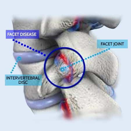
The human spine has twenty-four movable bones, known as vertebrae. These vertebrae stack on top of one another to create the spinal column. The spinal column supports the body, allows movement, and protects the spinal cord and spinal nerves.
As mentioned, the coming together of facets connects or joins vertebrae to establish spinal motion segments for mobility. The coming together of a facet from the vertebra above and connecting to the vertebra's facet below forms the facet joint. Facet joints are the smallest in the neck and most abundant in the lower back.
A properly aligned spinal joint allows for effortless motion in the neck, upper back, mid-back and lower back. Movement and mobility afforded to a facet joint are due to the articular cartilage and synovial membrane lining.
Daily stresses, traumatic events, prolonged sitting, and poor postures can result in wear and tear of these joints and their associated structures, including the facet joint’s soft tissues. The wearing out or degenerative changes of a facet joint are referred to as facet hypertrophy. In facet hypertrophy, the facets expand with arthritic bones or spurs to counter the wear and tear resulting from the degenerative changes.
Common Signs When Facet Joints Are Involved
Facet hypertrophy may affect how the spine moves and how nearby nerves respond. The signs can vary depending on whether the neck or lower back is involved.
Common signs include:
- Stiffness in the neck or lower back: Often more noticeable after rest or prolonged sitting
- Reduced ability to bend or turn: Movement may feel limited, especially with extension or rotation
- Local pain near the spine: Usually felt on one side or both sides of the affected segment
- Symptoms that change with position: Sitting, standing, or bending may increase or reduce symptoms
- Nerve-related sensations (in some cases): Tingling, numbness, or weakness in the arms or legs when nearby nerves are affected
Facet joint changes can exist without strong symptoms, especially in early stages. When discs, ligaments, or surrounding tissues are also involved, symptoms may become more noticeable.
How Facet Joint Changes Are Assessed
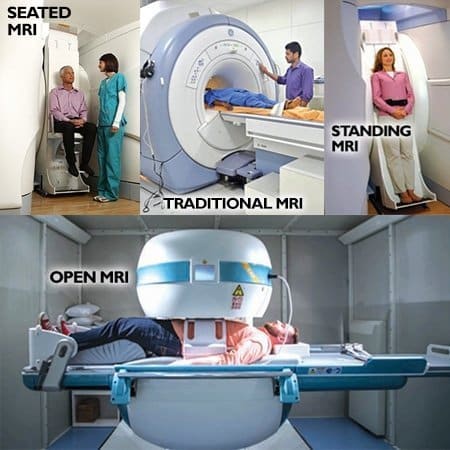
Facet hypertrophy is usually identified through a combination of history, physical assessment, and imaging when needed.
Assessment often includes:
- Review of symptoms and daily patterns: Looking at when stiffness or pain appears and how it changes with activity or rest
- Physical examination: Checking spinal joint movement, posture, and how different positions affect the spine
- Basic neurological checks: Used when there are signs of nerve involvement, such as tingling or weakness
When more detail is needed, imaging may be considered:
- MRI (Magnetic Resonance Imaging): Helps show discs, joints, ligaments, and nearby soft tissues
- CT scan (Computed Tomography): Provides a clearer view of bony changes such as joint enlargement or spurs
- X-rays: May be used to review overall alignment and joint spacing
Facet joint changes are often seen together with disc changes or ligament thickening. Imaging is usually considered when symptoms persist, change, or involve nearby nerves.
Radiofrequency Ablation (RFA) for Facet Joints: What to Know
Radiofrequency ablation (RFA) is a procedure sometimes used when facet joints are believed to be the source of symptoms. It involves applying controlled heat to a small nerve branch that carries signals from the joint.
The goal is to interrupt how signals are transmitted from the joint to the brain.
How the procedure works:
- A needle is guided near the targeted nerve
- Heat is applied to create a controlled lesion
- The nerve’s ability to transmit signals is reduced
What Research and Clinical Observations Show
Findings from clinical studies are mixed. Some research suggests limited short-term changes, while other studies show little difference when compared to non-invasive approaches.
- The MINT randomized trials found that adding radiofrequency denervation to an exercise program did not produce a clinically important improvement in chronic low back pain
- Sham-controlled trials have also shown similar outcomes between RFA and placebo procedures in some cases
What May Occur After the Procedure
Some individuals report temporary changes after RFA, including:
- Increased sensitivity in the first few weeks
- Muscle tightness around the treated area
- Altered nerve sensations during the healing phase
This is often linked to how the nerve responds after being exposed to heat and may settle over time
Points Often Discussed in Current Literature
Research has also described potential effects that may be considered when evaluating options:
- Temporary irritation of the targeted nerve
- Changes in nearby stabilizing muscles due to nerve involvement
- Variable duration of results across individuals
- The need for repeat procedures in some cases
Facet joint-related concerns often involve multiple structures, including discs, ligaments, and muscles. Because of this, non-invasive approaches are commonly explored before considering procedures that target a single nerve pathway.
When Is Spine Surgery Considered for Facet Joint Changes?
Spine surgery may be considered in certain situations when facet joint changes are part of a broader spinal condition. This is usually discussed when symptoms are persistent, progressive, or when multiple structures such as discs, joints, and ligaments are involved.
Some procedures that may be discussed include:
- Facet spacers or interlaminar implants: Small devices placed between spinal structures to create space and reduce joint loading
- Decompression procedures: Techniques aimed at increasing space around nerves when narrowing is present
- Spinal fusion: A procedure that connects two or more spinal segments to limit motion at a specific level
Each approach is designed to address specific structural findings, and the choice depends on how different parts of the spine are contributing to the overall condition.
Facet joint changes are often seen alongside disc changes and ligament thickening. Because of this, understanding how these structures interact is an important part of deciding whether any procedure is appropriate.
In many cases, non-invasive approaches are explored first, especially when symptoms are influenced by movement, posture, or load across the spine.
How Care Is Structured for Facet Joint Changes in KL

Facet hypertrophy in Kuala Lumpur is often linked to how the spine, discs, and surrounding tissues handle daily load. Because of that, care usually focuses on more than one area at the same time.
A structured approach may include:
- Chiropractic joint mobilization or adjustment
Gentle, focused methods directed at how spinal joints move and share load. - Physiotherapy and soft tissue work
Hands-on methods may be used to address muscle tightness, imbalance, and tissue strain around the spine. - Targeted rehabilitation exercises
Simple, guided exercises aimed at stability, coordination, and control of the spine. - Postural and daily habit guidance
Sitting, standing, and lifting patterns are often reviewed, especially for those with long sitting hours in KL.
Facet joint changes rarely occur alone. They are commonly seen together with disc changes, ligament thickening, or muscle imbalance. Because of that, care is usually planned to address how these structures work together rather than focusing on one area only.
What a Combined Spine Care Plan May Include
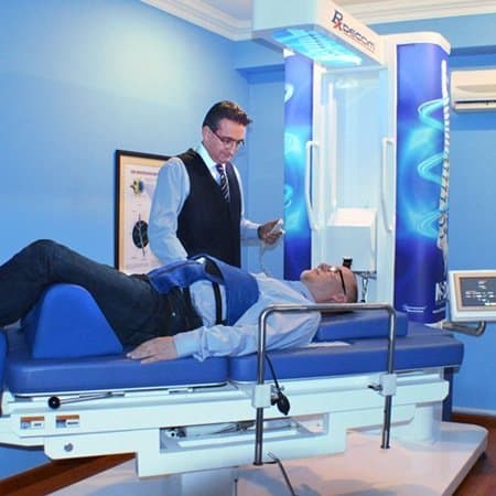
Facet hypertrophy is often linked to changes in discs, joints, muscles, and nearby tissues. Because of that, care is usually planned as a combination of methods rather than a single approach.
A structured plan may include:
- Chiropractic joint mobilization or adjustment: Gentle, focused techniques directed at how spinal joints move and share load.
- Spinal decompression (when appropriate): Used in some cases with the aim of reducing pressure within spinal segments.
- Hands-on physiotherapy: May include muscle work, trigger point techniques, and fascia-focused methods.
- Therapeutic modalities: Options such as high-intensity laser, shockwave, ultrasound, or electrotherapy may be used based on individual needs.
- Exercise-based rehabilitation: Guided programs using simple movements or equipment, aimed at strength, control, and stability.
Facet joint enlargement is rarely isolated. It is commonly seen together with disc changes, ligament thickening, or postural strain. Because of that, care plans are often adjusted over time based on how the spine responds.
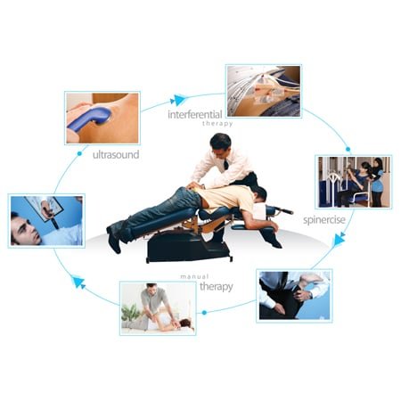
Physiotherapy Gym Programs in Bukit Damansara for Core and Facet Joint Support
At our Bukit Damansara center, CSC’s physiotherapy gym runs structured exercise programs for the muscles that support the facet joints. Sessions are planned around the load the facet joints tolerate, with attention to postural control.
Our physiotherapy-led approach includes low-impact strengthening, core activation, and mobility routines customized to the spine’s needs. Whether you're dealing with stiffness, nerve sensitivity, or postural overload, the sessions are progressed gradually so the joints are loaded within what they tolerate.
Facet Hypertrophy: What Matters Most
Facet hypertrophy involves changes in the small joints that guide how the spine moves. These changes rarely occur alone and are often linked to disc changes and ligament thickening at the same spinal level.
Because multiple structures may be involved, symptoms can vary and may shift depending on posture, activity, or daily habits. Understanding how joints, discs, and ligaments work together helps explain why symptoms are not always consistent.
Assessment is typically based on how the spine functions as a whole rather than focusing on a single structure. This allows for a clearer understanding of what may be contributing and how different parts of the spine respond during movement.
Planning a visit? You can review the current chiropractic price in Kuala Lumpur and physiotherapy fees in Kuala Lumpur before you come in.
Parents of a child or teenager with back stiffness or pain can read the guide to chiropractic care for children, which explains how assessment and care are matched to a child's age.
Facet Hypertrophy: Common Questions Answered
These are some of the most common questions people ask when trying to understand facet hypertrophy, how it develops, and what it may involve. The answers below are kept simple and focused to help you make sense of how the spine works and what to look out for.
What is facet hypertrophy in simple terms?
Facet hypertrophy refers to thickening or enlargement of the small joints at the back of the spine. These joints help guide movement, and changes in them may affect how the spine handles load and motion.
What causes facet joints to enlarge?
Facet joints may enlarge over time when discs lose height, posture habits change how load is distributed, or repeated stress affects the joints.
Can facet hypertrophy affect nerves?
Yes. When facet joints enlarge, they may reduce space around nearby nerves, which can lead to sensations such as tingling, numbness, or reduced strength in some cases.
Is facet hypertrophy the same as a slipped disc?
No. They are different, but often occur together. Disc changes may increase stress on facet joints, which can contribute to joint thickening over time.
What is the ligamentum flavum and how is it related?
The ligamentum flavum is a ligament that supports the spine from the back. It works closely with facet joints and may thicken or buckle when disc height reduces and joint loading changes.
Can facet hypertrophy exist without symptoms?
Yes. Some individuals may have joint changes without noticeable symptoms, especially in early stages.
How is facet hypertrophy assessed
Assessment usually includes reviewing symptoms, physical examination, and imaging such as MRI or CT when needed.
Does posture affect facet joints?
Yes. Long periods of sitting, standing habits, and repeated positions can influence how load is placed on the spine and its joints.
Why do symptoms change with position?
Different positions place different levels of stress on discs, joints, and nerves. This can change how symptoms are felt throughout the day.
Can facet hypertrophy occur in both the neck and lower back?
Yes. It is most commonly seen in the neck (cervical spine) and lower back (lumbar spine), where movement and load are highest.
What structures are usually involved together?
Facet joints, spinal discs, and ligaments often change together, especially when the spine has been under long-term stress.
When should someone consider getting it checked?
If stiffness, reduced movement, or nerve-related sensations continue or change over time, an assessment may help clarify what is involved.
Peer-Reviewed Medical References
- Kalichman L, Hunter DJ. Lumbar facet joint osteoarthritis: a review. Semin Arthritis Rheum. 2007;37(2):69-80.
- Manchikanti L, Boswell MV, Singh V, et al. Prevalence of facet joint pain in chronic spinal pain of cervical, thoracic, and lumbar regions. BMC Musculoskelet Disord. 2004;5:15.
- Eubanks JD, Lee MJ, Cassinelli E, et al. Prevalence of lumbar facet arthrosis and its relationship to age, sex, and race: an anatomic study of cadaveric specimens. Spine (Phila Pa 1976). 2007;32(19):2058-2062.
- Schwarzer AC, Aprill CN, Derby R, et al. Clinical features of patients with pain stemming from the lumbar zygapophysial joints. Is the lumbar facet syndrome a clinical entity? Spine (Phila Pa 1976). 1994;19(10):1132-1137.
- Revel M, Poiraudeau S, Auleley GR, et al. Capacity of the clinical picture to characterize low back pain relieved by facet joint anesthesia. Proposed criteria to identify patients with painful facet joints. Spine (Phila Pa 1976). 1998;23(18):1972-1976.
- Cohen SP, Raja SN. Pathogenesis, diagnosis, and treatment of lumbar zygapophysial (facet) joint pain. Anesthesiology. 2007;106(3):591-614.
- Juch JNS, Maas ET, Ostelo RWJG, et al. Effect of radiofrequency denervation on pain intensity among patients with chronic low back pain: the Mint randomized clinical trials. JAMA. 2017;318(1):68-81.
These references describe facet joint changes, facet-related pain and its assessment, and radiofrequency denervation in general. None of them evaluates a care program at Chiropractic Specialty Center. Each one can be checked through its free PubMed record.
The Author of Facet Hypertrophy: Causes, Symptoms and Assessment
Facet Hypertrophy: Causes, Symptoms and Assessment was written by Yama Zafer, D.C., who has 30+ years in chiropractic and physiotherapy and founded Chiropractic Specialty Center in Kuala Lumpur in 2006.
Last Updated: Facet Hypertrophy and Non-Invasive Care
Facet Hypertrophy and Non-Invasive Care was last reviewed and updated on September 25, 2026.
I have spinal stenosis on L4 L5. My feet are numb, and I have calf pain when walking.
I have done the injection at SJMC (Subang Jaya Medical Centre) but refuse to get the recommended surgery.
Declining the surgery is a legitimate position with stenosis, provided the reasons for reconsidering it are clear, and there is one thing to rule out first that changes the whole picture.
Calf pain on walking with numb feet has two quite different explanations, and they are treated by different specialists. Narrowing in the spine produces symptoms that build as you walk, are relieved specifically by sitting or bending forward, and allow you to walk considerably further pushing a trolley or going uphill than on level ground. Reduced blood supply to the legs produces calf pain that comes on at a fairly consistent distance every time, is relieved simply by standing still, and is not affected by whether you lean forward. If your pattern fits the second description, this needs a vascular assessment, and pulses in the feet plus a simple pressure comparison between arm and ankle will answer it quickly. Numb feet should also prompt a check for diabetes if that has not been done recently.
Assuming it is the stenosis, here is the honest position on your choice. The natural course of lumbar stenosis in people who do not have surgery is more favorable than commonly assumed: a substantial proportion remain stable or improve over years, and serious deterioration is not the usual outcome. Surgery generally gives faster relief of leg symptoms, with the difference between operated and non-operated groups narrowing over the longer term. So declining it is reasonable, and it is a decision that can be revisited rather than a door that closes.
What conservative management should consist of, since it needs to be more than avoidance. Use flexion deliberately rather than accidentally: a recumbent bike, walking with a trolley or wheeled walker, and short seated rests taken before symptoms force them all let you accumulate activity that upright walking will not. Build volume in small increments and track walking distance weekly, because that number is your real measure and it is more useful than a pain score. Restore hip extension, since tight hip flexors force the lower back into the extended position that closes the canal with every step. Strengthen the trunk and gluteal muscles. And address sustained standing and leaning back, which are the two everyday positions that provoke this most.
The injection at SJMC will have given temporary relief at best; that is what they do, and a good response to it is also useful information, since it confirms the level being blamed is the one causing your symptoms.
The thresholds at which the surgical question should be reopened are worth writing down: walking distance falling steadily despite a genuine conservative program, or weakness developing in a leg or a foot beginning to drag. New numbness around the genitals or anus, new difficulty passing urine, or loss of bladder or bowel control needs urgent medical attention the same day. The last one needs the same day rather than an appointment.