Slipped Disc Management Using Safe Methods in Malaysia

Slipped disc care in KL often begins with understanding how spinal discs affect the nerves, movement, and daily function. A slipped disc—also described as a disc bulge, protrusion, herniation, or extrusion—can contribute to neck issues, back issues, and symptoms traveling into the arm or leg, commonly referred to as sciatica. These changes develop gradually as spinal discs lose resilience from posture strain, repetitive loading, or natural aging.

At Chiropractic Specialty Center®, slip disc care in KL focuses on structured, non-invasive methods that combine physiotherapy, gentle and focused chiropractic methods, and technology-assisted spinal decompression. Our integrative approach is designed to assist individuals experiencing disc-related neck pain, lower back problems, or sciatica.

This article also presents real case findings demonstrating how coordinated physiotherapy, chiropractic care, and spinal decompression methods are used to address disc conditions without injections or surgery.

Table of Contents

Bulging Disc vs Herniated Disc: Understanding the Difference

Most hear terms such as slipped disc, disc bulge, herniated disc, or extruded discs but are unsure what they actually mean. The short video below explains how spinal discs function and how structural changes inside the disc can lead to bulging or herniation. Using a simple spine model, the explanation shows how the annulus fibers weaken, how the nucleus shifts, and why disc changes can contribute to neck pain, lower back pain, or sciatica.

Key Takeaways: Understanding Slipped Disc Management

      • Slipped discs develop gradually. Disc bulges, protrusions, herniations, and extrusions occur when spinal discs lose resilience due to repetitive strain, posture habits, aging, or reduced disc nutrition.
      • Symptoms may affect the neck, back, arms, or legs. Changes within spinal discs can influence nearby nerves and joints, sometimes contributing to neck stiffness, back discomfort, or symptoms traveling into the arm or leg (sciatica).
      • Non-surgical spine care is often explored first. Conservative approaches commonly combine physiotherapy rehabilitation, gentle chiropractic methods, spinal decompression systems, and guided strengthening exercises.
      • Clinical evaluation helps determine the best approach. Assessment may include physical examination, neurological testing, posture analysis, and imaging studies such as MRI when necessary.
      • Disc health depends on spinal movement and pressure balance. Daily posture habits, sitting time, lifting patterns, and spinal mechanics all influence how discs absorb load and receive nutrients.

When to Seek Professional Evaluation

Individuals experiencing persistent neck stiffness, back discomfort, leg symptoms, or changes in movement may benefit from a structured spinal evaluation. A careful assessment can help determine whether symptoms relate to spinal discs, joints, muscles, or nerve irritation.

At Chiropractic Specialty Center® in Kuala Lumpur, clinical evaluation may include movement assessment, neurological testing, and imaging review when available. Understanding how spinal structures interact helps guide appropriate non-invasive care strategies for disc-related conditions.


Learn Everyday Steps That Protect Your Spine

Short-term improvements are possible, but lasting change begins with understanding. Join our Free Spinal Disc Health Talk in Kuala Lumpur to see how posture awareness and small movement changes support disc recovery and overall comfort.

Clinical Observations Using Spinal Decompression Systems

A case study of two patients receiving non-surgical spinal decompression: With pre and post-care MRIs of the lumbar spine.

This case study was performed to evaluate the effects of spinal decompression system, on two patients one with a moderately extruded lumbar disc and the other with a severely extruded lumbar disc provided in a multidisciplinary center on an outpatient basis. The results have shown that it helped improve symptomatology and retracted severe spinal disc conditions.

The cases presented here demonstrate that protruded and extruded spinal discs may improve using the protocols without the need for invasive procedures. These results highlight a promising future for non-surgical advancements in physiotherapeutic care as utilized in the NSD protocols. Larger clinical studies are needed to show the effectiveness of this method on patients with varying characteristics.

Understanding Spinal Disc Conditions: Degeneration, Bulging, Herniation & Extrusions

Spinal issues are increasingly common, affecting individuals of all ages. Beyond common colds, spine-related conditions contribute to a significant number of lost workdays, particularly in individuals under 45 [1,2]. According to the American Academy of Orthopaedic Surgeons, the estimated annual direct cost for all spine-related conditions in 2002-2004 was $193.9 billion US dollars [3].

Research from the US[4], Europe [5], Japan [6], and India [7] cites and recommends the use of decompression system for conditions such as degenerative disc disease (DDD), facet conditions (facet hypertrophy, spinal bone spurs, or arthritis), post-surgical spine conditions, herniated discs, sciatica, and spinal stenosis, with some studies reporting 86% [8] to 92% [9] improvement.

Studies available on advanced non-surgical spinal decompression system have demonstrated improvements in up to 86-92%[9] of individuals experiencing discomfort. However, this study sought to explore the effects of decompression system on spinal discs utilizing pre and post MRI studies, objective physical findings, and patient-reported outcomes.

Full Educational Health Talk: Spinal Disc Problems, Sciatica & Spine Health

For readers who want a deeper understanding of spinal disc problems, the educational session below explains how spinal discs influence neck stiffness or discomfort, upper back issues, back pain, and spondylolisthesis.

In this extended health talk, Yama Zafer, D.C. (CSC’s leading chiropractor) discusses disc anatomy, disc pressure during sitting and standing, posture influences, sleep positioning, spinal movement, and the stages of disc damage—from disc bulge and protrusion to herniation.

The session also explains why symptoms may travel into the arm or leg and how daily habits influence spinal disc health.

Conservative Approaches Used for Spinal Disc Conditions

Management of slipped discs often involves a combination of coordinated methods rather than a single intervention. Conservative spine programs commonly focus on improving spinal movement, supporting surrounding muscles, and reducing excessive pressure within spinal discs.

Multidisciplinary care programs may include:

These approaches aim to support spinal mechanics and muscular balance while helping individuals improve movement patterns that influence disc health.

Slipped Disc Care with Decompression Therapy

Spinal decompression therapy systems in combination with physiotherapy, chiropractic care and exercises is a multi-prong approach designed to support spinal disc health. 


Case Study: Severe Spinal Disc Extrusion

This case study highlights the outcomes of decompression management for individuals experiencing significant spinal disc conditions. The following information has been published with patient consent. For further details on the care provided, please contact Chiropractic Specialty Center® at 03 2093 1000.

Case Study Overview

The following clinical cases illustrate how spinal decompression methods were used as part of a multidisciplinary spine care program. Both individuals presented with significant lumbar disc extrusion confirmed through MRI imaging.

The objective of this case analysis was to observe whether coordinated physiotherapy, chiropractic care, and decompression-based methods could influence symptoms, spinal function, and disc behavior over time. These cases provide observational insights into conservative management strategies for significant disc conditions.

Case Study: Severe Disc Extrusion – Patient 1

A 42-year-old male of German origin presented with persistent, severe discomfort in the lower back that extended down the posterior and postero-lateral areas of the right thigh and leg. On a visual discomfort scale of 1 to 10 (with 1 being minimal and 10 being the most severe), the patient reported a level 9 intensity.

The patient also exhibited mild right foot weakness and an absent Achilles reflex on the right. Due to the severity of his condition, provocative testing procedures such as SLR, Milgram’s, and Valsalva’s Maneuver were not performed. The following section summarizes findings from Magnetic Resonance Imaging (MRI) of his lumbar spine, as shown in Figure-1.

At the L4-L5 segment, the MRI indicated disc degeneration with a moderate reduction in disc height. There were also Modic Type-II changes at the inferior endplate of L4 and superior endplate of L5 vertebral bodies. The MRI further revealed a diffuse disc bulge slightly indenting the thecal sac.

At the L5-S1 segment, disc degeneration and a reduction in disc height were observed. Additionally, a large right para-central disc extrusion was identified, which significantly affected the spinal nerves and the spinal cord (thecal sac). The MRI findings indicated that this large disc extrusion compressed the right neural foramina and significantly affected the right lateral aspect of the thecal sac.

Care Management Plan & Frequency of Therapy – First Case

Following a comprehensive consultation, a personalized decompression program was initiated for the patient. The individual’s condition, symptoms, examination findings, and MRI impressions were entered into the Care Management Software of the RxDecom® (a spinal decompression system).

The initial phase of decompression management consisted of daily sessions (Monday-Friday) for three weeks, followed by a care schedule of three sessions per week for five weeks. The patient underwent a total of 30 therapy sessions following the NSD Therapy® protocol.

severe spinal disc herniation & extrusion

Response to Decompression Management &  Therapy – First Case

The patient showed excellent progress. By the final evaluation on April 27, 2010, there was significant improvement in overall function. The level of discomfort had reduced from a constant 9/10 to an occasional 1/10, with no recurring symptoms down the leg.

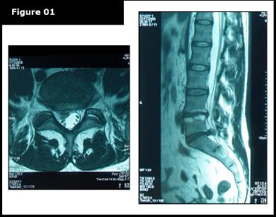
Orthopedic tests, including Valsalva’s Maneuver, Milgram’s test, and SLR, were all negative. Additionally, the Achilles reflexes were +2 bilaterally, indicating improved neurological function. To assess structural changes, a follow-up MRI scan was performed on April 29, 2010 (see Figure-2).

Proven slip disc treatment showing reversal of severe slipped disc

A board-certified radiologist conducted a comparative study between the new MRI and the previous MRI from November 11, 2008. The comparison showed a significant reduction of the extruded disc material, resulting in reduced compression on the spinal cord (thecal sac). Additionally, the neural foramina were intact bilaterally at L5-S1. However, at L4-L5, no significant structural changes were noted.

Second Case – Moderate to Severe Disc Extrusion

A 44-year-old male engineer of Persian descent sought care for severe lower back discomfort that extended to the left buttock, posterior thigh, and leg. The patient also reported numbness, tingling, and a burning sensation along the bottom of the left foot and big toe, as well as weakness affecting the entire lower left limb.

On a 1 to 10 scale for discomfort (1 being minimal and 10 being the most severe), the patient reported a level 10 intensity.

Upon examination:

  • The patient exhibited mild left foot weakness.
  • Achilles reflex was zero (0) on the left and +2 on the right.
  • Due to the severity of the condition, provocative orthopedic testing was not performed to avoid exacerbating symptoms.

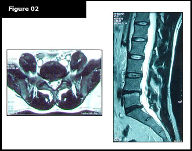
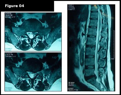
The patient had previously undergone two MRI scans, the first on December 29, 2009, and a second on January 23, 2010 (Figures-3 and 4).

repaired slipped disc with proven slipped disc treatment

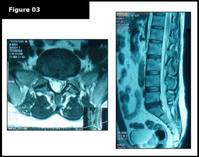
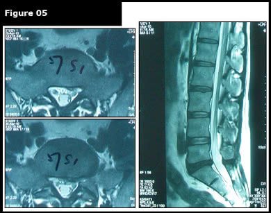
A comparative review of the MRI scans revealed a worsening of the disc bulge observed in December 2009. The condition had progressed to a moderate disc extrusion, which significantly compressed the spinal cord (thecal sac) and S1 nerve root.

extruded discs shown before proven slipped disc treatment

To address the condition, NSD decompression methods were recommended. Due to the severity of the disc condition, the patient underwent 25 therapy sessions, administered five times per week for five weeks.

Outcome of the Second Case 

The patient demonstrated continuous improvements throughout the course of therapy. By the final care session, the patient reported a complete resolution of lower back discomfort (0/10) and only an occasional mild sensation (1/10) down the leg on the visual analog scale.

The clinical evaluation performed on April 16, 2010, showed normal bilateral (+2) Achilles reflexes with no signs of foot weakness. Additionally, orthopedic tests including Straight Leg Raise (SLR), Milgram’s, and Valsalva’s Maneuver were all negative, indicating improved neurological function and stability.

A follow-up MRI scan of the lumbar spine on April 19, 2010 (Figure-5) was conducted to assess structural changes. A comparative analysis between the post-therapy MRI and the pre-therapy MRI (dated Jan. 23, 2010) revealed improvements, supporting spinal decompression management & approaches in promoting spinal recovery.

after image of proven slipped disc treatment

Discussion on Spinal Discs and How Non-Operative Measures With Spinal Decompression Provided These Favorable Outcomes

Discs are considered the largest avascular structure in the human body [10, 11, and 12]. They obtain life-sustaining nutrients such as water, oxygen, glucose, and amino acids through the process of diffusion, taking place across the endplates [13]. The diffusion of nutrients is passive, resulting from the vessels in the subchondral bone adjacent to the endplate’s hyaline cartilage [14].

The diffused nutrients are utilized by the chondrocyte-like cells of the nucleus and inner annulus to synthesize the proteoglycan molecule [14] a protein-like molecule needed for disc maintenance and management. A disruption in the flow of these nutrients dramatically impacts the spinal disc, leading to a decrease in proteoglycan production and an eventual deterioration of the disc [14].

Fact: Increased Intradiscal Pressure is the Leading Cause of Spinal Disc Disorders

With degeneration, discs lose their osmotic pressure [15, 16], making the spinal disc unable to maintain its hydrostatic behavior [17]. When a disc loses its hydrostatic behavior, the axial load-bearing activities will significantly increase the intradiscal pressure [17], leading to loss of height [18, 19] and fluids rapidly [20].

The average intradiscal pressure (non-load bearing) is 70mm Hg [21]. Any time the intradiscal pressure exceeds diastolic blood pressure, the infusion of nutrients and oxygen (imbibition) into the disc stops [21]. Also, increased intradiscal pressure, especially in a degenerated disc, leads to the degradation of the Aggrecan molecule (clusters of proteoglycans). The degraded Aggrecans are then readily leached out. Leaching or flushing out Aggrecans worsens proteoglycan deficiency [19, 21]. With the loss of fluids and proteoglycans, a degenerated disc is susceptible to bulging or, perhaps worse, tearing of fibers leading to disc herniations and extrusions.

What is the Root Cause of Spinal Disc Damage?

According to medical research, the underlying cause of disc degeneration, disc bulge, disc herniation, and disc extrusion is a lack of nutrients resulting from increased intradiscal pressure. Research conducted by neurosurgeons Gustavo and Martin [22] showed a significant reduction in intradiscal pressure due to distractive tensions applied. They stated that the intradiscal pressure dropped to negative levels in the range of -100 to -160mm Hg [22].

The care methods provided to the two patients in this report followed the specific NSD protocols of spinal decompression. The NSD system of therapy focuses on spinal decompression therapy through RxDecom®, a targeted decompression modality designed to restore disc hydration and function. The RxDecom® induces imbibition, promoting nutrient exchange that helps reduce intradiscal pressure and supports the recovery of spinal discs.

In addition to decompression therapy, the care approach included targeted chiropractic mobilizations and focused physiotherapy. Physiotherapy procedures such as ultrasound therapy, interferential current therapy, manual therapy, rehabilitative therapy, and strengthening exercises contributed to spinal stabilization and muscular support.

Conclusion: Understanding Conservative Management of Spinal Disc Conditions

Spinal disc disorders such as bulges, protrusions, herniations, and extrusions are complex conditions influenced by mechanical stress, disc nutrition, and spinal movement patterns. Research has shown that increased intradiscal pressure and reduced nutrient exchange can contribute to disc degeneration and structural changes.

The clinical cases presented in this report demonstrate how coordinated non-operative approaches—including physiotherapy rehabilitation, chiropractic joint methods, and decompression-based strategies—were used in managing significant disc conditions.

Although these observations involve a limited number of cases, the findings highlight the importance of continued research into conservative spine care approaches. Larger clinical studies are required to further evaluate long-term outcomes and determine how these methods may influence spinal disc recovery over time.

Author:

“Slipped Disc Management Using Safe Methods” is written by Yama Zafer, D.C., he holds degrees in physiotherapy and chiropractic from Cleveland Chiropractic University, Kansas City, and has nearly 30 years of experience in integrative non‑invasive spine and joint care; read more about Y. Zafer on his official bio page.

Reference

For those interested in research, we have provided a list of articles used in preparing this case study. Additionally, we have included links to two references. You may copy and paste them into Google Scholar to search for additional related research articles. Should you need further clarifications or information about our slipped disc management and care options, please contact our main center at 03 2093 1000.

Peer-Reviewed Medical References:

  • Occup Hazards. 1992;11:29–33.

  • Gou H, Tanaka S, Halperin EW, Cameron LL. Back pain prevalence in US industry and estimates of lost workdays. Am J Public Health. 1999 Jul;89(7):129–35.

  • American Academy of Orthopaedic Surgeons. Orthopaedic Knowledge Update. Chapter 2. Rosemont (IL): AAOS; 2008.

  • Practical Pain Manag. 2003 Mar–Apr.

  • Eur Musculoskelet Rev. 2008;3(2).

  • J Saitama Kenou Rehabil. 2006 Nov;6. Kawagoe, Japan.

  • Presented at: 45th Annual Indian Association of Physiotherapy Conference; 2007 Feb; Kolkata, India.

  • Am J Pain Manag. 1997 Apr;7(2).

  • Orthop Technol Rev. 2003.

  • Clin Orthop Relat Res. 1977;(129):101–14.

  • Roberts S, Urban JP, Evans H, Eisenstein SM. Biochemical and structural properties of the cartilage end-plate and its relation to the intervertebral disc. Spine (Phila Pa 1976). 1989 Feb;14(2):166–77.

  • Urban JP, Roberts S. Biochemical and physiological processes in the intervertebral disc. Biochem Soc Trans. 2002 Dec;30(6):839–44.

  • Connect Tissue Res. 1980;8:101–19.

  • Asian Spine J. 2009 Mar;3(1):39–44.

  • Adams MA, Dolan P, Hutton WC, Porter RW. Diurnal changes in spinal mechanics and their clinical significance. J Bone Joint Surg Br. 1990 Mar;72(2):266–70.

  • Nachemson A. Influence of age, spinal level, composition, and degeneration. Spine (Phila Pa 1976). 1988 Feb;13(2):139–225.

  • Adams MA, McNally DS, Dolan P. Stress distribution inside intervertebral discs: the role of hydrostatic pressure. J Bone Joint Surg Br. 1996 Nov;78(6):965–72.

  • Rajasekaran S, Bajaj N, Tubaki V, et al. Disc degeneration in asymptomatic subjects: a comparison between age groups. Eur Radiol. 2001 Feb;11(2):263–9.

  • Nachemson A. Disc pressure measurements. Spine (Phila Pa 1976). 1983 May–Jun;8(3):225–45.

  • Goldring MB, Goldring SR. Osteoarthritis. Arthritis Res Ther. 2003;5(3):120–30.

  • Calif J Altern Med. 1999 Dec.

  • Matsui H, Okano T, Tsuji H. Significance of neural compression in lumbar disc herniation. J Neurosurg. 1994 Sep;81(3):350–3.

Last Updated:

Last updated March 12, 2026: Slipped Disc Management Using Safe Methods in Malaysia.