


Herniated Disc Care in Kuala Lumpur
Herniated disc care in Kuala Lumpur often begins with understanding how the disc has changed and whether nearby nerves, joints, or supporting tissues are involved. A herniated disc, sometimes called a slip disc or ruptured disc, happens when the inner disc material moves outward through weakened outer fibers.
Depending on the level affected, this may change how the neck, back, arms, or legs feel and move. This page explains what a herniated disc means, what may contribute to it, and how structured non-surgical approaches such as chiropractic, physiotherapy, and rehabilitation are commonly used to support spinal movement and daily function.
Key Takeaways: Herniated Disc, MRI Findings & Non-Surgical Care
- A herniated disc happens when the inner disc material moves through weakened outer fibers and may affect nearby nerve pathways.
- The exact disc level and direction of the change are best understood through a structured clinical assessment and, when appropriate, an MRI.
- Symptoms may be felt in the neck, back, arm, buttock, or leg, depending on the level involved.
- Disc changes in the neck are commonly seen at C4-C5, C5-C6, and C6–C7, while lower-back changes are more often seen at L3-L4, L4-L5, and L5–S1.
- Guided physiotherapy, gentle chiropractic methods, rehabilitation, and movement planning are often considered before surgical options.
Need More info on Spinal Disc Care
If you’re experiencing symptoms of herniated disc, our team at Chiropractic Specialty Center® in Kuala Lumpur is here to assist. We provide non-invasive care plans tailored to your needs, focusing on safety and effectiveness. Reach out to us today to schedule a consultation and learn more about how we can support your spinal health: CSC in KL
Video: What Is the Difference Between a Bulging Disc and a Herniated Disc?
Many people come across terms such as bulging disc, prolapsed disc, protruded disc, herniated disc, ruptured disc, extruded disc, or fragmented disc, along with the commonly used term slip disc. The differences between these disc changes are not always clear.
A bulging, prolapsed, or protruded disc usually means the outer disc wall extends outward while still remaining contained, which helps keep the inner disc material in place. These are commonly considered contained disc changes.
By comparison, a herniated, ruptured, extruded, or fragmented disc means the inner disc material has moved through weakened or torn outer fibers, allowing part of the gel-like inner portion, known as the nucleus pulposus, to move outward.
The short video below explains how spinal discs work and how these structural changes may lead to either a disc bulge or a herniation. Using a simple disc model, it shows how the outer fibers change, how the inner material shifts, and why nearby nerves may be affected depending on the spinal level involved.
Key Moments From the Video
This short educational guide explains how spinal discs function, how the outer annular fibers change over time, and how disc bulges, herniations, and extrusion stages may affect nearby nerves in the neck, back, arms, or legs.
Timestamps:
- 00:00 Introduction
- 00:35 Disc function
- 00:46 Nucleus pulposus & annulus fibrosus
- 01:28 How annular tears develop
- 01:45 Bulge vs herniation
- 02:09 Stages from bulge to extrusion
- 02:43 Common symptoms and sciatica patterns
This video section complements the explanations below by showing how disc changes progress visually and why symptoms may vary depending on the spinal level involved.
Why Accurate Assessment Comes Before Herniated Disc Care
A herniated disc happens when the outer disc fibers tear, allowing part of the gel-like inner disc material, known as the nucleus pulposus, to move outward toward the spinal canal or nearby nerve pathways. Because the affected disc level lies several centimeters beneath the skin, muscles, and surrounding soft tissues, identifying the exact level involved is one of the most important first steps before any care is considered.
A careful clinical assessment helps determine which spinal level is affected, how nearby nerves and joints may be involved, and how the spine is functioning during everyday movements such as sitting, standing, bending, and walking. Symptoms felt in the neck, back, arm, or leg do not always directly reflect the exact location of the disc tear, which is why a structured assessment is so important.
Another key part of the evaluation is identifying the direction and pattern of the herniation. For example, the disc change may be central, paracentral, foraminal, or far lateral, and each pattern may affect different nerve pathways.
Understanding this clearly helps guide the next phase of care and helps avoid approaches that may not be suitable for the affected level.
Why MRI and Disc Location Findings Matter Before Care Planning
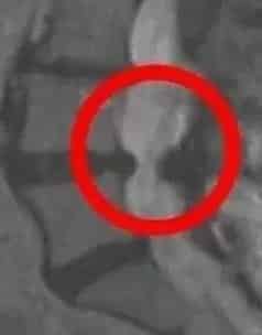
After the physical assessment, MRI is often the most useful imaging method for confirming the disc level involved and showing the extent of the annular tear. MRI provides a detailed view of the disc, surrounding nerves, and the spinal canal, helping show whether the inner disc material has moved toward sensitive neural structures.
Understanding the depth, location, and direction of the herniation is one of the most important parts of planning the next stage of non-surgical care. A central herniation may behave very differently from a foraminal or far lateral disc change, and these differences often help explain why one person may notice changes in the arm or leg while another mainly notices neck or back symptoms.
My understanding of herniated disc care is shaped not only by decades of clinical work in chiropractic and physiotherapy, but also by personal experience, having first dealt with a herniated disc more than three decades ago. That experience led to years of focused study, ongoing review, and careful observation of how different herniation patterns affect movement and daily function.
The section below explains how care is then structured once these findings are clearly identified.
What Can Contribute to a Herniated Disc Over Time?
A herniated disc rarely develops without an underlying reason. In many cases, it happens when ongoing mechanical stress is placed on a disc that has already started to weaken over time.
One commonly discussed factor is prolonged sitting, especially when combined with poor posture, repeated forward bending, or long hours in a fixed seated position.
MRI-based research has shown that as little as four hours of uninterrupted sitting may reduce disc height at the L4–L5 level, which helps explain why this region is frequently involved in lower-back disc changes Brief positional changes during the same time period were associated with fewer measurable disc-height changes in the study.
Repeated strain, sudden twisting movements, lifting-related stress, falls, or previous spinal injuries may also contribute to progressive weakening of the outer annular fibers. In many individuals, the structural change develops gradually over time before symptoms are first noticed.
As the outer annular fibers lose resilience, the inner disc material may become more likely to migrate outward during daily loading, bending, sitting, or lifting, which may eventually contribute to a herniation
Reference: Billy GG, Lemieux SK, Chow MX. Changes in lumbar disk morphology associated with prolonged sitting assessed by magnetic resonance imaging. PM R. 2014;6(9):790–795.
What Signs May Suggest a Herniated Disc?
A herniated disc may change how the spine, arms, or legs feel and move, depending on the spinal level involved and whether nearby nerves are affected.
Common signs may include:
- Sensations extending into the arm or leg
- Tingling or numbness
- Changes in strength or grip
- Reduced spinal flexibility
- Symptoms resembling sciatic nerve irritation
- Increased discomfort during prolonged sitting, bending, or coughing
The pattern often depends on the location of the herniation. A cervical disc may affect the neck, shoulder, or arm, while a lumbar disc may contribute to changes in the lower back, buttock, or leg.
How Herniated Disc Care Is Structured After Assessment
Once the level, depth, and direction of the herniation have been identified, care is usually planned around the disc itself and the surrounding tissues that may also be involved.
Because the affected disc commonly lies several centimeters beneath the skin and surrounding soft tissues, nearby structures such as the paravertebral muscles, facet joints, annular fibers, and supporting ligaments (thickening of ligaments flavum) may also need to be addressed.
Depending on the findings, ultrasound may be used for more superficial soft tissues, while laser-based methods may help reach deeper structures such as the facet joints and, in many individuals, the disc level itself. Electrotherapy may also be considered where muscular guarding is contributing to restricted movement.
Where appropriate, gentle soft-tissue methods and fascial release may be used to improve tissue mobility around the affected segment. In selected cases, spinal decompression and Cox flexion-distraction therapy may be incorporated to help reduce disc pressure and support spinal movement.
Throughout the care phase, the focus remains on controlled, non-rotatory spinal methods, with progression based on stability, imaging findings, and the individual’s functional response.
How Gentle Chiropractic and Physiotherapy Methods Are Selected for Disc Care
Once the exact disc level, direction of the herniation, and surrounding nerve involvement are clearly identified, the next step is selecting movement and manual methods that match the affected segment.
For disc-related concerns, the focus is usually on gentle, controlled, non-rotatory, and level-specific approaches that work with the affected spinal level rather than placing additional stress on it.
Depending on the findings, this may include gentle chiropractic methods such as Activator approaches, Thompson drop-assisted techniques, flexion-distraction, or block-assisted SOT positioning, together with guided physiotherapy, rehabilitation exercises, and movement planning.
The aim is to help reduce repeated mechanical stress on the affected disc while improving posture, walking tolerance, spinal stability, and day-to-day movement function.
Approaches that involve forceful twisting, sudden traction pulls, or aggressive axial loading may require additional caution when a herniated disc is already present, especially if nearby nerve pathways or, in the neck, the spinal cord may be involved.
This is why care planning should always be guided by MRI findings when indicated, neurological signs, and how the affected level responds during reassessment.
How Physiotherapy and Rehabilitation Support Disc Recovery Over Time
Physiotherapy and rehabilitation play an important role in helping the spine recover after a disc herniation, particularly once the affected level and movement restrictions have been clearly identified.
The goal is not simply movement for movement’s sake, but condition-specific, age-appropriate rehabilitation that works with the affected spinal level and surrounding tissues.
This may include guided walking plans, posture correction, movement retraining, stability exercises for the supporting muscles of the spine and hips, and progressive mobility work designed around the direction and level of the herniation.
Rehabilitation is also important for reducing the repeated mechanical stresses that may have contributed to the disc change in the first place, such as prolonged sitting, poor posture, weak supporting muscles, or repeated bending patterns.
As recovery progresses, physiotherapy may help improve walking tolerance, sitting tolerance, spinal stability, sleep posture awareness, and long-term movement habits that support everyday function.
The exact program should always be guided by how the symptoms respond over time and adjusted according to reassessment findings.
Which Neck Disc Levels Are Most Commonly Affected?
In the cervical spine, herniated discs are most commonly seen at C4-C5, C5-C6, and C6-C7, as these levels are exposed to frequent movement and repeated daily loading. These segments often experience the greatest stress during prolonged computer work, forward head posture, repeated looking down at mobile devices, and sudden neck movements.
Among these, C5-C6 and C6-C7 are especially common because they are involved in much of the neck’s flexion, extension, and rotational movement throughout the day.
The uppermost cervical segments, including the occiput-C1 and C1-C2 levels, are different because they do not contain intervertebral discs. These levels rely on joints, ligaments, cartilage, and surrounding soft tissues for movement and stability, which is why disc-related changes are not discussed at these segments.
For readers looking for more specific information, detailed explanations are available for each cervical level, including C2-C3, C3-C4, C4-C5, C5-C6, C6-C7, and C7-T1, where disc changes may be more commonly seen.
How Neck-Level Changes May Affect Symptoms
The level involved often helps explain the pattern of symptoms.
For example:
- C4-C5 may influence the neck, upper shoulder, and upper arm
- C5-C6 may affect the shoulder, upper arm, forearm, and thumb-side hand
- C6-C7 may contribute to changes extending toward the triceps, forearm, and middle fingers
This is why identifying the exact cervical level is an important part of the assessment and MRI review process.
Could Neck Symptoms Be Coming From Muscles and Posture Instead?
Not every neck-related symptom begins at the disc level. In some individuals, tightness within the rhomboids, trapezius, and surrounding fascial tissues may contribute to restricted neck movement, postural imbalance, and symptoms that may feel similar to cervical joint or disc-related irritation.
The educational video below explains how upper-back muscular tightness, trigger points, and postural strain may influence neck movement and why it is important to distinguish muscular causes from disc or joint-related findings.
Video Key Moments
- 00:00 Muscle vs disc-related neck symptoms
- 01:14 Trapezius & rhomboid anatomy
- 02:18 How to distinguish joint vs muscle tightness
- 04:10 Trigger point location
- 10:45 W-T-L posture control method
- 13:46 Isometric neck strengthening
This video complements the cervical disc section above by helping readers understand when the source may be muscular rather than disc-related.
Which Lower Back Disc Levels Are Most Commonly Affected?
In the lower back, herniated discs are most commonly seen at L4-L5 and L5-S1, as these levels absorb much of the body’s weight and are exposed to repeated bending, lifting, sitting, and rotational stress during daily activity.
These lower lumbar segments are often the most vulnerable because they sit at the transition point between spinal mobility and weight-bearing load. Disc changes at L3-L4 may also occur, although less commonly.
Because you already have dedicated pages for L1-L2, L2-L3, L3-L4, L4-L5, and L5-S1, this section creates an excellent opportunity for natural internal linking to each level-specific article.
The exact level involved often helps explain the pattern of symptoms. For example, disc changes at L4-L5 and L5-S1 are frequently associated with symptoms extending into the buttock, thigh, calf, or foot, which may overlap with sciatic nerve patterns.
How Lower Back Disc Herniation May Lead to Sciatic Nerve Symptoms
Sciatica is not a diagnosis by itself, but rather a pattern of symptoms that may occur when a lower lumbar disc change affects nearby nerve roots that contribute to the sciatic nerve.
The sciatic nerve is formed by nerve roots from the lower lumbar and sacral spine, commonly involving L4, L5, S1, and S2. When a disc herniation occurs at levels such as L4-L5 or L5-S1, the nearby nerve root may become irritated, leading to symptoms that extend into the leg.
These symptoms may include:
- sensations running into the buttock or leg
- tingling or numbness
- calf or foot symptoms
- changes in leg strength
- discomfort that increases during sitting or bending
Because the exact nerve level involved matters, this is another important area where MRI and clinical assessment help determine whether the symptoms are arising from the disc itself or from associated lower spinal structures.
Sciatica Video: Why the Lower Back Is Often the Starting Point
Sciatica is a symptom pattern rather than a diagnosis by itself. In many cases, symptoms extending into the buttock, thigh, calf, or foot may begin with a disc change at the lower lumbar levels, particularly L4-L5 or L5-S1, where nearby nerve roots contribute to the sciatic nerve.
The video below explains why symptoms felt in the leg often begin with disc changes in the lower back rather than the leg itself.
Key Moments From the Video
- 00:00 Why sciatica is a symptom, not a diagnosis
- 01:15 L4 to S2 nerve anatomy
- 02:30 Disc-related causes of sciatic symptoms
- 04:00 Muscle spasm and fascia response
- 05:45 Daily habits and posture
- 07:20 When stretching may or may not be suitable
- 11:00 Rotary movement cautions
- 12:30 Surgical vs conservative considerations
- 14:00 Daily habit adjustments
This video complements the lumbar disc section above by helping readers understand why symptoms in the leg often begin with structural changes in the lower back.
Lower Back Disc Considerations: Supporting Lumbar Function
Herniated discs in the lower back are most commonly seen at L4-L5 and L5-S1, where the spine absorbs much of the body’s weight during sitting, standing, bending, and lifting. These levels are exposed to repeated daily loading, which is why they are more frequently involved in disc-related structural changes.
The lumbar spine consists of five vertebrae, from L1 to L5, with the lower segments carrying the greatest mechanical stress. In particular, the L3-L4, L4-L5, and L5-S1 levels are often more vulnerable because they sit in the highest load-bearing region of the spine.
Simple daily habits may help reduce repeated stress on these segments. These may include:
- Limiting repeated bending and twisting at the waist
- Avoiding prolonged uninterrupted sitting
- Maintaining a neutral seated posture
- Using appropriate lower-back support when sitting for long periods
- Being cautious with lifting mechanics
A cloth-wrapped cold pack applied to the lower back for 15-20 minutes at a time may also be considered where localized irritation is present.
What Happens Inside a Herniated Disc and Why Movement Must Be Guided
A herniated disc happens when the outer fibers of a spinal disc weaken or develop small tears, allowing part of the inner gel-like material to move outward. This inner portion, known as the nucleus pulposus, normally helps the disc absorb pressure and support smooth spinal movement.
The outer wall of the disc, called the annulus fibrosus, acts as a strong fibrous ring that helps keep the inner material contained while distributing load during sitting, bending, lifting, and rotation.
When the outer fibers lose strength over time, the inner disc material may begin to shift beyond its usual boundary. Depending on the level involved, this may affect nearby nerve pathways and change how the neck, back, arm, or leg feels during daily movement.
These changes are more commonly seen in the lower back at L4-L5 and L5-S1, and in the neck at C5-C6 and C6-C7, where daily mechanical stress is often highest.
Because movement plays an important role in how the spine responds, exercises and rehabilitation methods should always be matched to the disc level and the direction of the herniation.
Certain movements may not be suitable in some situations, particularly:
- Repeated twisting through the spine
- High-impact loading
- Forceful bending movements
- Unsupervised rotational exercises
The goal is not to avoid movement altogether, but to ensure that movement strategies work with the affected spinal level rather than placing additional stress on it.
This is why careful assessment, imaging findings, and guided rehabilitation planning are essential before introducing exercise, decompression, or movement-based care.
Is Surgery Always Needed for a Herniated Disc?
In many cases, spine surgery is not the first step considered for a herniated disc. The most appropriate approach depends on the disc level involved, the severity of nerve involvement, imaging findings, and how symptoms are affecting daily movement and function.
A structured non-surgical plan is often considered first and may include guided physiotherapy, gentle chiropractic methods, spinal decompression where appropriate, and rehabilitation strategies designed around the affected level.
Imaging, particularly MRI when indicated, helps clarify the exact disc level, the direction of the herniation, and whether nearby nerve pathways are involved. This helps determine whether a non-surgical approach may be suitable or whether further medical evaluation is needed.
When symptoms include progressive weakness, significant changes in walking, or bowel or bladder changes, prompt medical assessment becomes especially important.
The decision should always be guided by the full clinical picture rather than based on symptoms alone.
Recovery Timeline: How Long a Herniated Disc May Take to Settle
The time it takes for a herniated disc to settle can vary widely from one person to another. It often depends on the disc level involved, the extent of the structural change, the presence of age-related degenerative changes, daily movement habits, sleep, posture, and how consistently lifestyle modifications are followed.
In many cases, noticeable improvement in symptoms may begin within 4 to 8 weeks, especially when aggravating activities such as prolonged sitting, repeated bending, and poor posture are addressed early.
More meaningful structural recovery and symptom stabilization may continue over 3 to 6 months, particularly when movement guidance, rehabilitation, and daily habit adjustments are followed consistently.
Several factors may influence the timeline, including:
- The size and type of the herniation
- Whether nearby nerves are involved
- The degree of disc dehydration or degeneration
- Age-related endplate and disc nutrition changes
- Walking, sleeping, and sitting habits
- Consistency with movement and ergonomic modifications
Because every disc change behaves differently, the timeline should be guided by symptoms, functional changes, and follow-up reassessment rather than by a fixed timeframe alone.
Research & Reference Note
The time course for disc-related symptom improvement varies depending on the size and type of the herniation, nerve involvement, age-related disc changes, and daily movement habits. Published reviews and meta-analyses report that many lumbar disc herniations show meaningful regression or symptom improvement within 3 to 6 months, with the main resorption process often occurring within the first 6 months of conservative care.
Suggested published references
- Zou T, Liu XY, Wang PC, et al. Incidence of Spontaneous Resorption of Lumbar Disc Herniation: A Meta-analysis.Clin Spine Surg. 2024.
- Zhong M, Liu JT, Jiang H, et al. Incidence of spontaneous resorption of lumbar disc herniation: a meta-analysis.Pain Physician. 2017.
When a Herniated Disc May Improve Through Natural Resorption
In some cases, a herniated disc may improve over time without surgical intervention. Research has shown that spontaneous shrinkage or resorption of disc material can occur, particularly in extruded or sequestrated lumbar disc herniations.
This means the body may gradually reduce the size of the herniated material through natural biological processes.
These processes may include:
- Gradual dehydration and shrinkage of the disc material
- Retraction of the displaced portion
- Natural inflammatory cleanup by immune cells
- Gradual reduction of local pressure around nearby nerve pathways
Imaging studies have shown that meaningful reduction may occur over 3 to 6 months, with some cases continuing to change over a longer period.
The likelihood of spontaneous regression may depend on:
- The type of herniation
- The size of the disc fragment
- Age-related disc changes
- Surrounding blood supply and tissue response
- Daily activity and posture habits
Recovery without formal care is possible, but the timeline and extent of improvement vary from person to person. Factors such as the type of herniation, age-related disc changes, posture, sleep quality, and daily movement habits may all influence how the disc responds over time.
Condition-specific, age-appropriate non-surgical care, together with posture correction, movement guidance, and lifestyle modifications, may help create more favorable conditions for recovery and may improve the likelihood of symptom improvement.
While spontaneous improvement is possible, ongoing monitoring is important, especially when symptoms include weakness, walking changes, or significant leg symptoms.
Research & Reference Note
Yes, published imaging studies and systematic reviews show that spontaneous regression or resorption of lumbar disc herniations is well documented, particularly in extruded and sequestrated disc herniations. Reported pooled rates range from approximately 66% to over 70%, with higher rates seen in more severe extruded or free-fragment disc types.
Suggested published references
- Zou T, Liu XY, Wang PC, et al. Incidence of Spontaneous Resorption of Lumbar Disc Herniation: A Meta-analysis.Clin Spine Surg. 2024.
- Xie L, et al. Prevalence, clinical predictors, and mechanisms of resorption in lumbar disc herniation: a systematic review. 2024.
- Rashed S, et al. Predictive factors for spontaneous regression in lumbar disc herniation. J Neurosurg Spine. 2023.
Full Video Guide: How Disc Changes Lead to Sciatica, Leg Symptoms & Daily Movement Changes
Symptoms that travel from the lower back into the buttock, thigh, calf, or foot often begin with structural changes in the spinal disc rather than in the leg itself. This in-depth educational session explains how disc bulges, protrusions, and herniations may affect nearby nerves, why sitting and sleep posture matter, and how everyday habits may influence disc health over time.
The video also covers MRI findings, walking mechanics, sleep position, exercises to avoid, and how lower-back disc changes may contribute to sciatica-like symptoms.
Key Moments From the Video
- 00:00 Back and neck symptom patterns
- 02:43 Cervical, thoracic, and lumbar spine anatomy
- 04:06 The spine’s 9 major functions
- 06:22 Disc anatomy: nucleus pulposus vs annulus fibrosus
- 09:07 Why healthy discs tolerate load well
- 13:10 Sleep and posture effects on the spine
- 18:30 MRI: healthy vs degenerative disc comparison
- 20:50 Natural disc repair and recovery mechanisms
- 28:49 Disc bulge, protrusion, and herniation stages
- 34:53 Can a herniated disc exist without symptoms?
- 38:00 Sitting and disc pressure
- 51:05 Sleep positions for spinal support
- 57:13 Exercises to avoid with disc-related symptoms
- 1:04:12 Walking, footwear, and joint loading
- 1:12:46 How disc changes may contribute to sciatica
- 1:33:12 Non-surgical care vs surgery considerations
- 1:42:40 Rotation, sports, and lower-back injury prevention
Video Takeaway: What This Session Helps You Understand
This video explains how spinal disc changes may begin long before symptoms become obvious and why leg symptoms often start in the lower back rather than in the leg itself.
It also helps readers understand the relationship between MRI findings, disc hydration, sleep posture, walking habits, sitting pressure, and sciatica-like symptoms.
For anyone trying to understand whether a bulging, protruded, or herniated disc may be involved, this session provides one of the most complete educational overviews on the page.
Why Disc Symptoms May Return After Improvement
Yes, disc-related symptoms may return if the daily habits and mechanical stresses that contributed to the original disc strain remain unchanged. A spinal disc that has previously been affected may remain more vulnerable to repeated loading if posture, sitting habits, sleep quality, and movement patterns are not improved over time.
Common contributing factors may include prolonged uninterrupted sitting, repeated bending or twisting, poor posture, reduced sleep quality, low activity levels, deconditioning of the supporting muscles, and long-term sedentary habits. These factors may continue to place repeated stress on the same spinal level, particularly in the lower back at L4–L5 and L5–S1.
Long-term lifestyle adjustments may help reduce the likelihood of recurrence. These may include posture correction, regular walking, reducing uninterrupted sitting time, improving sleep habits, ergonomic changes, and maintaining spinal stability through guided exercise and age-appropriate movement strategies.
Even after symptoms improve, ongoing habit changes often remain important because the goal is not only to help the disc settle, but also to reduce the mechanical factors that may contribute to future flare-ups.
Can Walking Help With a Herniated Disc?
Gentle walking is often one of the better-tolerated forms of movement because it encourages circulation and spinal motion without the sustained pressure often seen during prolonged sitting.
Walking is usually most helpful when done in a controlled way:
- Comfortable supportive footwear
- Level ground
- Neutral upright posture
- Looking forward rather than downward
- Avoiding steep hills early on
- Short, regular intervals
For many individuals, walking may be easier to tolerate than prolonged sitting.
Can Sitting Make a Herniated Disc Worse?
Prolonged uninterrupted sitting may increase pressure on spinal discs, particularly in the lower back.
This may become more noticeable when sitting with a rounded or hunched posture.
If sitting is unavoidable, it often helps to:
- Maintain an upright posture
- Keep the lower back supported
- Take regular standing and walking breaks
- Avoid prolonged forward bending
Frequent posture breaks are often more important than the chair itself.
Can a Herniated Disc Affect Nerves Long Term?
If nearby nerve pathways remain significantly affected for a prolonged period, strength, sensation, walking tolerance, or limb function may change.
This is why symptoms such as:
- Progressive weakness
- Increasing numbness
- Walking difficulty
- Bowel or bladder changes
should be assessed promptly.
The exact risk depends on the level involved and the degree of nerve or spinal cord involvement.
Can Exercise Make a Herniated Disc Worse?
Certain exercises may place additional stress on the affected level, especially when they involve:
- Heavy axial loading
- Repeated twisting
- Forceful bending
- Unsupervised high-impact movements
Exercise is still important, but it should be guided according to the disc level involved and how symptoms respond.
Movement should support spinal stability rather than increase load on the affected segment.
Which Sleeping Position Is Usually Better for a Herniated Disc?
Many people tolerate sleeping best on the back or side, depending on the level involved and how symptoms behave overnight.
When side sleeping, placing a pillow between the knees may help keep the spine in a more neutral position.
The most important factor is usually achieving uninterrupted restorative sleep, as sleep quality may influence how the body responds during recovery.
How Disc Changes May Also Affect Nearby Ligaments and Joints
Long-standing disc changes may sometimes be seen together with changes in nearby spinal structures, including the ligamentum flavum and the facet joints.
The ligamentum flavum is a ligament that forms part of the back wall of the spinal canal. Over time, repeated spinal loading, prolonged sitting, bending, and postural stress may contribute to thickening of this tissue in some individuals.
When this happens together with facet joint enlargement or disc-related changes, the available space around nearby nerve pathways may become reduced.
This may help explain symptoms such as:
- sensations extending into the leg
- tingling or numbness
- reduced walking tolerance
- stiffness during standing or bending
MRI is often helpful in identifying whether these surrounding structures are also involved, especially when symptoms do not appear to arise from the disc alone.
Because disc changes and surrounding tissue changes may coexist, care planning should be guided by imaging findings, movement assessment, and the exact level involved.
Frequently Asked Questions About Herniated Disc Symptoms, MRI & Non-Surgical Care
Many search for quick answers about herniated disc symptoms, MRI findings, sciatica, movement guidance, and when surgery may or may not be considered. The questions below are based on common search behavior and are aligned with the key sections already covered on this page.
A herniated disc happens when the inner gel-like portion of the disc moves through weakened outer fibers and begins to affect nearby tissues or nerve pathways.
No. A bulging disc usually remains contained within the outer disc wall, while a herniated disc involves the inner material moving through weakened or torn outer fibers.
Yes. Lower-back disc changes may affect nearby nerve roots and lead to leg symptoms and discomfort, much like sciatica where sensations extending into the buttock, thigh, calf, or foot.
Yes. Neck disc changes may affect nearby nerve pathways and influence not only how your neck and upper back feels, but also how the shoulder, arm, forearm, hand, or fingers feel and move.
Yes. Herniations at L4–L5 or L5–S1 may affect nerve roots that contribute to the sciatic nerve pain.
Common symptoms may include tingling, numbness, reduced strength, altered walking tolerance, stiffness, and sensations extending into the arm (upper extremity) or leg (lower extremity).
Prolonged uninterrupted sitting may increase mechanical stress on spinal discs, especially in the lower back.
MRI is often useful when the exact level, direction, and depth of the disc change need to be identified.
In many cases, surgery is not the first step considered. Clinical findings, MRI, and movement response help guide the next step.
No. Many cases are first managed with guided non-surgical approaches and careful reassessment.
Repeated twisting, forceful bending, high-impact loading, and unsupervised rotational exercises may place additional stress on the affected level.
Yes. Tightness in surrounding muscles and fascia may sometimes create symptoms that resemble disc-related findings.
Yes. Disc changes may sometimes be seen together with nearby ligament (ligaments flavum) or facet joint changes.
Prompt medical evaluation is important when symptoms include progressive weakness, major walking changes, or bowel or bladder changes.
Yes. If nearby nerve pathways are involved, walking tolerance, leg control, or balance may be affected.
Cervical disc changes affect the neck and may influence the arm, while lumbar disc changes are more likely to affect the lower back and leg.
The timeline can vary depending on the size and type of the herniation, the level involved, daily habits, and whether nearby nerves are affected. Many notice improvement within 4 to 8 weeks, while more meaningful recovery and stabilization may continue over 3 to 6 months. Progress is best guided by symptom changes, movement tolerance, and follow-up reassessment rather than by a fixed timeline alone.
Yes, in some cases it may improve over time through a natural process called spontaneous resorption, where the body gradually reduces the displaced disc material. Age, disc type, posture, and daily habits may influence the timeline. Condition-specific non-surgical care and lifestyle adjustments may help support a more favorable recovery environment.
Yes, a herniated disc can return, especially if the daily habits and mechanical stresses that contributed to the original disc problem remain unchanged. Prolonged sitting, repeated bending, poor posture, reduced sleep quality, and low activity levels may all increase the likelihood of recurrence. Long-term lifestyle modifications, posture correction, walking, and condition-specific exercise may help reduce the chances of future flare-ups.
Yes, when selected according to the disc level, MRI findings, and neurological signs, gentle chiropractic methods of dis care and physiotherapy may be suitable as part of a structured non-surgical slip disc plan. The focus is usually on controlled, non-rotatory, and level-specific approaches together with rehabilitation and movement guidance that work with the affected spinal segment rather than adding further stress.
Physiotherapy helps support recovery by improving spinal stability, walking tolerance, posture, and the strength of the muscles that support the affected level. A structured rehabilitation plan may also help reduce repeated mechanical stress from sitting, bending, and poor movement habits that may contribute to future flare-ups.
Addressing Spinal Disc and Ligament Changes
Factors That May Contribute to Ligamentum Flavum Thickening
Changes in the ligamentum flavum may develop due to prolonged spinal strain from:
- Postural misalignment
- Extended sitting periods
- Repetitive bending at the waist
- Twisting movements
Over time, these factors may contribute to structural changes in spinal ligaments. Conditions such as disc degeneration, scoliosis, and spinal misalignment may coincide with ligamentum flavum changes. Early assessment and structured care strategies can help manage these concerns efficiently.
Non-Invasive Approaches for Managing Post-Surgical Spinal Concerns
Individuals who have undergone surgical spinal procedures and continue to experience structural challenges may still benefit from non-surgical strategies. Integrated approaches, including spinal decompression, movement-based rehabilitation, and physiotherapy, have shown promising outcomes in post-surgical spinal care.
Timely intervention using structured, evidence-based methods may help maintain spinal function, promote mobility, and reduce the likelihood of future complications.
Yama Zafer, D.C. – Herniated Disc Care: Non-Invasive Options in Malaysia
“Herniated Disc Care in Kuala Lumpur” is written by Yama Zafer, D.C., with a background in physiotherapy and chiropractic from Cleveland Chiropractic University in Kansas City, has dedicated nearly 30 years to providing non-invasive spine and joint care; read more about Y. Zafer.
Peer-Reviewed References
- Billy GG, Lemieux SK, Chow MX. Changes in lumbar disk morphology associated with prolonged sitting assessed by magnetic resonance imaging. PM R. 2014;6(9):790–795. doi:10.1016/j.pmrj.2014.02.014.
- Chou R, et al. Nonpharmacologic therapies for low back pain. Ann Intern Med. 2017;166(7):493–505.
- Delitto A, et al. Low back pain clinical practice guidelines. J Orthop Sports Phys Ther. 2012;42(4):A1–A57.
- Deyo RA, Mirza SK. Herniated lumbar intervertebral disk. N Engl J Med. 2006;354(3):311–320.
- Foster NE, et al. The global burden of low back pain. Lancet. 2018;391(10137):2368–2378.
- van Middelkoop M, et al. Exercise therapy for chronic low-back pain. BMJ. 2010;340:c64.
- Zou T, Liu XY, Wang PC, et al. Incidence of Spontaneous Resorption of Lumbar Disc Herniation: A Meta-analysis. Clin Spine Surg. 2024.
- Zhong M, Liu JT, Jiang H, et al. Incidence of spontaneous resorption of lumbar disc herniation: a meta-analysis. Pain Physician. 2017.
- Xie L, et al. Prevalence, clinical predictors, and mechanisms of resorption in lumbar disc herniation: a systematic review.2024.
- Rashed S, et al. Predictive factors for spontaneous regression in lumbar disc herniation. J Neurosurg Spine. 2023.
Last Updated
Last updated on March 31, 2026: Neck Care in Kuala Lumpur: Signs & Non-Surgical Options
Recap: What This Page Helps You Understand
A herniated disc happens when the inner disc material moves through weakened outer fibers and begins to affect nearby spinal structures. Depending on the level involved, this may influence how the neck, back, arm, or leg feels and moves during daily activity.
This page explains how disc changes develop, how MRI and clinical assessment help identify the exact level involved, and why symptoms may sometimes begin away from the spine itself, such as in the arm, buttock, or leg.
It also outlines how non-surgical care is typically structured through guided physiotherapy, gentle chiropractic methods, rehabilitation, movement planning, and imaging-led decision making when appropriate.
Understanding the exact disc level, surrounding tissues, and movement patterns is often the key to planning the next step safely and accurately.
Share with others: