


Spondylolisthesis: Causes, Grades and Non-Invasive Management
Spondylolisthesis is the forward slip of one vertebra over the bone below it. It is most common in the lower back, at L4-L5 and L5-S1, and it is graded by how far the bone has moved.
Many cases of spondylolisthesis are small and stable, and many are found by chance on an X-ray taken for another reason. What shapes care is how the slip behaves: whether the bone moves when the spine bends, whether a nerve is pressed, and which positions ease the symptoms. With spondylolisthesis, bending forward often feels easier than leaning back, because bending forward opens the space the nerves pass through.
This guide to spondylolisthesis, its grades and non-invasive care, is published by Chiropractic Specialty Center, Bukit Damansara (KL) main center: WhatsApp+60 17 269 1873 Call+603 2093 1000
Key Takeaways for Spondylolisthesis
| Key point | What to know |
|---|---|
| Types | The two most common types are degenerative, from wear in the disc and joints, and isthmic, from a small stress crack at the back of the vertebra that usually starts in childhood. |
| Grades | Most slips are grade 1 or 2, and many cause few or no symptoms. |
| Over time | Long-term studies show that most slips change little over the years, and a slip that grows does not always cause more symptoms. |
| Positions | Bending forward often feels easier than leaning back, because it opens the space the nerves pass through, while long standing with the back arched tends to load the slip. |
| Care | Care usually starts without surgery: exercise for the deep trunk muscles, gentle non-rotatory manual care and changes to daily habits. |
Spondylolisthesis: Page Guide & Key Sections
- Types of Spondylolisthesis and What Causes Them
- Spondylolisthesis Grades: How Far the Bone Has Slipped
- Spondylolisthesis Assessment at Two KL Centers
- Signs and Symptoms of Spondylolisthesis
- Spondylolisthesis, Spondylolysis and Spondylosis: How They Differ
- How Spondylolisthesis Is Checked: X-Rays, MRI and CT
- Does Spondylolisthesis Get Worse Over Time?
- How Spondylolisthesis Is Managed Without Surgery
- Daily Habits That Ease the Load on a Slipped Vertebra
- When Surgery Is Discussed
- Explore Sciatica, Slipped Disc and Lower Back Pain Topics
- Spondylolisthesis FAQ
- Peer-Reviewed References
- The Author of Spondylolisthesis: Causes & Non-Invasive Management
- Last Updated: Spondylolisthesis: Causes, Grades and Non-Invasive Management
Types of Spondylolisthesis and What Causes Them
The system most doctors use to sort slips by cause was published by Wiltse, Newman and Macnab in 1976. It has five types:
- Degenerative. The disc and the small joints at one level wear down and let the vertebra drift forward. It is about three times more common in women than in men, it becomes more common with each decade after 40, and it is most often found at L4-L5.
- Isthmic. A stress crack, called spondylolysis, forms in the pars interarticularis, a thin bridge of bone at the back of the vertebra. The crack usually starts in childhood, tends to run in families and is most often found at L5-S1. Sports that arch the back again and again, such as gymnastics, add strain to this bridge.
- Dysplastic. The joints at the back of the vertebra formed differently before birth and do not hold the bone in place well. This type is less common.
- Traumatic. A sudden injury breaks a part of the vertebra other than the pars.
- Pathologic. A bone disease or tumor weakens the vertebra.
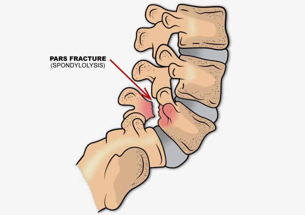
Degenerative and isthmic slips make up most cases. In a US community study that used CT scans on 188 adults aged 40 to 80, a pars crack was found in 11.5%, and about one in five people with a crack on both sides had no slip at all (Kalichman and colleagues, 2009).
Spondylolisthesis Grades: How Far the Bone Has Slipped
The Meyerding scale grades a slip by how far the upper vertebra has moved forward, measured as a share of the width of the vertebra below it (Koslosky and Gendelberg, 2020).
| Grade | How far the bone has slipped | What it usually means |
|---|---|---|
| Grade 1 | Up to 25% | The most common grade. Symptoms are often mild or absent. |
| Grade 2 | 26% to 50% | Still a low-grade slip. Care usually starts without surgery. |
| Grade 3 | 51% to 75% | A high-grade slip that is followed more closely. |
| Grade 4 | 76% to 100% | A high-grade slip. Surgery is discussed more often at this grade. |
| Grade 5 | More than 100% | Called spondyloptosis: the vertebra has slipped fully off the one below. It is rare. |
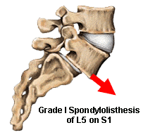
The grade describes the X-ray, not the person. Two people with the same grade can have very different symptoms, so the examination matters as much as the picture.
Spondylolisthesis Assessment at Two KL Centers
A first visit for spondylolisthesis at Chiropractic Specialty Center looks at how the slip behaves: which movements ease or bring on symptoms, how the hip and trunk muscles work, and whether bending X-rays have been taken. Existing X-rays, MRI reports and scan images are worth bringing. Directions and hours are on the Bukit Damansara contact page and the Bandar Sri Damansara contact page, or you can call or message either center:
- Bukit Damansara (KL, main center): Call +603 2093 1000 or WhatsApp +60 17 269 1873 to see the Bukit Damansara / KL center.
- Bandar Sri Damansara (KL): Call +603 6262 5777 or WhatsApp +60 12 455 6939 to see the Bandar Sri Damansara center.
Signs and Symptoms of Spondylolisthesis
Many people with spondylolisthesis have no symptoms at all. In that CT study, having a slip or a pars crack was not linked with having lower back pain (Kalichman and colleagues, 2009). When symptoms do appear, these are the common ones:
- An ache in the lower back that builds with long standing or leaning back, and eases with sitting or bending forward.
- Tight hamstrings, the muscles at the back of the thighs, mostly in young people with an isthmic slip.
- Pain, tingling or numbness that travels down one leg when a nerve root is pressed, often called sciatica.
- Heavy, tired legs after walking a set distance that ease after sitting or leaning forward. This points to a narrowed spinal canal, called spinal stenosis.
- A change in posture, or a step that can be felt along the lower spine, mostly with high-grade slips.
Leg weakness that keeps getting worse should not be ignored. It calls for a careful assessment by a competent provider, and conservative care can still be a reasonable route for as long as it keeps producing results.
For someone with spondylolisthesis and back or leg pain, new or worsening numbness around the genitals, between the genitals and anus, or around the anus, new difficulty passing urine, or loss of bladder or bowel control needs urgent medical attention: a thorough assessment by a competent clinician the same day. These changes can have several causes. Some may call for an invasive procedure, but being assessed does not mean that surgery is necessarily needed.
Spondylolisthesis, Spondylolysis and Spondylosis: How They Differ
The three names sound alike but describe different things:
- Spondylolisthesis is a slip: one vertebra has moved forward over the one below.
- Spondylolysis is a crack: a stress fracture in the pars, with or without a slip. In a study that followed the same people for 45 years, those with a crack on only one side never developed a slip (Beutler and colleagues, 2003).
- Spondylosis is wear: age-related thinning of the discs, thickening of the joints and bone spurs, without a slip. Our spondylosis guide covers it in detail.
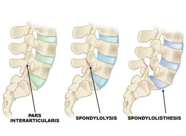
The three can appear together. Wear at L4-L5 can lead to a degenerative slip, and a pars crack at L5-S1 can lead to an isthmic slip.
How Spondylolisthesis Is Checked: X-Rays, MRI and CT
- Standing side-view X-ray. Shows the slip and its grade.
- Bending X-rays. Taken leaning forward and then leaning back, they show whether the vertebra moves between the two positions. A slip that moves is called unstable, and one that stays put is called stable. This split helps guide care (Vanti and colleagues, 2021).
- MRI. Shows the discs, the nerves and ligaments such as the ligamentum flavum. It is used when leg symptoms suggest a pressed nerve or a narrowed canal.
- CT scan. Shows bone in fine detail and can find a pars crack that a plain X-ray misses. In the same study, CT found pars cracks in about twice as many adults as older X-ray studies had.
Does Spondylolisthesis Get Worse Over Time?
For most people, spondylolisthesis changes slowly or not at all. A study that X-rayed 500 first-grade children and followed them into adulthood found pars cracks in 4.4% at age six and in 6% of adults, and a slip rarely grew after the teen years (Fredrickson and colleagues, 1984). When the people with pars cracks were checked again 45 years after the study began, their health scores matched those of the general population, and no one's slip had reached 40% (Beutler and colleagues, 2003).
Degenerative slips follow a similar pattern. A Japanese study followed 145 people with a degenerative slip for 10 to 18 years without surgery. The slip grew in about one in three, but the change did not track with symptoms, and 76% of those who started without nerve problems still had none after 10 years (Matsunaga and colleagues, 2000). People who already had nerve symptoms, such as leg pain on walking, did less well, which is why nerve symptoms that keep getting worse deserve a careful assessment.
How Spondylolisthesis Is Managed Without Surgery
Non-surgical care is the first step for most people with a degenerative slip, with or without nerve symptoms (Kalichman and Hunter, 2008), and it is also the usual starting point for low-grade isthmic slips (Vanti and colleagues, 2021). The aim is to do more of what helps the slipped level and less of what strains it, alone or with help from a chiropractor, physiotherapist or medical doctor. What helps is strength in the deep trunk and hip muscles, free movement in the hips, and positions that keep the lower back from arching for long periods. What strains it is long standing with the back arched, repeated back-bending, and lifting with the back instead of the hips.
Exercise for the deep trunk muscles
In a trial of 44 people with long-term back symptoms and an X-ray showing spondylolysis or spondylolisthesis, one group spent 10 weeks training the deep abdominal muscles together with the multifidus, the small muscles beside the spine, and then used them during the postures and tasks that had been bringing on symptoms. That group reported lower symptom and disability scores, and the change held at 30 months. The group that received usual care showed no real change (O'Sullivan and colleagues, 1997).
Gentle, non-rotatory manual care
Chiropractic care at CSC uses non-rotatory methods, with no twisting of the spine, and the slipped level itself is not pushed into a backward bend. Manual care is aimed at the stiff joints above and below it and at the hips, so that movement is shared more evenly and the slipped level is not doing all the work. Methods include instrument-assisted adjusting with an Activator and sustained positional release.
Physiotherapy
Physiotherapy centers on graded, supervised exercise, stretching of tight hip flexors and hamstrings, and practice in bending and lifting with the hips. Tight hip flexors tilt the pelvis forward and arch the lower back, the position that tends to load a slip.
NSD Therapy®
NSD Therapy® is a non-surgical spine care method invented by Yama Zafer, D.C. It brings together spinal decompression therapy, physiotherapy and non-rotatory chiropractic care, and it is the main approach used for spondylolisthesis at CSC. Which parts are used, and how, is decided after the assessment.
What a plan needs in order to work
For any care of a slip to have a good outcome, four things have to happen. The slip and how it behaves must be diagnosed accurately. The plan must be built around those findings. The plan must be carried out with as few errors as possible. And it must be topped with changes in posture, habits and daily activity. Meeting those conditions takes a provider with in-depth knowledge and experience of slips like this one.
Daily Habits That Ease the Load on a Slipped Vertebra
- Break up long periods of standing. Resting one foot on a low step flattens the arch in the lower back.
- Bend and lift with the hips and knees, and keep the load close to the body.
- Choose low-impact activity such as walking, cycling or swimming, and hold off on deep backward bends until the slip has been assessed.
- Keep a healthy weight, since extra weight adds load to the lower spine.
- Sleep on the side with the knees bent, or on the back with a pillow under the knees.
- Young athletes whose sport arches the back again and again should have back symptoms that keep coming back checked.
When Surgery Is Discussed
Surgery is a last step, not a first one. It is usually considered only when a fair trial of conservative care has not helped, when nerve symptoms keep getting worse, when walking distance stays very short, or when a high-grade slip keeps growing on repeat X-rays. The common operations are decompression, which frees pressed nerves, and fusion, which joins the slipped vertebra to the one below. A slip seen on a scan is not, by itself, a reason for surgery. A keyhole incision is still an incision, and working a camera and tools through it at different angles can disturb the tissue around it.
Planning a visit? You can review the current chiropractic price in Kuala Lumpur and physiotherapy fees in Kuala Lumpur before you come in.
Spondylolisthesis FAQ
What is spondylolisthesis?
Spondylolisthesis is a forward slip of one vertebra over the one below it, most often in the lower back at L4-L5 or L5-S1. The two common causes are wear with age (degenerative) and a stress crack at the back of the vertebra (isthmic). It is graded from 1 to 5 by how far the bone has moved, most slips are grade 1 or 2, and many are found by chance on an X-ray taken for another reason.
What are the symptoms of spondylolisthesis?
Many people have none. When symptoms appear, the common ones are a lower back ache that builds with standing or leaning back, tight hamstrings, leg symptoms from a pressed nerve, and heavy legs after walking that ease with sitting. Bending forward often eases the ache, because it opens the space the nerves pass through. New numbness around the genitals or anus, new difficulty passing urine, or loss of bladder or bowel control needs urgent medical attention the same day.
How is spondylolisthesis diagnosed?
A standing side-view X-ray shows the slip and its grade. Bending X-rays, taken leaning forward and then leaning back, show whether the vertebra moves between the two positions, which separates a stable slip from an unstable one and helps guide care. MRI shows the nerves and discs when leg symptoms suggest a pressed nerve or a narrowed canal, and CT shows fine bone detail such as a pars crack. The images are read together with the examination.
What are the grades of spondylolisthesis?
The Meyerding scale grades a slip by how far the upper vertebra has moved forward, measured as a share of the width of the vertebra below: grade 1 is up to 25%, grade 2 is 26% to 50%, grade 3 is 51% to 75%, grade 4 is 76% to 100%, and grade 5 means the vertebra has slipped fully off the one below. Grades 1 and 2 are the most common. The grade describes the X-ray, not the person, so the examination matters as much.
Can spondylolisthesis be managed without surgery?
In most cases, yes. Non-surgical care is the usual first step, and it centers on exercise for the deep trunk and hip muscles, gentle non-rotatory manual care and changes to daily habits. In a trial of people with a slip or a pars crack, ten weeks of deep trunk muscle training lowered symptom and disability scores, and the change held at 30 months. Surgery is considered when a fair trial of conservative care has not helped or nerve symptoms keep getting worse.
What is the difference between spondylolisthesis, spondylolysis and spondylosis?
Spondylolisthesis is a slip of one vertebra over another. Spondylolysis is a stress crack in the pars, the thin bridge of bone at the back of the vertebra, which may or may not lead to a slip. Spondylosis is age-related wear of the discs and joints, without a slip. The three can appear together: wear at L4-L5 can lead to a degenerative slip, and a pars crack at L5-S1 can lead to an isthmic slip.
Can spondylolisthesis be prevented?
Not every case can be prevented, since isthmic cracks often start in childhood and run in families. Strong trunk and hip muscles, a healthy weight and lifting with the hips all lower the load on the lower spine, while long standing with the back arched and repeated back-bending raise it. Young athletes whose sport arches the back again and again, such as gymnasts, should have back symptoms that keep coming back checked.
Peer-Reviewed References
- Wiltse LL, Newman PH, Macnab I. Classification of spondylolisis and spondylolisthesis. Clin Orthop Relat Res. 1976;(117):23-29.
- Koslosky E, Gendelberg D. Classification in brief: the Meyerding classification system of spondylolisthesis. Clin Orthop Relat Res. 2020;478(5):1125-1130.
- Fredrickson BE, Baker D, McHolick WJ, Yuan HA, Lubicky JP. The natural history of spondylolysis and spondylolisthesis. J Bone Joint Surg Am. 1984;66(5):699-707.
- Beutler WJ, Fredrickson BE, Murtland A, et al. The natural history of spondylolysis and spondylolisthesis: 45-year follow-up evaluation. Spine (Phila Pa 1976). 2003;28(10):1027-1035.
- Kalichman L, Kim DH, Li L, et al. Spondylolysis and spondylolisthesis: prevalence and association with low back pain in the adult community-based population. Spine (Phila Pa 1976). 2009;34(2):199-205.
- Matsunaga S, Ijiri K, Hayashi K. Nonsurgically managed patients with degenerative spondylolisthesis: a 10- to 18-year follow-up study. J Neurosurg. 2000;93(2 Suppl):194-198.
- O'Sullivan PB, Phyty GD, Twomey LT, Allison GT. Evaluation of specific stabilizing exercise in the treatment of chronic low back pain with radiologic diagnosis of spondylolysis or spondylolisthesis. Spine (Phila Pa 1976). 1997;22(24):2959-2967.
- Kalichman L, Hunter DJ. Diagnosis and conservative management of degenerative lumbar spondylolisthesis. Eur Spine J. 2008;17(3):327-335.
- Vanti C, Ferrari S, Guccione AA, Pillastrini P. Lumbar spondylolisthesis: STATE of the art on assessment and conservative treatment. Arch Physiother. 2021;11(1):19.
These references describe spondylolisthesis and its non-surgical care in general. None of them evaluates care at Chiropractic Specialty Center.
The Author of Spondylolisthesis: Causes & Non-Invasive Management
Spondylolisthesis: Causes & Non-Invasive Management was written by Yama Zafer, D.C., who has 30+ years in chiropractic and physiotherapy and founded Chiropractic Specialty Center in Kuala Lumpur in 2006.
Last Updated: Spondylolisthesis: Causes, Grades and Non-Invasive Management
Spondylolisthesis: Causes, Grades and Non-Invasive Management was last reviewed and updated on October 3, 2026.
I have severe back pain with footdrop. Been a couple months. Need help, thanks....
Foot drop should not be ignored, and two months is long enough that it needs a proper assessment now.
Foot drop means the nerve supplying the muscles that lift your foot is not working properly. How well the muscle recovers can depend on how long the nerve stays pressed, so the assessment should include a strength check, reflexes and, where needed, an MRI of the lower back. A competent provider, conservative or surgical, can do this. Conservative care can still be a reasonable route for as long as it keeps producing results. If the weakness keeps getting worse, or care is not helping, a surgical opinion is the next step. When you are seen, say the words foot drop, since they are understood immediately.
Two things worth establishing before that appointment.
How complete it is. Sitting with your leg out, try to lift the front of your foot upward against gravity, then against light resistance from your hand, and compare with the other side. Being unable to lift it at all is different from lifting it weakly, and being able to lift it against resistance is different again. Note also whether it has been getting worse over the two months or has been stable, since that distinction matters as much as the severity.
Whether it is coming from your back or from your knee. This is worth checking because the two are treated differently. The nerve that lifts the foot passes close to the surface just below the outer side of the knee, where it can be compressed by habitual leg crossing, prolonged squatting or kneeling, or significant weight loss. One test separates them: with your foot relaxed, try turning the sole inward against resistance. If turning inward is also weak, the problem is at the nerve root in your back. If turning inward is strong while lifting the foot and turning outward are weak, the compression is more likely at the knee. Given that you also have severe back pain, the root is the more likely source, but this is worth checking and worth mentioning.
Practically, while you are arranging this: be careful on stairs and curbs, since a foot that does not clear the ground catches easily and falls are common with this. Keep the ankle moving through its full range daily so the joint does not stiffen while the muscle is weak, and keep working the muscle even though it is weak, since it responds better than an unused one.
If your symptoms steadily get worse, or the care you are receiving is not helping, please arrange a medical review. New numbness around the genitals or anus, new difficulty passing urine, or loss of bladder or bowel control needs urgent medical attention: a thorough assessment by a competent clinician the same day.
Hi, I have spondylolisthesis at level two, but my leg is weak and in severe pain. The doctor is saying that maybe they will do surgery on my back. So do you have any suggestions? Thanks
The leg weakness is the part of your message that matters most, and it should not be ignored.
Pain alone with a spondylolisthesis, even a grade two slip, is usually a reason to try conservative management before considering an operation. Weakness means a nerve is not just irritated but losing some function, and how well it recovers can depend on how long it stays pressed. That is why weakness calls for a careful assessment now by a competent provider, conservative or surgical, rather than later.
Conservative care can still be a reasonable route for as long as it keeps producing results, and your doctor's surgical opinion is part of the picture. Two things are worth establishing quickly.
Whether the weakness is genuine and which nerve it involves. Test it rather than estimating. Try walking on your heels with the toes lifted, and then on your toes, comparing the two sides. Try rising onto the toes of the affected leg alone, ten times, and see whether it fails earlier than the other side. Try straightening the knee against resistance, and notice whether the knee feels like it might give way on stairs. Each of those maps to a different nerve root and tells the surgeon something specific.
Whether it is getting worse. Repeat the same tests every few days and write down the result. Weakness that is stable is a different situation from weakness that is progressing, and this is the single most important piece of information for the decision. If it is getting worse, tell your provider right away.
Three questions worth taking to your doctor. Which nerve root is being compressed and does it match my weakness. Is the plan a decompression alone or a decompression with fusion, and why. And what is expected to happen to the weakness with and without surgery.
If your symptoms steadily get worse, or the care you are receiving is not helping, please arrange a medical review. New numbness around the genitals or anus, new difficulty passing urine, or loss of bladder or bowel control needs urgent medical attention: a thorough assessment by a competent clinician the same day.
On conservative care, two points so you have the full picture. It has a real role here, both now while the weakness is being tracked and after any operation, since strength and function have to be rebuilt whichever route you take. And the slipped segment itself should not be manipulated into extension. Where manual care is appropriate, it is directed at the surrounding segments and at restoring hip movement, and extension of the lower back is the position that closes the space and provokes this pattern.
Hello, my mum was diagnosed with spondylolisthesis here in Nigeria. What kind of suggestions will you offer on how to treat her, please.
Before anything can be suggested for your mother, three details decide almost everything, and they should be on her report or obtainable from the doctor who made the diagnosis.
The grade. Spondylolisthesis is graded by how far one vertebra has slipped forward on the one below, from grade one, meaning up to a quarter, through to grade four. The large majority of cases are grade one or two, and those are usually managed without surgery. A high-grade slip needs closer follow-up, and a surgical opinion becomes part of that conversation if symptoms keep getting worse or care is not helping.
The type. In older adults it is usually degenerative, most often at L4-L5, where the joints and disc have worn and allowed the segment to shift. In younger people it is more often isthmic, at L5-S1, arising from a stress fracture in a small bridge of bone, frequently dating back to adolescence. The two behave differently and are not managed the same way.
Whether the slip is stable. X-rays taken bending forward and bending backward show whether the vertebra moves between the two positions. A stable slip and a mobile one call for different approaches, and this is the single most useful additional test to ask for.
What her symptoms are also matters more than the diagnosis itself. Mechanical back pain alone, worse on standing and leaning back, is the most favorable picture. Heaviness and aching in both legs that comes on after walking a certain distance and is relieved by sitting or leaning forward indicates the canal has narrowed, which is common with degenerative slips. Pain, numbness, or weakness down one leg in a defined pattern indicates a nerve root.
For a stable, low-grade slip, conservative management is well established and follows a consistent pattern. Extension of the lower back, meaning leaning or arching backward, is the movement that closes the space and provokes symptoms, so activity is arranged to limit sustained extension. Strengthening focuses on the deep abdominal muscles and the gluteals, which control the segment from the front and behind, and hip flexor tightness is addressed because it pulls the lower back into the position that aggravates it. Walking distance is rebuilt gradually rather than pushed. Where the canal is narrow, walking is often easier on an incline or pushing a trolley, because both put the spine into slight forward bend, and that can be used deliberately to build tolerance.
One point on treatment technique: the slipped segment itself is not manipulated into extension. Appropriate manual care is directed at the segments and joints around it and at restoring hip movement.
Surgery is considered where there is progressive weakness, where walking distance is severely limited by canal narrowing despite a proper course of conservative care, or where the slip is progressing on repeat imaging. It is not indicated by the diagnosis alone.
If your symptoms steadily get worse, or the care you are receiving is not helping, please arrange a medical review. New numbness around the genitals or anus, new difficulty passing urine, or loss of bladder or bowel control needs urgent medical attention: a thorough assessment by a competent clinician the same day.
Cervical spondylosis
Cervical spondylosis is the general term for age-related change in the neck: discs losing height and water content, joints at the back of the spine thickening, and bone spurs forming at the margins. It is a description of what a scan shows, not an explanation of why a particular person hurts, and that distinction matters more than anything else in the diagnosis.
The reason is that these changes are close to universal with age. They appear on the scans of large numbers of people who have no neck symptoms whatsoever, and the degree of change on imaging correlates poorly with the amount of pain a person has. So being told you have cervical spondylosis does not tell you what to do about it. What matters is which of three quite different situations you are in.
The first is mechanical neck pain. Stiffness, an ache across the neck and shoulders, worse late in the day and after sustained postures, no symptoms in the arms. This is the most common presentation and it generally responds well to restoring segmental movement, correcting the sustained positions that provoke it, and strengthening the deep neck flexors and the muscles that hold the shoulder blades, which are almost always weak in this group.
The second is nerve root involvement. Pain, numbness, or weakness traveling into one arm in a defined pattern, often with a lost reflex. Where narrowing at the exit opening has irritated a root, this still usually settles with conservative care over weeks to months, but treatment needs to be selected for it rather than applied generally.
The third is spinal cord involvement, and it needs the most care. The signs are clumsy hands, dropping objects, difficulty with buttons and keys, unsteady walking, symptoms in both arms or in the legs, or a change in bladder control. Where those are present, forceful manipulation of the neck is not appropriate, and a careful assessment with current imaging should come before any treatment, because the goal there is to prevent further loss. Gentle conservative care can still be reasonable if the findings allow it and it keeps producing results.
If you can say which of those three your symptoms fit, a far more specific answer is possible. If any of the third group applies, make sure the assessment and imaging come first.