


Schmorl’s Nodes: What They Are and What They Mean
A Schmorl’s node is a small indentation where disc material has pressed upward or downward through the endplate into the body of the vertebra next to it. They appear in a large proportion of ordinary spines, frequently in people with no symptoms, and they are most often reported on imaging taken for an unrelated reason.
A node can be tender for a period when it first forms, particularly if the surrounding bone shows an active inflammatory change, and then quietly stop mattering. Whether it is new, whether the adjacent bone is reacting, and how the segment behaves under load are the questions that carry clinical weight, and all three come from the examination rather than from the phrase on the report.
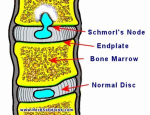
Schmorl’s nodes are changes in the spinal endplates where part of the disc presses upward or downward into the nearby vertebral bone. They are commonly seen on MRI or X-ray imaging and are often found incidentally, even in people who do not notice any symptoms.
When symptoms are present, they are more often linked to surrounding disc wear, endplate irritation, or how the affected spinal segment responds during daily movement and loading.
In some individuals, these changes may influence spinal flexibility, posture, and the way nearby muscles and joints adapt over time.
A structured non-invasive approach focuses on how the spine moves, how pressure is distributed across the discs and endplates, and whether nearby joints, muscles, or posture habits are adding unnecessary stress to the affected level.
At Chiropractic Specialty Center (CSC), chiropractic, physiotherapy, and guided rehabilitation strategies are used to assess spinal mechanics and help improve day-to-day movement function.
This guide to Schmorl’s nodes, what they are and how they relate to disc bulges and protrusions, is published by Chiropractic Specialty Center, Bukit Damansara (KL) main center: WhatsApp+60 17 269 1873 Call+603 2093 1000
Key Takeaways: What to Know About Schmorl’s Nodes
- A Schmorl’s node is an intravertebral disc herniation, where part of the disc moves upward or downward into the vertebral endplate rather than backward toward the spinal canal.
- Many Schmorl’s nodes are incidental findings on MRI or X-ray and may not cause noticeable symptoms.
- When symptoms occur, they are more often linked to endplate irritation, inflammation, surrounding disc wear, or changes in how the spinal segment moves.
- Endplate changes may affect disc nutrition, which in some cases may be associated with gradual disc degeneration over time.
- Movement-focused chiropractic, physiotherapy, and rehabilitation strategies may help improve spinal mechanics, posture, and day-to-day movement tolerance.
Quick Summary of Schmorl’s Nodes and Disc Changes
Use the table below for a quick overview of what Schmorl’s nodes are, how they differ from slipped disc changes, and what they may mean for spinal movement and disc health.
| Topic | Summary |
|---|---|
| What it is | A vertical disc herniation into the vertebral endplate |
| Radiology term | Intravertebral disc herniation |
| Direction of movement | Upward or downward into adjacent vertebral bone |
| Different from slip disc | Does not usually extend backward toward nerves or the spinal canal |
| How it is found | Most commonly seen on MRI, CT, or X-ray |
| Symptoms | Often none; when present, may involve stiffness, inflammation, or movement-related pain |
| Possible long-term effect | May be associated with endplate irritation, reduced nutrient diffusion, and gradual disc wear in some cases |
| Related disc changes | Bulge, protrusion, prolapse, herniation, extrusion |
| How care is structured | Focus on posture, spinal mechanics, movement, and loading patterns |
Is a Schmorl’s Node a Type of Disc Herniation Into the Vertebral Endplate?
A Schmorl’s node is often described in radiology literature as an intravertebral disc herniation, meaning part of the spinal disc moves upward or downward through the vertebral endplate and into the adjacent vertebral bone.
Unlike a more typical disc bulge, protrusion, or herniation that extends backward toward the spinal canal, a Schmorl’s node involves vertical migration through the upper or lower endplate.
In simple terms, it is a disc change that moves into the bone above or below the disc space rather than outward toward the nerves.
These findings are commonly seen on MRI and may be incidental, although acute nodes may sometimes show surrounding inflammation or bone marrow edema.
This distinction is important because the direction of disc migration influences how symptoms and movement changes may present.
How Schmorl’s Nodes Relate to Disc Bulges, Protrusions, and Slip Disc Changes
Although a Schmorl’s node is different from a typical slipped disc, both involve structural changes within the intervertebral disc.
A standard disc bulge, protrusion, prolapse, or herniation usually extends backward or sideways, where it may come closer to the spinal canal, spinal cord, or exiting nerves.
A Schmorl’s node, by comparison, extends vertically through the vertebral endplate.
In some cases, endplate changes may influence nutrient exchange between the vertebral bone and the disc. Because spinal discs depend on nutrient diffusion through the endplates, changes in this area may be associated with gradual disc wear and later degenerative disc changes over time.
For this reason, Schmorl’s nodes may sometimes be seen alongside other disc findings such as bulges, protrusions, or degenerative disc changes in the same spinal region.
This section is an excellent place to naturally link to your pages on disc bulge, disc protrusion, prolapse, extrusion, and slipped disc care.
Consult Our Team for Personalized Spinal Care
If you suspect you have Schmorl’s nodes or are experiencing spinal pain, our team at Chiropractic Specialty Center is here to help. We offer comprehensive assessments and non-invasive care plans tailored to your needs. Contact us today to schedule a consultation and take the first step toward better spinal health.
Understanding Schmorl’s Nodes and Spinal Mobility Considerations
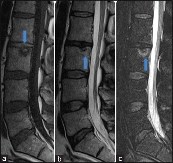
Schmorl’s Nodes refer to structural variations in spinal disc mechanics, where cartilaginous structures from spinal discs extend into vertebral surfaces. These spinal adaptations may be detected through routine spinal imaging assessments and are often associated with structural mobility variations.
For individuals seeking structured spinal mobility assessments, CSC provides targeted movement-based spinal support strategies tailored to individual postural and movement-based needs.
What Are Schmorl’s Nodes?
Schmorl’s Nodes, also known as Schmorl’s Nodules, are structural indentations or protrusions that may develop within vertebral surfaces. These variations occur when spinal disc cartilage extends into vertebral end-plates, influencing spinal mechanics and movement-based function.
- Schmorl’s Nodes are frequently identified in routine spinal imaging assessments, particularly in individuals experiencing spinal mobility concerns.
- Although Schmorl’s Nodes may be present without symptoms, structured spinal movement assessments may help maintain long-term spinal mobility and postural stability.
For personalized spinal mobility assessments, contact CSC for structured movement-based spinal support strategies.
Schmorl’s Nodes in Younger Individuals
Although Schmorl’s Nodes are often associated with structural mobility considerations in aging individuals, they may also be identified in younger adults and teenagers. Research suggests that:
- Approximately 70% of individuals diagnosed with Schmorl’s Nodes may have a genetic predisposition to spinal disc structure variations.
- Vertical spinal disc mechanics adaptations, often associated with vertebral end-plate considerations, may influence movement efficiency.
For individuals with Schmorl’s Nodes identified in early adulthood, structured spinal mobility recovery programs may provide targeted movement-based spinal support strategies to encourage postural stability and joint mobility.
Schmorl's nodes are sometimes reported on a teenager's spine imaging, and the guide to chiropractic care for children explains how a young person's back pain is assessed.
Assessing Schmorl’s Nodes in Spinal Imaging
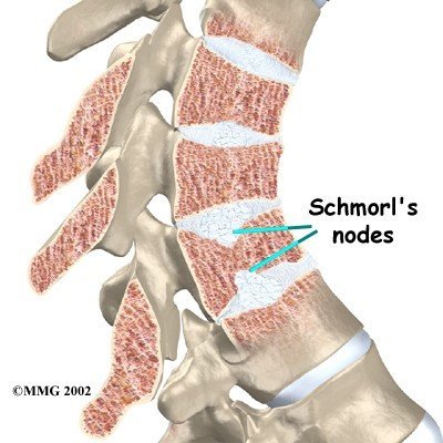
Schmorl’s Nodes are typically identified through routine spinal imaging assessments, including:
- Spinal X-rays: May detect structural indentations in vertebral surfaces.
- CT Scans or MRI Assessments: Provide detailed imaging of spinal disc mechanics and vertebral end-plates.
These structural variations may appear as notches or protrusions extending into vertebral bone structures.
For individuals experiencing spinal mobility considerations, a comprehensive movement-based spinal assessment may provide targeted spinal recovery strategies to maintain long-term mobility and joint function.
Schmorl’s Nodes and How They May Affect Movement
Schmorl’s Nodes are structural indentations or variations in vertebral end-plates, where cartilaginous spinal disc components extend into vertebral surfaces. These spinal adaptations may not always present with movement-based concerns; however, in some cases, they may influence spinal mobility and joint function.
For structured spinal mobility assessments, contact CSC today for targeted spinal recovery solutions.
Are Schmorl’s Nodes Associated With Spinal Pain?
In most cases, Schmorl’s Nodes are not associated with movement-based pain. However, in some individuals, structural variations in spinal mechanics may influence movement efficiency and postural balance.
Potential Factors Influencing Spinal Mobility Considerations:
- Large Schmorl’s Nodes may contribute to spinal mobility variations.
- Structural adaptations in vertebral end-plates may influence soft tissue and joint mechanics.
- Changes in spinal mechanics may be associated with postural and movement-based considerations.
For structured movement-based spinal support, contact CSC for a spinal mobility assessment.
Spinal Disc Integrity and Nutrient Flow Considerations
Spinal discs require continuous nutrient diffusion to maintain structural function and mobility. Unlike other soft tissues, spinal discs do not have a direct blood supply; instead, they receive essential nutrients through vertebral end-plates.
The process of imbibition, where spinal discs absorb nutrients from vertebral surfaces, may be influenced by structural variations in vertebral end-plates. Schmorl’s Nodes may contribute to structural mobility variations, potentially influencing nutrient transfer efficiency.
For movement-based spinal support strategies, contact CSC for structured spinal mobility assessments.
Schmorl’s Nodes and Spinal Mobility Adaptations
In some cases, Schmorl’s Nodes may contribute to postural and joint stability considerations, particularly when associated with:
- Structural changes in spinal disc mobility.
- Postural adaptations influencing movement-based recovery strategies.
- Variations in vertebral mechanics affecting spinal flexibility.
For structured spinal movement strategies, contact CSC today for targeted postural recovery solutions.
How Care and Prevention Are Structured for Schmorl’s Nodes
When a Schmorl’s node is associated with stiffness, movement limitation, or nearby disc and endplate changes, the focus should be on restoring balanced spinal mechanics rather than concentrating on the imaging finding alone.
The goal is to improve how the vertebral endplates, spinal discs, joints, and surrounding muscles work together during everyday movement.
A structured care plan may include:
- Gentle chiropractic and physiotherapy-guided movement work
- Mobility exercises for the affected spinal region
- Flexibility works for surrounding muscles and joints
- Posture and sitting-position review
- Trunk control and spinal stability exercises
- Gradual return to daily activities and exercise
Preventive strategies are equally important, especially for individuals exposed to repeated spinal loading from long sitting hours, lifting, sports, or poor posture habits.
Helpful preventive measures may include:
- Avoiding prolonged slouched sitting
- Reducing repetitive heavy loading
- Improving lifting mechanics
- Taking regular movement breaks
- Maintaining spinal flexibility and core strength
The aim is to reduce excessive vertical loading stress on the vertebral endplates while maintaining normal spinal mobility and function.
Convenient Locations for Spinal Movement Recovery Programs
CSC offers targeted spinal recovery programs across multiple locations in Kuala Lumpur , including:
- Damansara Heights (Bukit Damansara): Near Mont Kiara for targeted spinal mobility recovery solutions.
- Bandar Sri Damansara: Serving Sungai Buloh, Desa ParkCity, and Kepong residents with targeted spinal recovery strategies.
For structured movement-based spinal support programs, contact CSC today for targeted spinal mobility assessments.
Encouraging Spinal Movement Confidence Through Structured Support Strategies
CSC provides structured spinal recovery solutions for individuals managing joint mobility variations, postural adaptations, and spinal integrity considerations.
For targeted spinal mobility strategies, contact CSC today to schedule a structured movement-based spinal support assessment.
Lifestyle Strategies to Support Spinal Health and Prevent Schmorl’s Nodes
Maintaining spinal integrity and postural stability is essential for long-term joint mobility and movement efficiency. Individuals diagnosed with Schmorl’s Nodes or those seeking preventive spinal strategies may benefit from targeted lifestyle modifications to support spinal mechanics and overall function.
Key Lifestyle Strategies for Spinal Integrity:
- Optimize Postural Habits
- Maintain neutral spinal alignment while sitting, standing, or lifting.
- Avoid prolonged slouched positions that place unnecessary stress on vertebral end-plates and spinal discs.
- Engage in Movement-Based Activities
- Incorporate low-impact exercises such as walking, swimming, or controlled flexibility movements.
- Avoid high-impact activities that place excessive stress on spinal structures.
- Limit Excessive Spinal Loading
- Reduce repetitive heavy lifting, particularly in individuals with spinal disc integrity considerations.
- Use proper lifting mechanics by engaging core stability muscles.
- Support Nutritional and Hydration Needs
- Maintain a balanced diet rich in nutrients that support joint and bone health.
- Stay adequately hydrated to encourage spinal disc function and mobility.
- Incorporate Strength and Flexibility Training
- Engage in core stability exercises to maintain spinal integrity and postural balance.
- Avoid overloading spinal joints with excessive weight-bearing activities.
- Minimize Sedentary Lifestyle Factors
- Take regular movement breaks if seated for extended periods.
- Adjust ergonomic workstation setups to encourage proper spinal alignment.
For individuals seeking structured spinal mobility strategies, CSC provides targeted movement-based recovery programs tailored to postural balance and spinal function.
For personalized spinal support assessments, contact CSC today for comprehensive movement-based spinal solutions.
Planning a visit? You can review the current chiropractic price in Kuala Lumpur and physiotherapy fees in Kuala Lumpur before you come in.
Common Questions About Schmorl’s Nodes and Disc Changes
Most discover a Schmorl’s node on MRI or X-ray and immediately wonder whether it is serious, whether it is a slipped disc, and whether it can affect long-term spinal health.
Below are clear answers to the most common questions people ask about Schmorl’s nodes, imaging findings, movement changes, and how they relate to disc bulges, protrusions, and slip disc concerns.
What is a Schmorl’s node?
A Schmorl’s node is a vertical disc herniation where part of the spinal disc moves upward or downward through the vertebral endplate and into the adjacent vertebral bone. Unlike a typical slipped disc, it does not usually extend backward toward the spinal canal or nearby nerves.
Is a Schmorl’s node the same as a Slipped Disc (Sip Disc)?
Not exactly. Both involve disc changes, but the direction is different. A slip disc (slipped disc) usually bulges backward or sideways, while a Schmorl’s node extends vertically into the vertebral endplate above or below the disc space. Learn more about slip discs and what can be done for them without surgery.
Do Schmorl’s nodes always cause symptoms?
No. Many Schmorl’s nodes are incidental MRI or X-ray findings and may not cause noticeable symptoms. When symptoms are present, they are more often linked to inflammation, endplate irritation, or nearby disc wear rather than the imaging finding alone.
Can a Schmorl’s node be seen on MRI?
Yes. MRI is one of the best imaging tools for identifying Schmorl’s nodes, especially when there is surrounding bone marrow edema, inflammation, or acute endplate changes.
Can Schmorl’s nodes be linked with disc degeneration?
In some individuals, changes in the vertebral endplate may affect nutrient exchange between the vertebral bone and the disc. Over time, this may be associated with gradual disc wear or degenerative disc changes in the same spinal region.
Can Schmorl’s nodes occur together with disc bulges or protrusions?
Yes. In some cases, Schmorl’s nodes may be seen alongside disc bulges, protrusions, prolapses, or other slipped disc changes within the same spinal level or nearby segments. This is why imaging findings should always be reviewed together with symptoms and movement assessment.
References:
- Kim H, Raorane H, Sharma S, Wu P, Jang I-T. “Schmorl’s Node: What Is It, Symptoms, Management, and More.” Osmosis, 2020. Patient-education source, not a peer-reviewed study.
- Surucu Kara I, Çalmasur A, Orbak Z, Karavas E, Soyturk M. “Schmorl’s node and vitamin D deficiency: cause or coincidence.” Bone Abstracts, 2017;6:P083.
- Williams FMK, Manek NJ, Sambrook PN, Spector TD, MacGregor AJ. “Schmorl’s nodes: common, highly heritable, and related to lumbar disc disease.” Arthritis & Rheumatism. 2007;57(5):855-860.
- “Schmorl Node: A Cause of Acute Thoracic Pain: A Case Report and Pathophysiological Mechanism.” International Journal of Spine Surgery. 2020;14(3):441. This is a single case report, not a study of treatment.
- Healthline. “Schmorl’s Nodes: Causes, Diagnosis, and Treatment.” Reviewed by Angelica Balingit, MD, 2024. Patient-education source, not a peer-reviewed study.
- PubMed. “Percutaneous vertebroplasty in painful Schmorl nodes.” 2005;16328689.
- Egyptian Journal of Radiology and Nuclear Medicine. “Lumbar spine Schmorl’s nodes; prevalence in adults with back pain, and their relation to vertebral endplate degeneration.” 2019.
The Author of Schmorl’s Node: What It Means on an MRI Report
Schmorl’s Node: What It Means on an MRI Report was written by Yama Zafer, D.C., who has 30+ years in chiropractic and physiotherapy and founded Chiropractic Specialty Center in Kuala Lumpur in 2006.
Last Updated: Schmorl’s Nodes: What They Are and What They Mean
Schmorl’s Nodes: What They Are and What They Mean was last reviewed and updated on March 30, 2026.
Hi
I have significant mid and lower back pain for many years. I have tried multiple chiropractors, physical therapy, and acupuncture unsuccessfully. The only thing that seems to help temporarily is taking anti-inflammatory medication, which I don’t prefer since it makes me drowsy and has other side effects.
I don’t have any traumatic accidents that caused the pain, I believe it’s due to excessive sitting and poor posture over the years. What do you recommend? I am in dire need of a permanent solution to get rid of this pain; it’s disabling me from doing many daily activities.
One line in your message is the most informative thing in it, and it points somewhere different from where you have been looking.
Anti-inflammatory medication being the only thing that helps, alongside years of pain in both the mid and lower back, and multiple mechanical treatments failing, is the pattern that should prompt investigation for inflammatory back disease rather than more manual therapy. A marked response to anti-inflammatories is one of the recognized features used to identify it, and the repeated failure of chiropractic, physical therapy, and acupuncture fits, because those address mechanical problems and this is not one.
Check yourself against the other features. Morning stiffness lasting more than thirty minutes, timed rather than estimated. Pain that improves with movement and worsens with rest, which is the reverse of mechanical pain. Waking in the second half of the night with pain that drives you out of bed. Alternating buttock pain. Onset before the age of forty, building gradually rather than after an injury. And any personal or family history of psoriasis, inflammatory eye conditions, or inflammatory bowel disease.
If several of those fit, ask your doctor specifically to investigate for axial inflammatory back disease. The tests are inflammatory markers in the blood, imaging of the sacroiliac joints, and a specific genetic marker, and the appropriate specialty is rheumatology. This condition is regularly missed for years in exactly the way you describe, with people cycling through manual therapies that were never going to address it. The treatment is entirely different, and effective medication exists.
Involvement of the mid-back over years is worth mentioning specifically to your doctor too, since persistent thoracic pain is less commonly mechanical than lumbar pain and deserves its own consideration.
One practical point on the medication. Anti-inflammatories do not typically cause drowsiness. If yours does, check what you are actually taking, because combination products often include a muscle relaxant or an antihistamine alongside the anti-inflammatory, and those are what cause the drowsiness. A plain anti-inflammatory may suit you better, and that is worth asking a pharmacist about.
If the inflammatory investigations are negative, then the mechanical route still applies, but with a different emphasis from what you have tried. Passive treatment repeated over years does not change capacity. Progressive strengthening, increased in small steps and continued for months, is the part that does, and it is the component most often absent from the treatments people list when they say nothing has worked.
Fever, unexplained weight loss, or pain that is constant and unaffected by position call for a medical review. If your symptoms steadily get worse, or the care you are receiving is not helping, please arrange a medical review. New numbness around the genitals or anus, new difficulty passing urine, or loss of bladder or bowel control needs urgent medical attention: a thorough assessment by a competent clinician the same day.
I have a question; I'm 55 and have been to three different doctors for my back. The first one told me I had metastatic cancer, so I went through all the testing and pet scan and was told I didn't have cancer. The second doctor told me I had a fracture. At my last appointment with him, I was told I had an infection in the fracture, and It went away with no antibiotics. I felt for a while I might be getting better, or at least I was hoping. I started having a lot of pain again and went to a third doctor, and he told me I have Schmorl's nodes. I tried water therapy and was given a tens unit to help with the pain. My back seems to go back and forth from appearing like it might be feeling a little better to terrible pain, especially nerve pain. I'm at my wit's end, and I don't know what to do. I've never taken pain medication and don't want to.
Three doctors have not necessarily given you three contradictory answers. There is a good chance they have described the same thing at different stages, and understanding that is the first step out of where you are.
When an endplate fractures and disc material presses into the bone, the surrounding bone marrow swells. On MRI that swelling produces a bright area, and that appearance is one of the recognized mimics in spinal imaging. It can resemble infection. In some circumstances it can raise the question of a bone lesion. As it heals over months, the swelling fades and what remains is described as a Schmorl's node. So a sequence of cancer suspected, then fracture, then infection, then Schmorl's nodes, is a familiar pattern when a single healing endplate injury is imaged repeatedly by different people who have not seen the earlier films.
One detail supports that strongly. A genuine infection in the spine does not resolve without antibiotics. If you were told there was an infection and it went away with no treatment, the far more likely explanation is that the appearance was marrow swelling from the fracture rather than infection at all. That should be some relief.
What you need now is not a fourth opinion but one person reviewing all of it together. Collect every scan you have had as images on a disc, not as reports, along with the dates, and ask for a single specialist review of the whole sequence with one question attached: is this the expected evolution of a healing endplate fracture, and is anything still active. Comparing the films in order answers in one appointment what four separate opinions could not.
There is a second item that may have been lost in all of this, and at 55 it matters more than the label. If you had a vertebral fracture without a significant fall or accident, that fracture itself is the finding that needs explaining. Ask for a bone density scan, and for vitamin D, calcium, and the blood tests used to look for secondary causes of bone loss. A fracture from ordinary loading is how osteoporosis frequently announces itself, and it is treatable. It would be an unfortunate thing to have missed while three opinions concentrated on naming the lesion.
On the nerve pain, that deserves separate attention. Write down where it travels to at its furthest point, whether that distance changes with position, and whether there is any weakness, any foot catching, or any change in sensation. A healing fracture can reduce the height of the opening a nerve exits through, and that is a different problem from the bone itself with a different solution.
On pain medication, your position is respected and it does not have to change. It is worth knowing that pain kept at a level where you cannot move much will slow the rehabilitation that eventually gets you out of this, and that there are options between nothing and strong medication. That is a conversation to have on your own terms rather than a recommendation.
Water therapy and a TENS unit both manage symptoms. Neither addresses bone healing or bone density, so they are reasonable alongside the above and not a substitute for it.
If your symptoms steadily get worse, or the care you are receiving is not helping, please arrange a medical review. New numbness around the genitals or anus, new difficulty passing urine, or loss of bladder or bowel control needs urgent medical attention: a thorough assessment by a competent clinician the same day. Fever or unexplained weight loss alongside back pain also call for a medical review.
I've never done any weight lifting in my life, and I've never had trauma or accident.
Neither weight lifting nor trauma is required, and their absence in your history is entirely usual rather than a puzzle.
Most Schmorl's nodes form during the growing years, when the endplates at the top and bottom of each vertebra are cartilaginous and comparatively weak. Ordinary loading during that period can push disc material vertically into the bone through a weak point in the endplate, and it does not take an injury or an unusual activity. Studies of families and twins point to a substantial inherited component, meaning endplate strength and disc composition vary between people from the outset. Someone who has never lifted anything heavy can have them, and someone who has lifted for thirty years may not.
One correction worth making, because you may see it written elsewhere: they are generally not congenital in the sense of being present at birth. They develop, most often during growth, which is a different thing and matters because it means they are not something you were born destined to have symptoms from.
There are two other routes worth knowing about, and one of them is checkable. Nodes can form later in life where bone has become weaker, particularly with reduced bone density, and less commonly in association with other conditions affecting bone. Since your nodes are scattered across several levels and there is no trauma and no heavy loading in your history, a bone density scan and a check of vitamin D and calcium status are reasonable to request. That is a straightforward test that either finds something correctable or removes a worry.
Two implications follow from all this. First, nothing you did caused them, and nothing you avoided would have prevented them. Second, and more usefully: having them is not a reason to avoid loading now. Avoidance reduces bone density and muscle strength over time, both of which protect the spine, so the goal is sensible loading with attention to technique rather than protection from it.
I have scattered Schmorl nodes L1-L5+L5-S1. I have mild desiccation with disc herniation with bulging discs. It's not supposed to be painful, but mine is extremely painful. We started with physical therapy, but that worsened my condition, so we will try Aquatic therapy. If all fails, I strongly believe that I should have surgery because if left untreated and it really gets bad, one could become paralyzed. Do you have any suggestions for me? I've basically been bedridden for 6 weeks and haven't been doing anything because it hurts so bad. Though I'm not complaining about my doctor, he wants to monitor me. Do you have any suggestions for me because I surely don't want to become paralyzed?
Thank you for your consideration.
The belief driving your plan deserves addressing first, because it is the part most likely to lead you somewhere you would not choose.
Scattered Schmorl's nodes, mild disc desiccation, and bulging discs across several lumbar levels do not describe a condition that progresses toward catastrophe if left untreated. That is worth stating plainly, because the fear that it will is common and it pushes people toward operations that this particular picture responds to poorly. Surgery is not preventive for degenerative change. It addresses a specific structure compressing a specific nerve, and it works best when one level clearly explains the symptoms and the examination agrees with the scan. The more levels involved, and the less any single one accounts for the pain, the worse surgery performs. Widespread findings with severe pain and no single matching level is, in outcome terms, close to the least favorable situation to operate on.
So the question is not whether to have surgery if everything else fails. It is whether there is a single identifiable target. That is answered by a neurological examination and, where a level is suspected, by a diagnostic block: an image-guided injection of local anesthetic at one level that temporarily removes your pain. If no target can be identified that way, an operation is unlikely to help you and may leave you worse.
Second, the physical therapy that made things worse. That is information rather than failure, and it usually means one of two things: the starting dose was too high, or the exercises loaded the direction that provokes you. It does not mean you cannot do rehabilitation. It means the entry point was wrong. Aquatic therapy is a sensible response for exactly that reason, since it reduces load while allowing movement, and it is often the way back into land-based work rather than a replacement for it.
Third, and this follows from pain being described as extreme while the imaging is described as mild: when pain is markedly out of proportion to the structural findings and flares with ordinary graded activity, the nervous system's own responsiveness is usually part of the picture. That is not a suggestion the pain is imagined. It is a recognized and treatable component, and it responds to graded exposure started far below the level that provokes, to consistent sleep, and to pacing, rather than to further structural investigation.
One thing worth checking if it has not been: whether an inflammatory cause has been excluded. Morning stiffness over half an hour, pain that eases with movement and worsens with rest, and pain waking you in the second half of the night all point that way and are investigated with blood markers.
Finally, have one clinician take the lead and write down what is being treated, what should improve, and by when. Multiple approaches tried in sequence without a baseline is how years pass without a clear answer.
I'm 24, and I have been diagnosed with superior endplate collapse/chronic stress fracture with Schmorl's node and Modic Type-II Changes in the D11 Vertebral body. Should I be worried about any consequences later in my life? Can I do weight training and regular jogging? Is there any way I can make everything back to normal?
Taking your three questions in order, because the answers are different in tone and it is worth being precise.
Should you worry about consequences later. Less than the wording of the report suggests. Two details in it are actually reassuring. Modic type two changes indicate fatty marrow change, which is the appearance of a settled, chronic state rather than active inflammation. Type one, with marrow swelling, is the pattern associated with an active and often painful process, and you do not have that. And a chronic stress fracture with an endplate change at T11 in someone your age generally reflects an injury that occurred during growth, has healed, and is now stable. Stable is the operative word. This is not a condition that progresses on its own in the way the phrase endplate collapse implies.
What does influence your next thirty years is not the appearance of that vertebra but the strength and conditioning of the spine around it, and that is within your control.
Can you do weight training and jogging. Yes to both, with attention to how rather than whether. Jogging is well tolerated by this kind of change and worth building gradually rather than starting at your old volume. For weight training, the pattern worth respecting is heavy compressive load applied through a rounded spine, which is the one combination associated with endplate injury and the one that is easy to avoid. In practice: hinge at the hips with a neutral spine, use a trap bar rather than a straight bar for deadlifts if you have the option, use chest-supported rows, and replace weighted sit-ups and loaded crunches with planks, dead bugs, and loaded carries, which train the trunk without repeatedly bending a loaded spine. Progress load slowly and do not chase single maximum lifts. T11 sits near the junction where the stiffer upper back meets the mobile lower back and takes high bending stress, so quality of position through that region matters more than the number on the bar.
Can everything go back to normal. Not on imaging, and it is better to hear that plainly. A healed endplate change does not remodel back to an unmarked vertebra, and any future scan will continue to describe it. But imaging appearance and function correlate poorly, and normal function, full activity, and being free of symptoms are realistic goals that many people with identical reports achieve. The target is what you can do, not what the film says.
One thing worth arranging at 24 with a stress fracture in your history: a check of vitamin D and calcium status, and an honest look at whether your nutrition has matched your training load, since inadequate intake relative to activity is a common and correctable contributor to stress injuries in this age group.
Get assessed rather than training through it if you develop new focal pain that does not settle, pain that wakes you at night, or any leg symptoms, numbness, or weakness.
Can a Schmorl's node along the superior endplate of L2 lead to spondylodiscitis? Having a hard time figuring out who to see after two visits to the ER found infection in my blood work with a fever and severe pain in stomach and back, and all the CT scans showed in the findings was the node. However, while the fever is gone, my lower back keeps having spasms and pain, so I see if there could be a correlation.
The most useful thing in this reply is a single point about imaging: CT is not the test that rules out a disc space infection, and a CT showing only the node does not settle the question. MRI with contrast is the investigation for this, and it detects the changes far earlier and far more reliably. If you have had documented infection in your blood alongside severe back pain, and no one has yet performed an MRI with contrast, that is the specific thing to ask for.
The combination you describe is the one that clinicians are taught to take seriously: a documented bloodstream infection together with new severe back pain. Bacteria circulating in the blood can settle in a disc and the bone beside it, and the back pain often begins before anything is visible on a CT scan. Fever settling does not exclude it, particularly if you were given antibiotics at any point, because partial treatment can suppress the fever while the infection in the spine continues.
There is a further complication worth knowing, because it explains why this is confusing. An acute Schmorl's node with active bone marrow change and an early disc space infection can look similar on imaging, and they are distinguished by the pattern on MRI together with blood markers rather than by the CT appearance. So the node on your scan is not necessarily the explanation for your symptoms, nor is it necessarily unrelated.
On who to see, the practical answer is not another emergency department visit. Ask to be referred to an infectious diseases specialist or a spine surgeon, and take the following with you: the CT reports, the blood results that showed infection including which organism if one was identified, and the dates of any antibiotics you were given. Ask for current inflammatory markers, meaning ESR and CRP, since those are the tests used to track this and a trend across dates is far more informative than a single value.
Continuing back spasm and pain weeks after a documented infection is a reasonable basis for that request, and it is worth making it firmly.
If fever returns or pain wakes you at night, get urgent medical attention the same day rather than waiting for an appointment, given the infection in your history. New numbness around the genitals or anus, new difficulty passing urine, or loss of bladder or bowel control also needs urgent medical attention: a thorough assessment by a competent clinician the same day.
Hi! I was 17 years old(2017) when I was diagnosed with multilevel schmorl's node formation in the lumbar region, and it caused a lot of pain. Sometimes the pain was unbearable. After continuous medications for one year, the pain disappeared. But after five years, the pain came back again. This time it is even more severe than before! Although I was taking all necessary precautions during these years. What should I do now?
Two things in your history deserve to be raised with your doctor, and neither may have come up the first time.
The first concerns the original diagnosis. Multiple Schmorl's nodes appearing across several lumbar levels in a seventeen year old is a specific pattern, not a random finding. It is the picture associated with endplate injury during growth, and where it occurs with vertebral wedging and an increased forward curve in the mid-back it has a name and a recognized course. Ask whether that was considered, and ask for the posture of your thoracic and thoracolumbar spine to be assessed rather than only your lumbar images reviewed. It matters because that condition has established management, and because it changes what your rehabilitation should focus on.
The second is more important, and it is the reason to see a doctor rather than a therapist first. Severe back pain that began in your teens, settled, and has returned worse in your early twenties should have inflammatory causes excluded before anything else. There is a category of inflammatory back conditions that typically begins before forty, and it is regularly missed for years because it is assumed to be mechanical. The features that distinguish it are specific and you can check them yourself. Morning stiffness lasting more than half an hour. Pain that improves with movement and gets worse with rest, which is the opposite of ordinary mechanical back pain. Pain in the second half of the night that wakes you and drives you out of bed. A marked response to anti-inflammatory medication. Alternating buttock pain. Any history of eye inflammation, psoriasis, or bowel inflammation in you or your family.
If several of those fit, ask your doctor specifically to investigate for inflammatory back disease, including blood markers of inflammation and imaging of the sacroiliac joints. This is worth pressing on, because the treatment is entirely different from the treatment for a mechanical problem, and earlier diagnosis matters.
On what to do now regardless of which of these applies. Your imaging is from 2017 and no longer describes your spine, so current imaging is the starting point rather than a re-reading of the old films. And there is a lesson in how the first episode was handled: a year of medication settled the pain but nothing in that year built the capacity of your back. When pain is managed and strength is not restored, the tissue is left less able to tolerate load than before, which is a common reason problems return in a worse form years later.
So the plan this time should include what the last one did not: a progressive loading program built up gradually rather than rest and medication alone. Precautions and avoidance feel protective and, over five years, tend to leave you weaker rather than safer.
Fever, unexplained weight loss, or pain that is constant and unrelated to position call for a medical review. If your symptoms steadily get worse, or the care you are receiving is not helping, please arrange a medical review. New numbness around the genitals or anus, new difficulty passing urine, or loss of bladder or bowel control needs urgent medical attention: a thorough assessment by a competent clinician the same day.
Thank you so much for the information here. I am 45 yrs old and; have schmorl's nodes, scoliosis, herniated discs, Si joint dysfunction, and scoliosis. I knew I had scoliosis since a child, but I recently became aware of other things due to pain and MRI scans. I have worked with children since I was a teen and; was working as a Child Development worker FT for the past 5 yrs, but have been on leave for several months due to my chronic pain. I see a physiotherapist, chiropractor, and massage therapist, but things are not getting a lot better; my work asks when I will be coming back. I don't know what to say. Do you recommend that I not go back to that line of work? Lifting children, & bending were regular tasks.
No one should tell you to leave your career from a comment box, and I am not going to. But the question can be reframed into one that is answerable, and there is something in your account that deserves attention before any decision about work is made.
You are seeing a physiotherapist, a chiropractor, and a massage therapist at the same time, and you are not improving. That pattern is worth examining, because three practitioners working in parallel usually means no single person owns the plan. Nobody can tell which input is helping, nothing is measured against a baseline, and there is no agreed point at which the approach changes if it is not working. Months can pass that way while everyone involved is doing reasonable work. The first change to make is not a fourth opinion. It is to have one clinician take the lead, write down what is being treated and why, define what should improve and by when, and set a review date at which the plan changes if the targets are missed.
The second point concerns what is likely missing. Manual treatment and massage manage symptoms. They do not build capacity. Returning to a job that involves repeatedly lifting children and bending requires a specific physical capacity, and that is developed by graded, progressive loading over months, not by treatment received passively. If nothing in your current care is progressively loading you, the gap between what your body can currently do and what the job demands is not closing, whatever the treatment feels like at the time.
That reframes your question usefully. Rather than should I return, ask: what exactly does the job require, what can I do now, and can the gap be closed over a defined period. Write the demands down concretely, the weight of a child lifted from the floor, how many times an hour, how long spent bent or kneeling, how long on your feet. Then have your capacity tested against those numbers and reassessed after a defined block of work, say twelve weeks. That gives you evidence rather than a guess, and it gives your employer something specific.
On what to tell your work now: an interim answer is legitimate and usually better received than an open-ended one. Telling them you are following a structured program with a review at a set date, and that you will give a definite answer then, is honest and gives them something to plan around. Ask also what modifications are possible, because a graded return, reduced hours, a lifting restriction, encouraging children to climb up rather than be lifted, and adjusting table and chair heights can change the demands substantially. Many people who cannot return to the full role can return to a modified one and build from there.
On the diagnoses themselves, one thing is worth saying. Schmorl's nodes, disc changes, and scoliosis known since childhood are findings that appear on a great many scans, including those of people with no pain. Scoliosis you have had for over thirty years is not a new cause of pain at 45. So the list on your report does not necessarily identify what is hurting you, and treating every item on it is a common reason care goes in circles. The sacroiliac joint is the one item on your list that is diagnosed by examination rather than imaging, and it is worth confirming it has actually been tested rather than inferred.
Finally, pain that has kept you off work for several months has effects of its own that are treatable and often untreated: disrupted sleep, loss of conditioning, and a reasonable wariness of the movements that hurt. Those respond to graded exposure and to addressing sleep directly, and leaving them out is another reason progress stalls. If a full medical review has not been done since this began, that is also worth having, to confirm nothing else is contributing.
I have t11 and t12 l3,l4 damage. I also have schmorl's nodes. I am 59. Should I avoid heavy or moderate weight lifting exercises? I currently do mostly abdominal and light back and leg exercises.
Blanket avoidance is usually the wrong answer at 59, and it is worth explaining why before getting to the modifications.
Schmorl's nodes are areas where disc material has pushed vertically into the bone of the vertebra above or below rather than backward toward the nerves. They are common, appear on a substantial share of spine scans, and in most people are old, stable, and unrelated to current symptoms. On their own they are not a reason to stop lifting.
One question decides whether that applies to you, and it is worth asking whoever holds your images: is there marrow edema at any of those levels, or are the margins well defined and corticated. Edema indicates a recent or active change, and loading through that level should be reduced while it settles. Well-defined margins indicate old, healed lesions, which do not need to be worked around.
The second thing worth clarifying is the word damage. It is not a radiological term, and what your report actually says at T11-T12 and L3-L4 matters, because degenerative change, a disc herniation contacting a nerve, and a healed endplate lesion have quite different implications for training.
Assuming no active lesion and no nerve involvement, here is the substance of it. Avoiding load is not a neutral choice at your age. Bone density and muscle mass both decline without resistance training, and both protect the spine. Stopping lifting reduces the tissue's tolerance and makes ordinary daily loads relatively harder, which is a poor trade. What should change is not whether you load, but how.
The combination worth respecting is heavy compressive load applied through a fully rounded spine. That is the loading pattern most associated with endplate and disc injury, and it is also entirely avoidable. In practice that means hinging at the hips with a neutral spine rather than rounding the back, favoring a trap bar over a straight bar if you deadlift, using chest-supported rowing, and replacing loaded sit-ups and weighted crunches with anti-movement work such as planks, dead bugs, and carries, which train the trunk without repeatedly bending a loaded spine. Split squats and leg presses set up so the lower back stays neutral are usually well tolerated. T11-T12 sits at the junction where the stiffer thoracic spine meets the mobile lumbar spine and carries high bending stress, so technique through that region matters more than the absolute weight on the bar.
On intensity: moderate loads for moderate repetitions, progressed slowly, achieve nearly all the benefit. There is little reason to test single maximum lifts at 59, and that is where technique fails first.
Two things worth arranging. A bone density scan, because if bone density is low that genuinely does change the recommendation, particularly regarding loaded flexion and rotation. And a few supervised sessions to check the hinge pattern under load, since almost all of the risk here lives in technique rather than in the diagnosis.
Stop and be assessed rather than training through it if you get sudden focal pain during a lift that does not settle, pain that wakes you at night, or any new leg symptoms, weakness, or numbness.