

L1-L2 Disc Problems: Symptoms, MRI Findings and Care
L1-L2 is the first and second lumbar vertebrae and the disc between them, high in the lower back. The nerves emerging at this level supply the groin and the upper front of the thigh, so symptoms arising here appear there rather than in the calf or the foot, which is one useful clinical clue about the level.
Upper lumbar levels are involved far less often than L4-L5 or L5-S1, so a finding at L1-L2 is more frequently incidental than causative.
This clear CSC guide explains L1-L2 disc problems, symptoms, MRI findings, nerve signs, and care so readers can understand what the scan may mean and when further assessment is important.
L1-L2 is an upper-lumbar motion segment between the first and second lumbar vertebrae. It helps support the trunk and lies near the exiting L1 and traversing L2 nerve-root pathways. Disc, facet-joint, ligament, and nerve-space findings at this level can be clinically relevant, but upper-lumbar symptom patterns are variable and do not always follow a neat map. (Kim et al., 2010)
This page is general education. It cannot determine which structure explains an individual symptom, whether imaging is needed, or which form of care is suitable. MRI findings should be interpreted together with the history, examination, neurological findings, and any referral needs. (Brinjikji et al., 2015)
This guide to L1-L2 disc problems, their symptoms, MRI findings and why the two may not match, is published by Chiropractic Specialty Center, Bukit Damansara (KL) main center: WhatsApp+60 17 269 1873 Call+603 2093 1000
Key Points About L1-L2
| Question | Clear answer |
| Where is L1-L2? | It is the upper-lumbar segment between L1 and L2. |
| Which nerve pathways are nearby? | The L1 root exits through the foramen; the L2 root continues downward through the canal and lateral recess. |
| Do MRI findings always explain symptoms? | No. Degenerative and disc findings may be present without symptoms and require clinical correlation. |
| Which symptoms may be associated? | Patterns may include upper-lumbar or groin-region pain, front or upper-thigh sensations, or changes in hip-flexion strength, but other causes must be considered. |
| When is urgent medical assessment important? | With an L1-L2 disc problem, any new symptoms should be considered alongside your back or leg pain. New or worsening numbness around your genitals, between your genitals and anus, or around your anus needs urgent medical attention: a thorough assessment by a competent clinician the same day. New difficulty passing urine needs the same urgent assessment, even if bladder and bowel control remain intact, and so does any loss of that control. Several conditions can cause these symptoms. Some may call for an invasive procedure, but being assessed helps identify the cause and does not automatically lead to surgery. Progressive weakness, major trauma, fever with spinal symptoms, or concern for fracture, infection or cancer also need prompt evaluation. |
Where L1-L2 Sits and What It Does
L1-L2 is within the upper lumbar spine, below the thoracolumbar junction. The vertebral bodies and disc share load, while the facet joints and surrounding ligaments guide and limit motion. The spinal canal lies behind the vertebral bodies, and the intervertebral foramen forms an opening at each side of the segment. (Fardon et al., 2014)
At the L1-L2 level, the exiting L1 nerve root passes through the L1-L2 foramen. The L2 nerve root travels downward within the canal before exiting at the level below. A foraminal or far-lateral finding may therefore be closer to the exiting L1 root, while a posterolateral or lateral-recess finding may be closer to the traversing L2 root. This is an anatomical relationship, not proof that the finding is causing symptoms. (Fardon et al., 2014)

Figure 1. The direction of an L1-L2 finding may place it closer to the exiting L1 or traversing L2 nerve-root corridor. Imaging still requires clinical correlation.
Common MRI Terms at L1-L2
MRI reports use standardized terms to describe anatomy. These words describe what is visible; they do not, by themselves, establish the source of pain, numbness, weakness, or reduced function. (Fardon et al., 2014)
- Disc degeneration or desiccation: The disc shows reduced water signal, height loss, or other age- and load-related changes.
- Disc bulge: Disc tissue extends broadly beyond the edge of the disc space. A bulge is not the same as a focal herniation.
- Disc protrusion: A focal herniation in which the base is wider than the outward portion of the displaced material.
- Disc extrusion: A focal herniation in which the outward portion is wider than its base, or the material extends beyond the disc space.
- Sequestration: A disc fragment has lost continuity with the parent disc.
- Annular fissure: A separation within the disc annulus. The radiology term annular fissure is generally preferred to wording that assumes a traumatic tear.
- Facet hypertrophy: The facet joints appear enlarged or degenerative and may contribute to reduced space near a nerve corridor.
- Ligamentum flavum hypertrophy: The ligament at the back of the canal appears thickened and may contribute to canal or lateral-recess narrowing.
- Foraminal or lateral-recess narrowing: The space near an exiting or traversing nerve root appears reduced; the clinical relevance depends on the whole presentation.
Related reading: disc bulge terminology and MRI meaning and MyChiro MRI guide.
Why L1-L2 MRI Findings and Symptoms May Not Match
Degenerative disc findings, bulges, protrusions, and annular fissures are common on imaging, including in people who report no symptoms. Their frequency generally increases with age. For that reason, the presence of an L1-L2 finding should not automatically be described as the cause of every symptom. (Brinjikji et al., 2015)
Upper-lumbar disc herniations can also produce variable patterns. Published surgical series describe front or anterolateral thigh symptoms, weakness, sensory changes, or broader patterns rather than one reliable symptom map. Similar groin or upper-thigh symptoms can arise from the hip, abdominal wall, pelvis, urinary system, or other neurological and musculoskeletal sources. (Kim et al., 2010)
Clinical correlation means comparing the scan with the history, examination findings, neurological changes, movement tolerance, and other relevant health information. It may also mean deciding that the scan finding is incidental or that another form of assessment is needed. (Brinjikji et al., 2015)
What an L1-L2 Finding May Mean - and What It Does Not Mean
| An L1-L2 finding may mean | It does not automatically mean |
| A structural change is visible on MRI. | The finding is the cause of every symptom. |
| A nearby nerve-root corridor may have less space. | The nerve is permanently damaged. |
| Several structures may be contributing. | One named method is appropriate for everyone. |
| Further assessment may be useful. | Imaging, decompression, injections, or surgery are automatically required. |
| The wording can guide questions for a clinician. | The scan predicts a fixed outcome or recovery timeline. |
Symptoms That May Be Associated With L1-L2
L1-L2 findings may be associated with upper-lumbar pain, groin-region symptoms, front or upper-thigh pain or tingling, or changes in hip-flexion strength. Some people describe difficulty with stairs, hills, or lifting the thigh. These are possible associations rather than a diagnosis based on symptom location. (Kim et al., 2010)
A far-lateral or foraminal finding may be closer to the exiting L1 root, while a posterolateral finding may be closer to the traversing L2 root. Central upper-lumbar findings can affect more than one neural structure because the upper-lumbar canal contains several descending nerve roots and lies nearer the conus medullaris than lower lumbar levels. (Kim et al., 2010)
Why Position and Activity Can Change Symptoms
Groin pain, abdominal-wall symptoms, hip pain, and upper-thigh changes also have many non-spinal explanations. New, severe, progressive, or unexplained symptoms should not be assumed to come from L1-L2 without an appropriate assessment.
Sitting, standing, bending, lifting, and walking distribute load differently across the disc, facet joints, ligaments, and nerve spaces. Some people notice symptoms during extension or prolonged upright activity; others are more sensitive to sitting or flexion. No single position is universally better for every L1-L2 presentation.
A useful assessment looks for repeatable patterns: which movements increase or decrease symptoms, whether strength or sensation changes, how walking is affected, and whether the response is local or travels into the groin or thigh. A temporary positional change does not, by itself, identify the responsible structure.
Facet and Ligamentum Flavum Findings
Facet hypertrophy describes enlargement or degeneration of the small joints behind the disc. Disc-height loss and facet-joint changes often coexist. Together, they may contribute to reduced foraminal or lateral-recess space, but the scan and the clinical picture may not match closely. (Fujiwara et al., 1999)
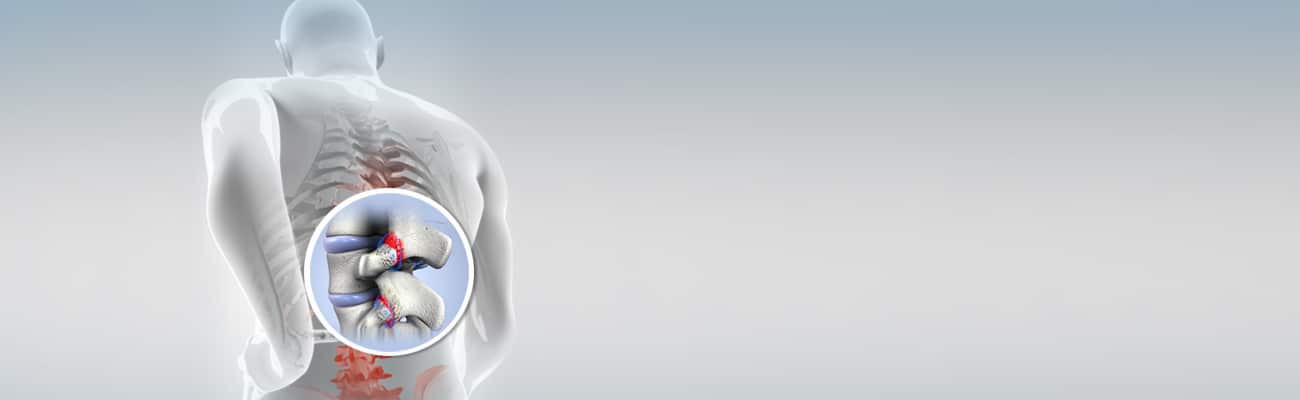
Ligamentum flavum hypertrophy describes thickening of the ligament at the back of the spinal canal. Thickening can contribute to narrowing, especially when combined with disc and facet changes. The finding is not automatically symptomatic and does not determine one particular form of care. (Sairyo et al., 2005)
How Nearby Lumbar Levels Relate to L1-L2
Lumbar MRI reports often list changes at more than one level. Nearby-level findings should be interpreted individually rather than assuming that one level mechanically causes another. The main value of the level-by-level guides is to clarify anatomy, terminology, and the nerve pathways that may be nearby.
- L2-L3 upper-lumbar guide
- L3-L4 lumbar guide
- L4-L5 disc and nerve guide
- L5-S1 disc and nerve guide
- coccydynia and tailbone guide
How an L1-L2 Concern Is Assessed
An assessment may include the history and timeline of symptoms, aggravating and easing factors, walking and stair tolerance, hip and lumbar movement, strength, reflexes, sensation, and screening for hip, abdominal, pelvic, or other non-spinal causes. The aim is not to force every symptom into an L1-L2 explanation.
Existing MRI or X-ray images may be considered alongside the written report. A new scan is added when its result will change the plan, such as an MRI to confirm a suspected disc problem.
The assessment should also identify the limits of chiropractic or physiotherapy scope and when medical, radiology, neurological, orthopedic, or emergency referral is appropriate.
Care and Self-Management Options
General options may include clear education, activity modification, progressive exercise, rehabilitation, and support for returning to normal activity. Manual approaches may be considered as part of a broader plan when appropriate, rather than as a stand-alone promise of structural correction. Care should be person-centered and adjusted to health history, goals, preferences, and response.
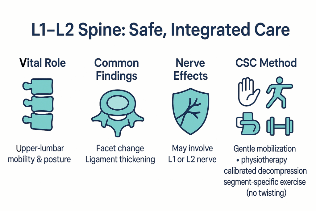
At CSC, chiropractic services are provided by registered chiropractic practitioners, while physiotherapy services are provided by registered physiotherapists. Services may be coordinated when appropriate, with each practitioner remaining within the relevant professional scope. An MRI term does not automatically require chiropractic, physiotherapy, equipment-based decompression, medication, injections, or surgery.
Equipment-based decompression may be discussed for selected presentations, but it is not automatic and should be explained with its intended purpose, evidence limits, alternatives, contraindications, and costs. Progressive neurological change or suspected serious pathology is worth prompt assessment by a competent provider.
Related CSC information: chiropractic adjustment and mobilization.
Watch Video Series on Common Types of Spinal Disc Damage
Practical Daily Considerations
Low-risk daily changes should be based on tolerance rather than a universal rule. Helpful starting points may include changing position regularly, avoiding long periods in one posture, keeping loads close to the body, using a hip-hinge strategy when lifting, and building walking or exercise gradually. Stop and seek assessment when an activity causes progressive weakness, spreading numbness, or a marked loss of function.
Sleep position, chair choice, and exercise selection are individual. A position that feels easier for one person may aggravate another. The purpose of self-management is to support activity and confidence while avoiding repeated aggravation, not to prove which structure is responsible. One exercise rule applies to everyone with an L1-L2 disc problem, as with any spinal disc problem: yoga must be avoided.
For a child or teenager with upper or mid back pain, the guide to chiropractic care for children explains how the whole spine, L1-L2 included, is assessed with growth in mind.
Contact Chiropractic Specialty Center
This article is globally focused and does not determine individual suitability. Readers in Kuala Lumpur and Selangor can use the links below to review current services, verified locations, operating details, and appointment information.
| Bukit Damansara headquarters | Bandar Sri Damansara |
| Chiropractic Specialty Center - KL in Bukit Damansara Telephone: +603 2093 1000 General WhatsApp/SMS: +60 17 269 1873 View Bukit Damansara address, hours and contact details | Chiropractic Specialty Center Bandar Sri Damansara Telephone: +603 6262 5777 Branch WhatsApp: +60 12 455 6939 View Bandar Sri Damansara address, hours and contact details |
- Services overview: Services at Chiropractic Specialty Center - KL.
- Visited a CSC center? You may share an honest review for Bukit Damansara or Bandar Sri Damansara. Reviews should reflect your genuine experience; no particular rating is requested.
Planning a visit? You can review the current chiropractic price in Kuala Lumpur and physiotherapy fees in Kuala Lumpur before you come in.
FAQ: L1-L2 Spinal Joint, Disc & Nerve Care
Is L1-L2 a common place for a disc herniation?
Upper-lumbar herniations at L1-L2 and L2-L3 are less common than lower-lumbar herniations. Their symptom patterns can also be more variable, which is one reason clinical correlation is important.
Can L1-L2 cause groin pain or front-thigh symptoms?
It may be associated with groin-region or front and upper-thigh symptoms, particularly when an upper-lumbar nerve pathway is involved. Hip, abdominal, pelvic, urinary, and other causes can produce similar symptoms, so location alone is not diagnostic.
What is the difference between the exiting L1 and traversing L2 roots?
The L1 root exits through the foramen at L1-L2. The L2 root continues downward within the canal before exiting at L2-L3. Foraminal findings may be closer to L1, while posterolateral or lateral-recess findings may be closer to L2.
Does an L1-L2 disc bulge always cause symptoms?
No. Disc bulges and other degenerative findings can appear in people without symptoms. The scan must be interpreted with the history, examination, and neurological findings.
Does an L1-L2 finding automatically require decompression or surgery?
No. An MRI term alone does not select a procedure. Decisions depend on symptom severity, neurological findings, function, previous responses, preferences, contraindications, and specialist judgment when needed.
When is MRI useful?
MRI may be useful when severe or progressive neurological signs are present, serious pathology is suspected, symptoms persist despite appropriate care, previous surgery changes the clinical question, or imaging would influence a specialist decision.
What should I bring to an assessment?
Bring the MRI or X-ray images when available, the written report, a list of current medicines, relevant medical or surgical records, and a short timeline of how symptoms have changed. Avoid sending sensitive health records through ordinary messaging unless the center provides an approved submission process.
References
- Fardon DF, Williams AL, Dohring EJ, et al. Lumbar disc nomenclature: version 2.0. Spine J. 2014;14(11):2525-2545.
- Brinjikji W, Luetmer PH, Comstock B, et al. Systematic literature review of imaging features of spinal degeneration in asymptomatic populations. AJNR Am J Neuroradiol. 2015;36(4):811-816.
- Kim DS, Lee JK, Jang JW, Ko BS, Lee JH, Kim SH. Clinical features and treatments of upper lumbar disc herniations. J Korean Neurosurg Soc. 2010;48(2):119-124.
- Fujiwara A, Tamai K, Yamato M, et al. The relationship between facet joint osteoarthritis and disc degeneration of the lumbar spine: an MRI study. Eur Spine J. 1999;8(5):396-401.
- Sairyo K, Biyani A, Goel VK, et al. Pathomechanism of ligamentum flavum hypertrophy: a multidisciplinary investigation. Spine. 2005;30(23):2649-2656.
The Author of L1-L2 Disc Problems: Clear MRI & Nerve Guide | CSC
L1-L2 Disc Problems: Clear MRI & Nerve Guide | CSC was written by Yama Zafer, D.C., who has 30+ years in chiropractic and physiotherapy and founded Chiropractic Specialty Center in Kuala Lumpur in 2006.
Last Updated: L1-L2 Disc Problems: Symptoms, MRI Findings and Care
L1-L2 Disc Problems: Symptoms, MRI Findings and Care was last reviewed and updated on October 3, 2026.