


L4-L5 Herniated or Slipped Disc: Symptoms, Causes and Care
An L4-L5 disc bulge or herniation often starts as low back pain that builds with sitting, bending or lifting. When a slipped disc at L4-L5 reaches a nerve, a cramp-like ache or tingling can run through the buttock and down one leg. A cough may feel like a knife in the back, and the muscles that lift the foot and big toe can weaken.
In my experience, most of this damage follows an injury, sudden or repeated, or strain repeated day after day. Long sitting, slouching, and bending, twisting or lifting at the waist all add to it, and most forward bending happens at this level. Age is one factor, but genetics, lifestyle, posture and past injuries play a part too.
Care starts with an examination that matches the scan to the nerve tests. It combines gentle, non-rotatory chiropractic care with physiotherapy and hands-on soft tissue work, then precise exercise once the back has settled. Every step is kept within what the patient can comfortably tolerate. The sections below cover healing without an operation, the nerve roots, the stages of disc damage, MRI terms, daily habits and surgery.
This guide, L4-L5 Disc Bulge, Herniation & Treatment Without Surgery, is published by Chiropractic Specialty Center, Bukit Damansara (KL) main center: WhatsApp+60 17 269 1873 Call+603 2093 1000
L4-L5 Disc Bulge and Herniation: Key Takeaways
| Point | What to know |
|---|---|
| Is it serious? | To some degree, always. On my scale of 0 to 10, a true bulge rates 1 or 2. A disc that reaches the nerves, such as the L5 root running past this disc, rates 3 to 5. |
| Can it heal without surgery? | No disc returns to its original state, with or without an operation. Surgery is still the last option, and progress is judged by the retested nerve findings, not by the scan. |
| Bulge, herniation or slip disc | Slip disc is the everyday name for every stage. A true bulge has swollen without touching the nerves; in a herniation the center has broken through the outer wall. |
| Scan versus symptoms | In an MRI study of 98 adults with no back pain, 52 percent had a disc bulge and 27 percent a protrusion (Jensen 1994). The examination shows whether a finding explains the pain. |
| What is avoided | Twisting, bending at the waist, lifting, deadlifts and rotational manipulation while the disc is irritable. Yoga must be avoided, since so many poses fold or twist the lower back at this level. |
| Sitting breaks | With acute back pain, stand up every 15 minutes. People without back problems take a break every 30 to 45 minutes. |
| Signs that need a hospital | New numbness around the genitals or anus, new difficulty passing urine, or loss of bladder or bowel control needs urgent medical attention at a hospital emergency department straight away, within hours. |
L4-L5 Herniated or Slipped Disc: Page Guide
- Can an L4-L5 Herniated Disc Heal Without Surgery?
- Arranging an L4-L5 Assessment in KL
- L4-L5 Symptoms: Back, Leg and Nerve Signs
- What Causes an L4-L5 Disc Bulge or Herniation
- Bulge, Herniation and the Stages of Disc Damage
- Stenosis, Facet and Ligament Changes at L4-L5
- L4-L5 Disc Bulge Treatment Without Surgery
- What Happens at the First Assessment
- How Long an L4-L5 Disc Bulge Takes to Settle
- Daily Habits: Sitting, Prayer, Sleep, Travel and the Gym
- When Surgery Is Considered
- Choosing a Treatment for an L4-L5 Disc Bulge
- How L4-L5 Compares with Other Lumbar Levels
- Video: Why L4-L5 Back and Leg Problems Keep Returning
- L4-L5 Disc Bulge and Herniation FAQ
- Peer-Reviewed and Clinical References
Can an L4-L5 Herniated Disc Heal Without Surgery?
Not fully, and an operation cannot make it whole either. In my experience no disc returns to its original state, and a herniated disc is no exception. As I tell patients: “No matter what I or the best surgeons in the world do, we cannot cure something permanently. There will always be some level of damage.”
A 2025 perspective article on chronic low back pain stresses matching the history and examination to the imaging before care is planned (Sima 2025). So I judge progress by the examination, not by a change on the scan. That means the strength, sensation and reflexes of the leg, and how long the person can sit, stand and walk.
That does not make surgery the next step. With a herniated or bulging disc it is the last option, as the surgery section explains.
At CSC, the active care plan ends when a person reaches what I call MMI, maximum medical improvement. That is the point where two to three more weeks of care bring no further change in the retested findings. Care then becomes as-needed, with preventive care, daily-living advice and home exercises. Over the years, how the disc copes depends on daily habits. It also depends on keeping up the maintenance care, exercises and stretches given at the end of the plan.
I also write about L4-L5 from experience:
In my early twenties I developed a severe L4-L5 disc herniation. Surgery was recommended. The specialists were honest about possible outcomes. A course of careful, non-surgical care produced meaningful improvement and ultimately redirected my entire career.
That experience is why I pursued chiropractic education at Cleveland University-Kansas City. It is also why, since 1996, I have focused on the full spectrum of disc, joint and nerve changes at L4-L5 and the neighboring lumbar levels.
When symptoms or imaging point to L4-L5, the segment deserves a detailed, findings-based assessment rather than routine force or a one-size-fits-all plan.
Arranging an L4-L5 Assessment in KL
When back or leg symptoms may come from L4-L5, a first visit checks strength, sensation, reflexes, walking tolerance and how movement changes the symptoms. Any MRI or X-ray is read alongside those findings, so bring the images themselves on CD or film, not only the report. Opening hours and directions for both centers are on the contact page. Either Kuala Lumpur center can arrange the visit:
- Bukit Damansara (KL, main center): Call +603 2093 1000 or WhatsApp +60 17 269 1873 to see the Bukit Damansara / KL center.
- Bandar Sri Damansara (KL): Call +603 6262 5777 or WhatsApp +60 12 455 6939 to see the Bandar Sri Damansara center.
L4-L5 Symptoms: Back, Leg and Nerve Signs
Some people notice only lower-back stiffness after prolonged sitting. Others develop clear leg symptoms. Typical patterns include:
- Lower-back stiffness or aching that builds with sitting, bending or standing and often eases when lying down
- Sensations that travel into the buttock, outer thigh, calf or foot (tingling, pulling, heat-like feelings or heaviness)
- Symptoms that become easier to trigger with longer periods of sitting, driving or walking
- Reduced confidence in walking or standing for extended periods
- In more advanced cases, a feeling of heaviness or reduced control in the foot or ankle
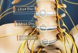
In my experience, a damaged L4-L5 disc often brings cramp-like sensations in the low back or buttock, sometimes running down the leg like sciatica. As nerve irritation grows, pain can turn into numbness and tingling, and then weakness. With a torn disc, turning over in bed can wake a person at night. Coughing can feel like a knife going through the back, and even laughing can hurt, because each movement makes the torn fibers vibrate.
To me, sciatica is not a diagnosis. It describes a symptom: an inflamed sciatic nerve. Pain or altered feeling runs from the lower back or buttock down the leg, usually one leg and sometimes both. In every case of sciatica I have seen, there was some disc involvement, from mild to moderate. At L4-L5, a narrowed nerve opening or thickened joints and ligaments can crowd the same nerve roots.
Nerve irritation at this level can come from pressure: disc material, facet overgrowth or a narrowed space. It can also come from chemical sensitivity around the disc and nerve root. That is why symptom intensity does not always match the size of a finding on an MRI.
Which Nerve Root: L5 or L4
Two nerve roots pass the L4-L5 disc. The L5 root crosses this level on its way down, and the L4 root leaves the spine through the opening at the side. Each one has its own map on the skin and its own muscles (Berry 2019).
- L5 root: pain, tingling or numbness along the outer shin to the top of the foot, and weakness lifting the big toe
- L4 root: symptoms along the inner shin, and weakness lifting the foot at the ankle
The examination tests these skin areas and muscles one by one. Where the disc material sits on the scan helps show which root is involved: in the middle, to one side or out in the side opening.
When Foot Drop or Progressive Weakness Needs a Careful Assessment
Foot drop means difficulty lifting the front of the foot, so walking on the heels becomes hard. Leg weakness that keeps getting worse should not be ignored. It calls for a careful assessment by a competent provider, and conservative care can continue only while the weakness itself is improving.
Video: Sciatica vs Slipped Disc, How to Tell the Difference
Pain that travels into the buttock, thigh, calf or foot usually starts in the spine, at a disc such as L4-L5. In this video I explain how to tell sciatica, nerve irritation and a slipped disc apart, and why the spinal source matters when choosing care.
To jump to a part of this video or read along, see the Sciatica vs Slipped Disc video page.
Key Moments for This Video on Sciatica and Spinal Disc Damage
- 00:00 Why sciatica is a symptom pattern, not a stand-alone diagnosis
- 00:27 How L4, L5, S1, and S2 roots form the sciatic nerve
- 02:36 Why many leg symptoms begin with a lumbar disc issue
- 05:03 Sitting, walking, posture, and daily load at the lower back
- 10:05 Why forceful cracking may need more caution in disc-related sciatica
- 10:57 When surgery may be discussed versus conservative-first care
What Causes an L4-L5 Disc Bulge or Herniation
Most tears and wear-related changes in the L4-L5 disc follow an injury, sudden or repeated, or repeated harmful strain. A sudden injury, such as a heavy lift, a fall or a sports injury, can tear the disc’s tough outer ring. Repeated strain works more slowly, over months or years, until the ring weakens and the soft center pushes outward.
In my experience, poor habits supply most of the strain on L4-L5. The ones I see most often are:
- long sitting, because the weight of the upper body bears down on the spinal joints and discs, and leaning forward adds to it;
- slouching over a desk, a laptop or a phone;
- bending and twisting at the waist, worst when the two come together;
- lifting in poor positions, and deadlifts and free weights at the gym;
- standing for long periods.
Poor posture works slowly. It strains the muscles first, then the ligaments. If it goes on long enough, it damages the joints and finally reaches deep into the disc.
Age plays a part, but it is not the whole story. I have found a dried-out disc (disc desiccation) on the scan of a seven-year-old girl, and healthy discs in people well into their 80s. Genetics, lifestyle, daily living, past injuries and posture all count too.
Teenagers can get low back pain too, from sport or a heavy school bag. The guide to chiropractic care for children explains how CSC assesses the lower back, L4-L5 included, in a growing spine.
Discs rarely herniate while they are healthy. In the research I have read, a healthy disc herniated in the laboratory only under forces far beyond anything in daily life. So degeneration comes first. Poor sitting and other improper loads raise the pressure inside the disc. That pressure keeps water and nutrients out and weakens the fibers of the outer ring until they buckle or tear. The guide to degenerative disc disease explains how a disc degenerates, step by step.
L4-L5 also carries a heavy share of the load. Most forward bending happens at L4-L5, which is why it and L5-S1 are where disc problems are found most often. The daily habits section shows how to cut that strain down.
Bulge, Herniation and the Stages of Disc Damage
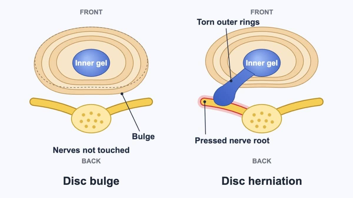
MRI reports use words such as disc bulge, protrusion, prolapse, herniation, extrusion and fragmentation. To me they are stages of one process, and slipped disc is the everyday name for all of them. A disc bulge is a progression of a degenerated or damaged disc. A herniation is a progression of a bulge, and an extrusion is a progression of a herniation. My video Why Spinal Discs Get Damaged shows them in order. This is how I define each stage:
- Degenerated or damaged disc: the fibers of the outer ring weaken and tear, and they can no longer hold the soft center in place.
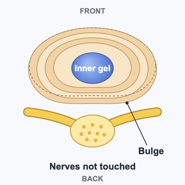
- Disc bulge: the fibers swell more than usual while the center stays put. A true bulge does not press on the spinal cord or the nerves. At L4-L5 it leaves the nerve sac and the L5 root alone. It is the mildest form of a contained slipped disc.
- Protrusion: the center moves outward, toward the spinal canal or the side openings where the nerves leave, but the outer fibers still hold it. It is a progression of the bulge and is still contained.
- Prolapse: a progression of the protrusion, barely contained. Only a few outer fibers keep the center, the nucleus pulposus, from rupturing into the canal.
- Herniation, also called a rupture: the last intact fibers tear and the center breaks through the outer wall, the annulus fibrosus. It is no longer contained, and at L4-L5 it can reach the nerve sac, the L5 root or the L4 root in the side opening.
- Extrusion: a progression of the herniation, where the center is pushed from the middle of the disc out to its edge. These are large herniations that need prompt assessment, which does not automatically mean surgery and can mean conservative care.
- Fragmentation and sequestration: the last and most dangerous stage, when part of the center breaks off and floats free in the spinal canal.
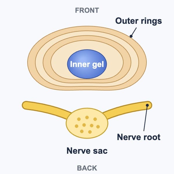
For me, the dividing line is contact. A true bulge has not pressed on or pushed aside the thecal sac, the sac around the nerves, or a nerve root. A disc that does either is no longer a bulge but a prolapse or a herniation, whatever the report calls it. The drawings below show a lumbar disc from above, with the front of the body at the top and the nerves behind the disc.
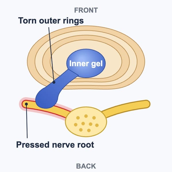
Not every disc moves at the same pace. Once an L4-L5 disc is damaged, the change can come quickly or take years. So some people notice a gradual onset, and others a sudden change after a period of repeated strain. Small tears can be missed on an MRI.
Many changes on a scan cause no symptoms at all. As I put it, the most common symptom of spinal disc damage is zero. In a 1994 study, 98 adults with no back pain had an MRI of the lower back. Of these, 52 percent had a disc bulge at one level or more, and 27 percent a protrusion (Jensen 1994). That is why the scan is read together with the examination.
Video: L4-L5 Disc Bulge vs Herniation, What the MRI Terms Mean
In this short video I explain how a spinal disc is built and how its fibers tear. I also show why a bulge comes before a herniation and an extrusion, and the symptoms a slipped disc can bring in the back, buttock and leg.
Key moments and an edited transcript of this video are on the Bulging Disc vs Herniated Disc video guide.
Key Moments of Disc Herniation and Bulge Video
- 00:00 Intro to disc bulge, disc herniation, and slipped disc
- 00:35 What spinal discs do and how they allow bending and twisting
- 01:01 Why disc damage begins before larger disc issues appear
- 01:28 How annular tears develop
- 01:45 Bulge versus herniation: which comes first and why
- 02:09 Bulge, herniation, and extrusion explained
- 02:43 Why disc location can affect sciatica and leg symptoms
PIVD, PID and Prolapse: What These Report Words Mean
PIVD stands for prolapsed intervertebral disc, and PID means the same thing. Both words are common on scan reports in Malaysia and other parts of Asia. They are umbrella words, a bit like saying “slipped disc.” They say that disc material has moved out of its normal place, but not how far or in what shape.
So when a report says “PIVD at L4-L5,” the next step is to find which stage it describes, from a bulge to a fragment. The other key detail is where the material sits: in the middle, to one side, or in the opening where the nerve exits. Its position often explains the symptoms better than its size, as the nerve root section shows.
Diffuse Bulge, Thecal Sac and Canal Size
Many L4-L5 reports say something like “diffuse disc bulge indenting the thecal sac.”
To me, a diffuse disc bulge is one that bulges in all directions. The thecal sac is the soft, fluid-filled tube that holds the nerve roots in the low back.
A disc that indents the sac has gone beyond a true bulge. By my definition it is a prolapse or a herniation, whatever the report calls it. On my scale of seriousness it rates 3 to 5, not 1 or 2. The word bulge is often used loosely, even in some scan reports, so the examination decides what the finding means.
What matters more is how much room is left. Studies that measure the spinal canal most often describe central stenosis with one of two numbers (Steurer 2011). The front-to-back width is under 10 millimeters, or the cross-section area is under 70 square millimeters. Many reports simply grade narrowing as mild, moderate or severe.
Someone with a narrow canal may walk well, while someone with a small finding may have strong leg pain. The exam at CSC checks walking, nerve tests and strength, then matches them to the MRI. When the canal is narrowed, care often uses gentle methods that bend the spine forward, such as flexion-distraction. It often adds spinal decompression and exercises built around the bent-forward positions that usually feel easier.
Stenosis, Facet and Ligament Changes at L4-L5
At L4-L5 the disc does not work in isolation. The facet joints, the ligamentum flavum and the overall stability of the segment all share the load. Over time, repeated stress can produce several changes together:
- Facet hypertrophy: enlargement or bony overgrowth of the small joints at the back of the segment
- Ligamentum flavum thickening: the yellow ligament between the laminae becomes thicker and less flexible
- Spinal stenosis: the available space for the nerves becomes narrower in the central canal, the lateral recess or the foraminal exit zone
- Spondylolisthesis: a forward shift of L4 relative to L5, graded by how far the vertebra has moved
L4-L5 is the level most often involved in degenerative lumbar stenosis (Lee 2015). When disc change, facet overgrowth and ligament thickening occur together, they can crowd the nerves. That can happen even if no single structure looks dramatic on its own.
Stenosis has its own pattern, called neurogenic claudication: leg pain, often in both legs, that comes on with standing or walking. It eases with sitting, lying down or bending forward, and gets worse with long standing or leaning back. In one study of 100 people with lumbar stenosis, 95% had back pain and sciatica and 91% had this walking pattern (Lee 2015).
In my experience, these changes and a disc bulge, herniation or extrusion often occur at the same time, side by side. The pages on Ligamentum Flavum Hypertrophy, Facet Hypertrophy and Spondylolisthesis explain each of these findings in more detail.
What Does Neural Foraminal Narrowing Mean?
Neural foraminal narrowing means the opening at the side of the spine, where a nerve root leaves the canal, has become smaller. At L4-L5 that opening is where the L4 root leaves the spine, as the nerve root section explains.
Reports name the side: left, right or bilateral, which means both sides. They may also name the cause, such as a foraminal disc protrusion, overgrown facet joints or ligament, or a slipped vertebra (Berry 2019).
A 2010 MRI grading system uses three grades (Lee 2010):
- Mild: the fat around the nerve root is lost on two opposite sides
- Moderate: the fat is lost all around the root, and the root keeps its shape
- Severe: the root itself is flattened or changes shape
As with canal size, the examination shows whether the narrowing explains the leg symptoms.
What Mild Disc Space Narrowing Means
Reports often call a thinner L4-L5 disc mild disc space narrowing or loss of disc height. The word mild grades the picture on the scan; it does not mean the change is unimportant.
As the disc loses height, more of its load shifts onto the facet joints, which can become arthritic and enlarge. The ligamentum flavum, a ligament inside the canal, also slackens and folds inward, narrowing the canal. The weakened disc is also more likely to tear, bulge and protrude.
At L4-L5, the level where stenosis is most common, a narrowed disc can affect the nerves before the back hurts much. It may show up first as numbness or weakness in the leg, or as trouble standing and walking for long.
What Is L4-L5 Spondylosis?
Spondylosis is a broad word for wear-related change in the spine, and it has more than one definition. Used broadly, it covers the discs, the vertebrae and the joints between them (Middleton 2009). On an L4-L5 report it can mean any mix of these:
- a thinner, drier disc
- bony spurs, called osteophytes, at the edges of the vertebrae
- arthritis of the facet joints
These changes are common in people without back pain, and age is only one of the factors behind them, as the causes section explains.
So, like PIVD, the word names the kind of change, not how much it matters, and the examination decides that. The spondylosis guide covers it across the spine.
L4-L5 Disc Bulge Treatment Without Surgery: Gentle, Non-Twisting Care
Treatment without surgery for an L4-L5 disc problem combines gentle chiropractic care, physiotherapy, hands-on soft tissue work and, later, precise exercise. I recommend chiropractic and physiotherapy together rather than one or the other, and the assessment shows which tissue needs which method.
A disc may already show a bulge, a herniation, a tear in its outer ring or more advanced change. The segment then copes less well with sudden twisting force, as does one whose facet joints and ligamentum flavum have thickened. Forceful twisting, or repeated fast rotational adjustments, may add stress to structures already under strain.
This does not mean every hands-on method is unsuitable. The choice of method follows a clear assessment of symptoms, movement findings and imaging when available.
I am not a big fan of rotary chiropractic manipulation. I use it only when I am 100 percent sure there is no spinal disc damage. Cracking and twisting should not be done on a spine with a damaged disc, even by a very experienced chiropractor. Conservative care has to be condition-based and appropriate, care that does no harm.
At CSC this is a firm rule while an L4-L5 disc is irritable: no bending or twisting. Care also leaves out rotational or aggressive physiotherapy, manual therapy and chiropractic manipulation. A twisting movement can make an existing disc problem worse. Stretching and exercise also wait until the early, irritable stage has passed.
Care is kept within what the patient can comfortably tolerate. Patients are asked to mention any discomfort straight away, since discomfort during care can lead to more inflammation and pain afterward. My motto has always been “No pain, more gain.”
Low-Force Options and What Studies Found
After a structured review of posture, movement, nerve findings and imaging, some people are guided toward methods that keep rotational stress low. One example is chiropractic manipulation that is non-rotatory using the Activator instrument, which delivers a quick, controlled impulse with the body in a neutral position.
A 1998 case report described a 26-year-old man with a lumbar disc herniation, leg pain and foot drop (Polkinghorn 1998). He had been advised to have surgery. His symptoms resolved within 90 days of non-rotatory chiropractic manipulation. One case cannot show how others will respond.
A 2012 systematic review of eight trials found clinically meaningful benefits with this method, similar to those of fast manual manipulation for spinal pain. The review also noted small samples, short follow-up and a lack of control or sham groups (Huggins 2012).
A broader plan can also include:
- controlled flexion-distraction
- carefully dosed non-surgical spinal decompression
- guided rehabilitation focused on stability and load tolerance
A study looked back at 160 people with lumbar disc problems (Adar 2024). Some had spinal decompression or motorized traction added to conventional physiotherapy. Both additions were more effective than conventional physiotherapy alone.
In a randomized trial, 40 people with chronic low back pain all did core stability exercises (Walewicz 2020). Those who also had radial shockwave had less pain and better function than those given a sham treatment.
A meta-analysis of randomized trials looked at high-intensity laser therapy for muscle and joint problems (Song 2018). It reduced pain and disability more than the control treatments. The trials differed widely, and the reviewers called for larger studies with longer follow-up. None of these is a universal solution, and technique choice is strongest when it matches the individual’s findings rather than a fixed protocol.
Alongside these, the CSC team of chiropractors and physiotherapists chooses from the following, each matched to the tissue that needs it:
- ultrasound
- electrotherapy
- cold therapy with the QMD cryothermal device, never its heat setting when a disc or facet joint is involved
- trigger point and myofascial therapy
- stretching and strengthening when the time is right
- balance and neuromuscular training
- devices for pain management
What Happens at the First Assessment in Kuala Lumpur
A first assessment for L4-L5 symptoms takes from 30 minutes to two hours, from arrival to leaving, depending on how complex the problem is. I do not rush it, because a problem that seems mild is sometimes more serious and needs other areas assessed too. Any existing MRI or X-ray is worth bringing as the actual CD or films. A written report alone helps, but the images show the disc shape, canal space, nerve openings and facet changes directly.
The visit begins with a detailed conversation about current symptoms, past medical history, what makes the problem better or worse and any other conditions.
It helps to write the history down beforehand. Note how the problem began and progressed, and everything tried so far: at home, after watching videos or with other practitioners. Write down the questions to ask as well, since they are easy to forget in the room. A family member who comes along helps too, because each person remembers different things.
The physical examination that follows includes:
- posture and movement analysis
- palpation, which means examining by touch
- range-of-motion testing
- neurological checks of strength, sensation and reflexes
- selected orthopedic tests, such as the straight leg raise, which checks for an irritated nerve root
Even when the main complaint is the lower back, the examination routinely screens the neck, because problems in the two regions often occur together. The examination also extends to the hamstrings, iliotibial band, hips, pelvis and legs.
Comfortable clothing is recommended. Any discomfort during the examination is reported at once, even if it seems small. Parking is available at both centers.
Lower back pain is among the most common reasons people come to CSC. From L4-L5 it often spreads into the buttock and the back or side of the thigh. The interview is long because many structures can produce the same pain, and each answer narrows the list:
- a muscle or ligament strain
- facet joints pressing together or enlarging
- disc degeneration
- a bulge, protrusion, prolapse, herniation, extrusion or fragment
- a segment that moves too much
- a vertebra that has slipped forward (with or without a pars defect) or backward
- a thickened ligamentum flavum
- narrowing of the canal or the foramen
- pressure on the exiting L4 or the traversing L5 nerve root
How CSC Chooses Between X-Ray and MRI Scans
Imaging is added when it will answer a question the examination leaves open. Usually one scan is enough, and an X-ray and an MRI are rarely needed together. The choice follows the findings:
- If they point to alignment, the soft tissues or a facet joint problem (facet joint syndrome), an X-ray may be all that is needed.
- If they point to the disc, from a bulge through to a fragment, an MRI is used. A disc diagnosis is then confirmed, not guessed.
- If a step is felt between the bony points of the spine, both an X-ray and an MRI are used. The step is a sign of a slipped vertebra, such as a grade 1 or 2 spondylolisthesis with a pars defect.
Spondylolisthesis at L4-L5: Three Extra X-Ray Views
In my experience, a slipped vertebra is much more common at L4-L5 and L5-S1 than higher up. It usually comes with disc and joint wear, facet hypertrophy, a thickened ligamentum flavum, and narrowing of the canal and the nerve openings.
When an X-ray shows any slip, forward or backward, CSC may add three side-view X-rays. They are taken standing upright, bent forward and leaning back, to see whether L4 shifts on L5 with movement. An MRI then shows the effect on the discs, joints, ligaments, nerve openings, canal and thecal sac.
Assessment and Care Fees, Explained Before Any Commitment
The first assessment is charged at a fixed fee regardless of whether it lasts 30 minutes or longer. Reading and reporting MRI or X-ray images, when they are brought on CD or film, is charged separately.
Insurance cover depends on the policy: some plans include chiropractic or physiotherapy and some do not. It is worth checking with the insurer, and the center can say what documents it provides for a claim.
How Long Does an L4-L5 Disc Bulge Take to Settle?
There is no fixed time. The pace depends on how severe the problem is, any other conditions, and how consistently the advice is followed. Without improvement by the fifth session, a reassessment is due. If there is still no improvement by the 10th session, stopping or continuing is a decision made together with the chiropractor.
Exercise is not part of the first sessions. It begins once the back is stable, most often between the 5th and 7th session and occasionally from the 3rd. The first set depends on how severe the problem is. Typical starting points are:
- Kegel exercises when the pelvic floor is weak
- wall-assisted squats
- the cat and camel movement
- glute, hamstring, calf and quadriceps strengthening
- core canister work for the deep muscles around the trunk
Doing the home measures in the plan and avoiding activities that repeatedly load or twist the segment gives care a better chance. Progress tends to slow when patients independently add aggressive YouTube exercises, rotational stretches, premature core strengthening or prolonged sitting without correction.
Reassessment occurs regularly. The tests from the first visit are repeated, pain is rated on a visual analog scale (VAS), and the patient’s own account is recorded. Where progress stalls, a further specialist opinion is discussed. When the retested findings stop changing, the active plan ends at maximum medical improvement, as the section on healing without surgery explains.
How Plans Differ for Older and Younger Adults
Stenosis and facet arthritis at L4-L5 are found more often in older adults, and for them the plan moves more gently:
- more emphasis on changing daily habits
- stretching introduced later
- extra neuromuscular stability work, such as a rocker board, a wobble board, balance on a gym ball and Kegel exercises
- decompression sessions that are lighter and shorter
- softer manual and myofascial techniques
A younger adult with an L4-L5 bulge usually follows the standard sequence: settling the irritated tissue first and strengthening later.
Daily Habits: Sitting, Prayer, Sleep, Travel and the Gym
I may see a patient for 30 minutes or two hours; the other 22 hours of the day decide how the disc is loaded. Long sitting often aggravates disc pain, so practical adjustments include:
- A chair with genuine lumbar support, sitting so the pelvis and lower back stay against the backrest with no gap
- A laptop connected to an external monitor set at eye level, so the head and trunk do not lean forward over the screen
- A break every 15 minutes with acute back pain, standing to walk for 10 to 20 seconds; without back problems, a break every 30 to 45 minutes
- No bending or twisting at the waist while the disc is irritable, and no lifting, or less than 2 kg when it cannot be avoided
- No aggressive rotational stretches lying on the back, forceful hamstring stretches that tug on an irritated sciatic nerve, or generic bending exercises
- No yoga: so many of its poses fold or twist the lower back that it must be avoided with an L4-L5 disc problem
For those who perform daily prayers, deep flexion in the ruku’ position may need temporary modification when symptoms are intense. The sujood position is generally better tolerated. In periods of significant pain, performing prayers while seated is a reasonable temporary adaptation provided extreme forward flexion is still avoided.
Posture and sleep matter most, and sleep above all. Aim for at least six hours, and seven to eight hours of unbroken sleep where possible. A short night is not made up with an afternoon nap. Lying down lowers the pressure inside the disc, and that is when it draws in fluid and nutrients.
Among the positions that do not hurt, I suggest the right side first and the left side second. Third is the back, with a bolster under the knees. On the side, a pillow keeps the neck in line with the spine, and a bolster sits between the knees. The left side is used if the right side brings on heartburn.
Ice can be used 2 to 3 times a day, at least 2 hours apart, for 10 to 15 minutes at a time. Each time, the pack is wrapped in a thin towel and a timer is set.
Long road trips are avoided where possible. When a trip cannot wait, the reclined front passenger seat is the easier place to sit. A driver stops every 45 to 60 minutes to get out, walk and stretch the calves.
Sport resumes after recovery. Anything that hurts at the time, or brings on more pain within 72 hours, is left out, since inflammation can take that long to appear. Swimming laps waits too; walking in water is gentler.
Gym training waits too: free weights are paused for 2 to 6 months, and the gym itself is discouraged early on. When the back is stable, a few machines come back in:
- hip adductor and abductor
- leg curl and leg extension
- light rowing
- lat pulldown
- for some, chest press
Deadlifts, leg presses and free weights stay out. On the treadmill the incline stays at zero, as walking uphill adds load to the lumbar discs.
When Surgery Is Considered for an L4-L5 Disc
Surgery has its place; I do not say it should never be done. With a herniated or bulging disc, though, it comes last, once the conservative route has been exhausted. As a first choice, I would not recommend steroid injections or surgery. Methods that repeatedly twist or aggressively mobilize an already inflamed L4-L5 segment can make the problem worse even while producing a temporary feeling of relief.
Structured non-surgical care commonly continues when:
- the nerve findings are stable
- weakness is not progressing
- imaging shows disc, facet or stenosis changes without urgent features
With an L4-L5 disc bulge or herniation, any new symptoms should be considered alongside the back or leg pain. New or worsening numbness around the genitals, between the genitals and anus, or around the anus needs urgent medical attention: a thorough assessment at a hospital emergency department straight away, within hours. New difficulty passing urine needs the same urgent assessment, even if bladder and bowel control remain intact, and so does any loss of that control. Several conditions can cause these symptoms. Some may call for an invasive procedure, but being assessed helps identify the cause and does not automatically lead to surgery.
An earlier surgical opinion is sought when leg weakness keeps getting worse despite carefully adjusted care (see the foot drop section). It is also sought when a well-run conservative program clearly fails after a reasonable number of sessions. In my practice, surgery is considered in three situations: a conservative plan that is not working, a severe case such as a sequestered disc or an infection, and the urgent symptoms above.
What Research Shows About Some Procedures and Conservative Approaches
Three randomized trials tested radiofrequency denervation, a procedure that uses heat to switch off small nerves around the joints (Juch 2017). The trials included people with chronic low back pain from the facet joints, the sacroiliac joints or a combination of sources. Adding the procedure to a standardized exercise program brought no clinically important extra benefit over exercise alone.
Surgery can create more space in selected cases of severe stenosis or worsening nerve loss. It also changes the mechanics of the segment and carries its own recovery demands.
Conservative care remains the usual starting point for most people who do not have progressive weakness or the urgent signs above.
What Is the Best Treatment for an L4-L5 Disc Bulge or Stenosis?
No single treatment is best for every L4-L5 disc bulge or stenosis. The choice depends on what the examination finds, how irritated the nerve is and how long the symptoms have lasted. Any plan can be checked against these points:
- An examination first: strength, sensation, reflexes and walking are tested, and any MRI is read against them (see the first assessment).
- Methods that suit an irritable disc: gentle, non-twisting methods come first, without forceful twisting or rotational cracking (see the treatment section).
- A plan for a narrowed canal: bent-forward positions usually feel easier, and an older adult’s plan moves more gently (see how plans differ for older adults).
- Realistic expectations: no disc returns to its original state, with or without surgery. The plan aims at how the back and leg cope with daily life (see whether it can heal without surgery).
- Measured progress: the first visit’s tests are repeated, and the plan is reviewed if they do not improve (see how long it takes).
- When to seek a surgical opinion: for example, leg weakness that keeps getting worse, or a well-run plan that clearly fails (see the surgery section).
Before choosing, call or WhatsApp the centers being considered. Describe the L4-L5 findings and ask how each would assess them.
How L4-L5 Compares with Other Lumbar Levels
Each lumbar segment handles load and nerve pathways differently. Problems higher up or at the lumbosacral junction can produce overlapping but distinct patterns:
- L1-L2 Disc Problems: Symptoms, MRI Findings and Care: upper lumbar issues that may affect the low back, groin or hip region
- L2-L3 Disc Bulge & Herniation: Signs, Symptoms and Treatment: may influence the front of the thigh and hip-flexor function
- L3-L4 Spine Care in Kuala Lumpur: can affect the front of the thigh, knee region and leg strength
- L5-S1 Herniated Disc and Disc Bulge: S1 Nerve Signs and Care: commonly involves the buttock, calf, heel and foot
Video: Why L4-L5 Back and Leg Problems Keep Returning
Many L4-L5 back and leg problems improve for a while, then return. That pattern is often linked to an incomplete assessment, short-term patching, or missing some of the structures involved. In this video I explain why lower back, disc, joint and nerve problems often recur. I also explain why long-term decisions should rest on the full mechanical and clinical picture.
Key Moments for Video on Recurrent Back Issues
- 00:00 Why neck and back problems often return
- 00:25 Why short-lived improvement does not always mean full recovery
- 00:45 How shortcuts can lead to recurrence
- 02:05 Why identifying the real drivers matters
- 02:35 Single-factor care versus multifaceted care
- 03:00 Why informed decisions matter before choosing the next step
Fees for the first assessment and for care are listed on the chiropractic price in Kuala Lumpur and physiotherapy fees in Kuala Lumpur pages. The fee for a plan is explained and agreed before care begins.
Related Posts
L4-L5 Disc Bulge and Herniation FAQ
Is walking good for an L4-L5 disc bulge?
Walking in short, comfortable spells is part of daily life during recovery, and it breaks up long sitting, which often aggravates disc pain. With acute back pain, stand up to walk every 15 minutes; without back problems, every 30 to 45 minutes. Uphill walking adds load to the lumbar discs, so the treadmill stays flat. If walking brings on leg pain that eases with sitting or bending forward, the canal may be narrow, which the assessment checks. Walking tolerance is also one of the tests repeated at each reassessment.
How serious is an L4-L5 herniated disc?
It depends on the tissues it affects, more than on the size of the disc on the scan. I rate seriousness on a scale of 0 to 10: 0 is not serious at all, 1 the mildest and 10 life-altering and critical. A true bulge, swollen only slightly beyond its normal borders, rates 1 or 2. A herniated disc rates 3 to 5 when it presses on the sac around the nerves or touches a nerve root. At L4-L5 that means the L4 root in the side opening or the L5 root running past the disc. Leg weakness that keeps getting worse, or the urgent signs in the last answer below, change the priority and call for prompt assessment.
Can a disc bulge come back after it settles?
It can. The disc and the joints around it work every day, so the same strain that caused the first episode can bring symptoms back. In my experience, a problem ignored for months or years is more likely to leave some lasting symptoms or occasional flare-ups, even after a good recovery. Keeping up the exercises and stretches from the end of the plan, breaking up long sitting and lifting with care all reduce that strain. The video on recurring back problems explains why problems return.
Is chiropractic safe with a herniated disc?
In my view, chiropractic care is safe when it is performed by a competent, registered chiropractor and is condition-based. Problems come from the practitioner’s choices, and not all chiropractors are equal, so what matters is the method. With an irritable L4-L5 disc, I leave out forceful twisting and rotational manipulation and use low-force, non-twisting methods instead, as the treatment section explains. Before any method is chosen, the assessment checks strength, sensation and reflexes, so that a nerve problem needing a different route is not missed.
Is a slip disc the same as spondylolisthesis?
No. A slip disc means disc material pushing out of its normal place. Spondylolisthesis means a vertebra slipping forward or backward over the one below it. Both are common at L4-L5, and they can occur together, often with facet and ligament thickening. The examination may find a step between the bony points of the spine. CSC then adds an X-ray and an MRI to see whether a vertebra has slipped, and how far.
When should L4-L5 symptoms be assessed urgently?
New numbness around the genitals or anus, new difficulty passing urine, or loss of bladder or bowel control needs urgent medical attention at a hospital emergency department straight away, within hours. Either sign is enough on its own, and bladder or bowel control does not have to be lost first. Several conditions can cause these signs, and being assessed does not automatically mean surgery. Leg weakness that keeps getting worse should not be ignored. It calls for a careful assessment by a competent provider, and conservative care can continue only while the weakness itself is improving.
How to Read This Guide
This guide is general information about L4-L5 disc problems and cannot assess any one reader’s situation. The cause of the symptoms, the meaning of a scan and the right next step are decided at an individual assessment.
Peer-Reviewed and Clinical References
- Adar S, Apaydın O, Dündar U, et al. Comparison of the Effectiveness of Traditional Motorized Traction and Non-surgical Spinal Decompression Therapy Added to Conventional Physiotherapy for Treatment of Chronic Low Back Pain. Cureus. 2024;16(9):e69610.
- Berry JA, Elia C, Saini HS, Miulli DE. A Review of Lumbar Radiculopathy, Diagnosis, and Treatment. Cureus. 2019;11(10):e5934.
- Huggins T, Boras AL, Gleberzon BJ, Popescu M, Bahry LA. Clinical effectiveness of the activator adjusting instrument in the management of musculoskeletal disorders: a systematic review of the literature. J Can Chiropr Assoc. 2012;56(1):49-57.
- Jensen MC, Brant-Zawadzki MN, Obuchowski N, et al. Magnetic resonance imaging of the lumbar spine in people without back pain. N Engl J Med. 1994;331(2):69-73.
- Juch JNS, Maas ET, Ostelo RWJG, et al. Effect of Radiofrequency Denervation on Pain Intensity Among Patients With Chronic Low Back Pain: The Mint Randomized Clinical Trials. JAMA. 2017;318(1):68-81.
- Lee S, Lee JW, Yeom JS, et al. A practical MRI grading system for lumbar foraminal stenosis. AJR Am J Roentgenol. 2010;194(4):1095-1098.
- Lee SY, Kim TH, Oh JK, et al. Lumbar Stenosis: A Recent Update by Review of Literature. Asian Spine J. 2015;9(5):818-828.
- Middleton K, Fish DE. Lumbar spondylosis: clinical presentation and treatment approaches. Curr Rev Musculoskelet Med. 2009;2(2):94-104.
- Polkinghorn BS, Colloca CJ. Treatment of symptomatic lumbar disc herniation using activator methods chiropractic technique. J Manipulative Physiol Ther. 1998;21(3):187-196.
- Sima S, Diwan A. Contemporary clinical perspectives on chronic low back pain: The biology, mechanics, etc. underpinning clinical and radiological evaluation. JOR Spine. 2025;8(1):e70021.
- Song HJ, Seo HJ, Lee Y, Kim SK. Effectiveness of high-intensity laser therapy in the treatment of musculoskeletal disorders: A systematic review and meta-analysis of randomized controlled trials. Medicine (Baltimore). 2018;97(51):e13126.
- Steurer J, Roner S, Gnannt R, Hodler J; LumbSten Research Collaboration. Quantitative radiologic criteria for the diagnosis of lumbar spinal stenosis: a systematic literature review. BMC Musculoskelet Disord. 2011;12:175.
- Walewicz K, Taradaj J, Dobrzyński M, et al. Effect of Radial Extracorporeal Shock Wave Therapy on Pain Intensity, Functional Efficiency, and Postural Control Parameters in Patients with Chronic Low Back Pain: A Randomized Clinical Trial. J Clin Med. 2020;9(2):568.
These references describe low back pain, disc herniation and lumbar stenosis and their care in general. None of them evaluates care at Chiropractic Specialty Center.
The Author of L4-L5 Disc Bulge, Herniation & Treatment Without Surgery
L4-L5 Disc Bulge, Herniation & Treatment Without Surgery was written by Yama Zafer, D.C., who has 30+ years in chiropractic and physiotherapy and founded Chiropractic Specialty Center in Kuala Lumpur in 2006.
Last Updated: L4-L5 Herniated or Slipped Disc: Symptoms, Causes and Care
L4-L5 Herniated or Slipped Disc: Symptoms, Causes and Care was last reviewed and updated on October 10, 2026.
What does L4-L5 failure of a right lateroforaminal herniation mean? what does C5-C6 is the site of a median protrusion mean?
What does L4-L5 is the site of a right lateral herniation mean?
These words describe where the disc material sits, and its position decides which nerve it can reach. At L4-L5, material inside the canal usually reaches the L5 root (outer calf to the top of the foot, with weakness lifting the foot or the big toe). Material out at the side opening, described as lateroforaminal or lateral, reaches the L4 root instead (front and inner thigh toward the inner shin, with weakness straightening the knee). Both of your L4-L5 phrases describe the right side.
In my definitions, a protrusion is disc material that has moved toward the canal or the side openings without breaking through the outer fibers, and a herniation is one where it has broken through. Failure is not a usual report word, so ask whoever wrote the report what it means rather than guessing.
A median protrusion at C5-C6 sits in the middle, pointing toward the spinal cord. Signs worth reporting promptly are clumsy hands, dropping things, trouble with buttons, unsteady walking or a change in bladder control. Arm or leg weakness that keeps getting worse calls for a careful assessment. New numbness around the genitals or anus, new difficulty passing urine, or loss of bladder or bowel control needs urgent medical attention at a hospital emergency department straight away, within hours.
The neck and lower back findings are usually separate problems, so check each on its own. For the midline protrusion at C5-C6, the question is whether the spinal cord is affected: clumsier hands, dropping things, trouble with buttons, coins or keys, changed handwriting, unsteady walking in the dark, or a change in bladder control. If any of these have appeared, ask for a neurological assessment with current imaging; forceful neck manipulation would not be appropriate.
For L4-L5, your symptoms show which root is involved: the front and inner thigh toward the inner shin with weakness straightening the knee points to L4, and the outer calf to the top of the foot with weakness lifting the foot or the big toe points to L5. Write down where the symptoms reach, whether that is spreading or retreating, and whether anything feels weak, and take it to the doctor who ordered the scan, together with the question of what failure means in your report.
Arm or leg weakness that keeps getting worse calls for a careful assessment. New numbness around the genitals or anus, new difficulty passing urine, or loss of bladder or bowel control needs urgent medical attention at a hospital emergency department straight away, within hours.
I just found out my boyfriend has L4-L5; there is no spinal canal stenosis or narrowing and there is a Small bulging disc at L5-S1; I would love to be informed more by anyone who can give me some details on the do's and don't he will need to help him prevent worsening. Much appreciated!!
A small bulge with no narrowing of the canal is a mild finding, and in my definition a true bulge does not press on the nerves. Still, disc damage tends to progress if the habits behind it continue, so the do’s and don’ts are worth following.
Do: walk daily in comfortable spells; break up sitting, every 15 minutes while his back pain is acute and every 30 to 45 minutes otherwise; sit with the lower back against a supportive chair; sleep at least six hours, and seven to eight unbroken where possible; and, once his back is stable, strengthen the glutes, legs and deep core with exercises matched to him.
Don’t: bend and twist at the waist, above all together; lift heavy loads in poor positions; do deadlifts or free weights while the back is irritable; or do yoga, which must be avoided with a disc problem because so many poses fold or twist the spine. He should also avoid anything that hurts at the time or brings on more pain within 72 hours.
If his symptoms keep getting worse, he should arrange an assessment. New numbness around the genitals or anus, new difficulty passing urine, or loss of bladder or bowel control needs urgent medical attention at a hospital emergency department straight away, within hours.
Hi my mother aged 62 generally complains for excessive leg pain (especially calf muscles ). MRI Lumbar spine Scan has given following impression (1) Mild Lumbar Spondylolysis (2) Minimal Scoliosis of spine towards right side(3) L3 l4 disc minimal bulge indenting thecal sac and bilateral traversing nerve roots (4) L4-5 disc moderate diffuse bulge compressing thecal sac stenosing bilateral leteral recesses& neural foraminae with impingement of bilateral traversing nerve roots.
Neuro surgeon advises for surgery to relieve nerves , can you please suggest any non surgical options and chances of success of non surgical options . patient is based in Bangalore . Any right doctor or practitioner of non surgical therapies in Bangalore is highly desirable
Calf pain as the main symptom at 62 calls for two non-spinal causes to be checked first. One is circulation: calf pain that comes on at about the same walking distance every time and eases just by standing still. A doctor can check the foot pulses and compare blood pressure at the arm and the ankle. The other is medication: some common medicines, statins among them, can cause aching in the calves, so mention any that were started around the time the pain began.
If it is the spine, her report describes bulges at L3-L4 and L4-L5 reaching the nerve roots on both sides. By my definition, a disc that indents the sac or reaches the nerves is no longer a simple bulge, so the report’s wording does not settle how serious it is. One test helps: if her leg pain eases when she sits or bends forward, and she can walk further pushing a trolley, that points to the spine.
In that case, forward-leaning positions such as a trolley or a recumbent bike let her stay active, and her walking distance, recorded weekly, is the best measure of change. Leg weakness, a foot that catches, or a calf that becomes swollen, red or hot needs prompt medical assessment. New numbness around the genitals or anus, new difficulty passing urine, or loss of bladder or bowel control needs urgent medical attention at a hospital emergency department straight away, within hours.
My L4 and L5 vertebrae are rubbing on the nerves. My spinal disc has degeneration and bulging. I’m in physical therapy for the discs for my current condition which may have resulted from a previous injury where my pelvis was crushed in a car accident. I’m in a lot of pain, feeling pain in my back. Leg and toes. I’m m also trying to lose weight to take pressure off the nerves in my lower back. Are there any other therapies or procedures that I can do to slow down my deterioration?
Your old pelvic injury deserves its own examination. A pelvis that was crushed and healed may not move as it did, which changes how load passes through the sacroiliac joints and the hips into the lower back. The sacroiliac joints often cause one-sided pain below the belt line, worse getting up from a chair or standing on that leg, and they do not show on a lumbar MRI. Ask for your sacroiliac joints and hips to be examined, and for X-rays of the pelvis and hips to be reviewed if that has not been done since the accident.
Note exactly which toes are affected: the top of the foot and the big toe point to L5, and the outer foot and little toe to S1. With a damaged disc, care should avoid twisting, and the hips and glutes deserve strengthening once the back is stable.
Reducing weight lowers the load on the spine and hips, and a recumbent bike or walking in water keeps you active when walking on land hurts. Leg weakness that keeps getting worse should not be ignored. It calls for a careful assessment by a competent provider, and conservative care can continue only while the weakness itself is improving. New numbness around the genitals or anus, new difficulty passing urine, or loss of bladder or bowel control needs urgent medical attention at a hospital emergency department straight away, within hours.
Can you call me for a appointment
Please do not post a phone number or personal details in a public comment. Appointments are made by calling or sending a WhatsApp message to either Kuala Lumpur center: Bukit Damansara, the main center, on +603 2093 1000 or WhatsApp +60 17 269 1873, and Bandar Sri Damansara on +603 6262 5777 or WhatsApp +60 12 455 6939. Opening hours are on the contact page.
When you call, say what the problem is, how long it has been there and whether symptoms travel into an arm or a leg. Bring any MRI or X-ray images on CD or film as well as the reports, and a list of your medicines.
Hi!
I am 20 years old. And I am suffering from an L4-L5 disc bulge. It was diagnosed last August -September period. I underwent physiotherapy. It's fine. But sometimes the pain triggers again and causes my left leg to pain harder. What should I do? The doctor prefers surgery, which I don't want to.
At 20, with a bulge that settled with physiotherapy and now flares now and then, it is fair to ask your doctor which finding makes surgery necessary. In my practice, surgery is considered when a conservative plan is not working, in severe cases such as a sequestered disc, or with urgent signs. New numbness around the genitals or anus, new difficulty passing urine, or loss of bladder or bowel control needs urgent medical attention at a hospital emergency department straight away, within hours.
A true bulge does not press on the nerves while you lie still for a scan, but hours of sitting, driving or bending load the disc and can push its fibers out onto a nerve. I call this a disc bulge in transition, and it explains leg pain that comes and goes with what you do.
Flares often return when the exercises stop once the pain settles. Break up sitting, every 15 minutes during a flare and every 30 to 45 minutes otherwise; avoid bending and twisting at the waist and heavy lifting; and, once the back is stable, keep up exercises matched to you, such as wall-assisted squats and glute, hamstring and core canister work. Avoid anything that hurts at the time or brings on more pain within 72 hours. Leg weakness that keeps getting worse should not be ignored. It calls for a careful assessment by a competent provider, and conservative care can continue only while the weakness itself is improving.
Last 30 days I have been suffering from Lower back pain.
Here are the impressions of my lumbar MRI:
• Straightening of lumbar spine noted.
• Anterior marginal osteophytes at all levels.
• Disc desiccation was noted at L4-L5 levels.
• L4-L5: Large postero-central disc protrusion causing marked compression in the central canal
• and left lateral recess.
• Visualized vertebrae are normal in height, alignment, MR morphology, outline and signal
• intensity.
• The yellow ligament is normal in thickness.
• Facet joints are normally visualized.
• Conus medullaris is normal in position and signal intensity pattern.
• Visualized soft tissues are normal in signal intensity.
• Bilateral sacroiliac joints show normal hyperintense signals, suggesting maintained joint
• spaces.
• The spinal Canal diameter is as follows:
• L1/2-10.5 mm, L2/3- 9.5 mm, L3/4-8.0 mm, L4/5-3 .5 mm and at L5/S1-11.0 mm.
Kindly guide my present condition
I am 43 years old with back pain. It all start during off-road traveling 30 days ago. Could this be due to a slipped disc and if so what can be done?
A large protrusion pressing on the central canal at L4-L5 is the size and position at which the nerves running down the canal can be affected, so the urgent signs matter: new numbness around the genitals or anus, new difficulty passing urine, or loss of bladder or bowel control needs urgent medical attention at a hospital emergency department straight away, within hours.
If none of those apply, care starts without surgery unless weakness is getting worse. With a disc like this, forceful twisting and cracking are avoided; care uses gentle, non-rotatory methods with physiotherapy, and exercise waits until the back is stable. Track each week how far your left leg symptoms reach: symptoms that retreat toward the back mean the nerve is less irritated.
On the rest of the report: straightening of the lumbar curve often reflects muscle guarding. Disc desiccation and bony spurs are not simply age-related; genetics, daily habits, past injuries and posture all play a part, and they are common in people without pain. Leg weakness that keeps getting worse should not be ignored. It calls for a careful assessment by a competent provider, and conservative care can continue only while the weakness itself is improving.
Assuming none of the urgent signs apply, the first weeks are about calming the irritated nerve, not stretching or strengthening: no bending or twisting at the waist, no lifting (or under 2 kg when it cannot be avoided), short comfortable walks, and a break from sitting every 15 minutes while the pain is acute. Find the position that eases your leg symptoms and use it.
Exercise begins once the back is stable, most often between the fifth and seventh session: gentle work such as Kegel exercises where needed, wall-assisted squats, cat and camel, and glute, hamstring, calf and core canister strengthening, built up gradually. Avoid anything that hurts at the time or brings on more pain within 72 hours.
Measure each week how far your leg symptoms reach and how long you can sit and walk. If the symptoms spread further down, or weakness appears, seek assessment rather than continuing. Forceful twisting or cracking is not appropriate with a large central protrusion. New numbness around the genitals or anus, new difficulty passing urine, or loss of bladder or bowel control needs urgent medical attention at a hospital emergency department straight away, within hours.
Hello,
I have shooting pain on both sides down to my feet. MRI was done and now I need help. Could you please provide an interpretation of the following MRI report? Is it indicative of a good or bad condition? Additionally, what steps should I take if the MRI report indicates a problematic condition?
General MRI Findings:
T10-11: Mild disc bulge. Non-compressive posterolateral ligamenta flava thickening.
T11-12: Mild disc bulge. Normal cord and spinal canal.
T12-L1: Mild disc bulge. Normal conus and spinal canal.
L1-2: Diffuse disc bulge. Normal spinal canal.
L2-3: Diffuse disc bulge. Mild neuroforaminal narrowing. Patent L2 exiting roots and DRG. Capacious central canal and lateral recesses.
L3-4: Diffuse disc bulge. Mild neuroforaminal narrowing. Patent L3 exiting roots and DRG. Capacious central canal and lateral recesses.
L4-5: Diffuse protrusion. Severe neuroforaminal narrowing. L4 exiting roots and DRG are impinged bilaterally.
L5-S1: Diffuse protrusion. Severe neuroforaminal narrowing. L5 exiting roots and DRG are impinged bilaterally.
Regards,
Fadzlee
The reassuring part of your report is that the central canal is described as roomy and the spinal cord and its lower end, the conus, are normal, so the main channel is not compressed. The findings from T10 to L3 are mild.
The significant part is at L4-5 and L5-S1: severe narrowing of the side openings where the nerves leave, with the nerve roots pinched on both sides. That explains pain down both legs. These openings narrow further when you lean back or stand for long and open a little when you bend slightly forward, which is why sitting often eases this pattern.
The exiting root at L4-5 is L4, which affects straightening the knee, and at L5-S1 it is L5, which affects lifting the foot and the big toe. Check both sides for weakness and note any change. Shooting nerve pain of this kind often responds poorly to ordinary painkillers, so ask your doctor about medicine for nerve pain. Leg weakness that keeps getting worse should not be ignored. It calls for a careful assessment by a competent provider, and conservative care can continue only while the weakness itself is improving. New numbness around the genitals or anus, new difficulty passing urine, or loss of bladder or bowel control needs urgent medical attention at a hospital emergency department straight away, within hours.
I'm 26 years old, and I'm suffering from disc bulge lumber and cervical spine. What can be done, and is this a condition you can help with?
At 26, bulges reported in both the neck and the lower back are worth taking seriously and reading carefully. In my definition, a true bulge does not press on the nerves on an MRI, and whether it explains your pain depends on whether your symptoms match a level on examination.
One other cause is worth ruling out at your age: inflammatory back pain, with morning stiffness lasting more than 30 minutes, pain that eases with movement and worsens with rest, and waking in the second half of the night. If several of these fit, ask your doctor about it, since its care is different.
If it is mechanical, long sitting with the head bent forward loads both regions, so break up sitting and set the screen at eye level. Care for disc problems avoids twisting and cracking, combines gentle chiropractic care with physiotherapy, and adds exercise once the spine is stable, including the deep neck and shoulder-blade muscles. Arm or leg weakness that keeps getting worse needs prompt assessment. New numbness around the genitals or anus, new difficulty passing urine, or loss of bladder or bowel control needs urgent medical attention at a hospital emergency department straight away, within hours.
Dear Dr Yama Zafer,
I am 65 years old and have just finished reading your detailed post on L4 L5 S1 disc herniation. Thanks for the beautiful elaboration. I have had mild to, at times, severe lower back pain on the left that runs into the left hip joint. My pain started about 6-7 months ago and I have a bit of side leaning to the right while walking.
However, for the last month, I have been having severe pain in the left hip joint radiating down to the left foot. Now standing and walking causes unbearable pain. A Recent MRI showed:
• L4-S1: An L4-L5 disc bulge with posterior-central disc extrusion and inferior migration causes severe spinal canal and bilateral neural foraminal stenosis (severe). The migrated components measure 6.5mm and impinge on the bilateral traversing nerve roots more on the left.
• L5-S1: A mild disc bulge causes mild spinal canal and bilateral moderate neural foraminal stenosis without impingement of nerve roots.
I would be highly thankful if you could kindly suggest a remedy.
Regards,
Brijesh Kumar
An extruded disc with a fragment that has moved downward, narrowing the canal and pressing on nerve roots on both sides, needs prompt attention. New numbness around the genitals or anus, new difficulty passing urine, or loss of bladder or bowel control needs urgent medical attention at a hospital emergency department straight away, within hours.
In my definitions, an extrusion is a large herniation that needs prompt attention, which does not automatically mean surgery. With pain that has grown over the past month and standing and walking now very difficult, a prompt spinal surgical assessment is the sensible next step, and the surgeon can weigh your examination against the scan. Before the visit, check for weakness lifting the front of the left foot, lifting the big toe and rising onto the toes, and note any change.
Ask also for your left hip to be examined, since hip arthritis can cause groin and front-of-thigh pain. Leaning away from the painful side is common with a disc herniation. Forceful twisting or cracking of the lower back is not appropriate with a fragment like this, and leaning slightly forward, for example on a trolley, usually makes walking easier. Leg weakness that keeps getting worse should not be ignored. It calls for a careful assessment by a competent provider, and conservative care can continue only while the weakness itself is improving.
I am 51 years. After recovering from covid, I had severe pain in my back while walking and standing. I was advised to rest. My MRI report says I have L4-L5 listesis (spondylolisthesis). I am on medication and light exercises. Pain persists. Please advice.
A slip at L4-L5 usually develops over years, so it was most likely there before your illness. A period of illness and rest weakens the trunk and hip muscles quickly, and that can make a slip that caused no trouble start to hurt.
Pain when standing and walking that eases when you sit or lean forward fits a slip with narrowing. Ask for the grade of the slip and for X-rays taken standing, bending forward and leaning back, which show whether L4 moves on L5.
Long standing and leaning back provoke this pattern, while forward-leaning positions such as a trolley or a recumbent bike let you stay active. Long rest weakens the muscles further. Once the back is stable, exercise starts gently and builds up, such as wall-assisted squats and glute and core canister work, and bending and twisting at the waist are avoided. Leg weakness that keeps getting worse should not be ignored. It calls for a careful assessment by a competent provider, and conservative care can continue only while the weakness itself is improving. New numbness around the genitals or anus, new difficulty passing urine, or loss of bladder or bowel control needs urgent medical attention at a hospital emergency department straight away, within hours.
My MRI shows my intervertebral disc heights at:
• L1-at 11 mm
• L2-at 9.4mm
• L3- at 1mm
• L4-at 7.4mm
• L5-at 9.9 mm
I can walk up to one minute. I have been suffering from this problem for the last 2 months. Please suggest what I should do now. I am residing in India.
Two months from onset to walking only one minute is a rapid change, and a disc space of 1 mm at L3 that appeared quickly calls for other causes to be checked before any treatment, infection in the disc and bone above all. Ask your doctor for blood tests for inflammation (ESR and CRP) and a full blood count, and mention any fever, chills, recent infection, weight loss, night sweats or pain at rest.
Ask whether your MRI was done with contrast, which helps tell infection from wear, and for the canal measurement at the narrowest level. Being able to walk only one minute warrants a prompt in-person assessment, with the images and not only the report.
Fever with back pain calls for a medical review. New numbness around the genitals or anus, new difficulty passing urine, or loss of bladder or bowel control needs urgent medical attention at a hospital emergency department straight away, within hours.
What do you recommend? What should one do if surgical fusion fails? For example, what to do for persistent pain after spine surgery that fussed the L4 and L5 with bars, plates, and screws?
Pain that persists after a fusion usually has a cause that can be found, and the cause decides the care. The common ones are a fusion that has not fully joined (X-rays taken bending forward and back, and sometimes a CT scan, show this), the hardware itself, the sacroiliac joints, the levels above and below the fusion, which take extra load, and irritation of a nerve root.
The sacroiliac joints are the most often missed: one-sided pain below the belt line, sometimes into the groin or the back of the thigh, worse getting up from a chair or standing on that leg. They are found by examination rather than by the scans usually taken after a fusion.
Ask for the source to be identified before any further operation is agreed. Gentle care for the hips, the sacroiliac joints and the muscles around them, with graded work on sitting and standing tolerance, does not involve manipulating the fused levels. If your symptoms keep getting worse, arrange a medical review. New numbness around the genitals or anus, new difficulty passing urine, or loss of bladder or bowel control needs urgent medical attention at a hospital emergency department straight away, within hours. Fever with back pain also calls for a medical review.
My question. I’m 71 years old with heart and kidney disease. My L4 vertebra is hanging over the L5. The neurosurgeon says spine surgery with nuts, bolts & plates is needed. I have rejected the surgical option for now.
What is the long-term outcome if nothing is done? Will I be a handle pain for now
Just how will it affect me other than pain over the long term?
I can handle the pain for now; please advise. How will it affect me other than pain over the long term?
The things to watch over the long term are walking distance, leg strength and balance. A slip of L4 on L5 can narrow the canal, which shows up as heaviness and aching in the legs that builds with walking and eases when you sit or lean forward. If that walking distance shortens over months, it affects fitness and independence, and weaker legs raise the risk of falls.
Record your walking distance on the same route once a month; it is the clearest sign of whether things are changing. Leaning slightly forward opens the canal, so a trolley, a wheeled walker or a recumbent bike lets you keep moving. Keeping your leg strength and balance up matters as much as the scan.
Given your heart and kidney disease, ask your neurosurgeon whether a decompression alone is possible rather than a fusion, and ask your doctor which pain medicines are safe for your kidneys. Leg weakness that keeps getting worse, or a foot that starts to catch, calls for prompt assessment. New numbness around the genitals or anus, new difficulty passing urine, or loss of bladder or bowel control needs urgent medical attention at a hospital emergency department straight away, within hours.
I have an L3-L4 right paracentral disc protrusion with severe stenosis of the right lateral recesses. Is this treatable without surgery?
Care usually starts without surgery unless weakness is getting worse, and your symptoms show whether the report explains them. A protrusion narrowing the right lateral recess at L3-L4 usually affects the L4 root: pain into the front and inner thigh toward the inner shin, weakness straightening the knee or a knee that gives way on stairs, and a reduced knee reflex.
Leaning back narrows the lateral recess and bending slightly forward opens it, so long standing and arching provoke this pattern, while sitting usually eases it. Care for a protruded disc avoids twisting and uses gentle, non-rotatory methods with physiotherapy. Strengthening, especially of the thigh muscles this root supplies, starts once the back is stable.
Ask the radiologist how much of the narrowing is disc and how much is bone or thickened ligament, since that changes the plan. Leg weakness that keeps getting worse should not be ignored. It calls for a careful assessment by a competent provider, and conservative care can continue only while the weakness itself is improving. New numbness around the genitals or anus, new difficulty passing urine, or loss of bladder or bowel control needs urgent medical attention at a hospital emergency department straight away, within hours.
I have a problem with L3-L4, l-l5, and L5-S1. The spinal canal dimensions are 9.3 and 9.8 mm, respectively, with the most recent MRI. I have undergone three sitting of Chiropractic, but the problem is not resolved. Further, the chiropractor asked for guidance from Neurosurgeon. I have radiating pain in the buttock, thigh, and right leg when walking. I could walk for 4 KM without pain in the past, but now everything has changed. Also, at present, I am undergoing IFT in a physiotherapy center.
Please advise what to do?
Studies that measure the lumbar canal most often call it narrowed below 10 millimeters front to back, so 9.3 and 9.8 mm describe real narrowing at two levels. Pain into the buttock, thigh and leg that comes on with walking and eases when you sit or bend forward fits that narrowing, called neurogenic claudication. If instead the calf pain comes at the same distance every time and eases just by standing still, ask a doctor to check the circulation in your legs.
Three sessions is too early to judge a plan. Without improvement by the fifth session the chiropractor should reassess, and by the 10th, whether to stop or continue is decided together. Your chiropractor asking for a neurosurgeon’s view is a careful step. Ask the neurosurgeon whether there is any weakness or reflex loss, and which findings would make surgery necessary.
Meanwhile, forward-leaning positions open the canal: walking with a trolley, a recumbent bike and short seated rests let you stay active, while long standing and leaning back close it. Record your walking distance each week. Leg weakness that keeps getting worse should not be ignored. It calls for a careful assessment by a competent provider, and conservative care can continue only while the weakness itself is improving. New numbness around the genitals or anus, new difficulty passing urine, or loss of bladder or bowel control needs urgent medical attention at a hospital emergency department straight away, within hours.
I have a problem with my L4 L5. I now experience nerve pain due to spinal nerve compression in my left leg. Can this be helped?
Nerve pain from a compressed lumbar root is often burning, electric or shooting rather than aching, it can be present at rest, and ordinary painkillers often do little for it. That is a feature of nerve pain, and your doctor can advise on medicine made for it.
What can be done starts with the examination. It shows which root is involved: L5 gives symptoms down the outer calf to the top of the foot, with weakness lifting the foot or the big toe, while S1 gives symptoms to the outer foot, with weakness rising onto the toes. It also shows whether any weakness is changing. With a damaged disc, care avoids twisting and cracking and uses gentle, non-rotatory methods with physiotherapy, and exercise starts once the back is stable.
Track how far the symptoms travel each day. Symptoms that retreat toward the back mean the nerve is less irritated, while symptoms spreading further down mean the opposite. Leg weakness that keeps getting worse should not be ignored. It calls for a careful assessment by a competent provider, and conservative care can continue only while the weakness itself is improving. New numbness around the genitals or anus, new difficulty passing urine, or loss of bladder or bowel control needs urgent medical attention at a hospital emergency department straight away, within hours.
I have the same symptoms. My doctor is requesting surgery because I have done a one-month recovery program, and my leg pain has lasted almost two years. Could you provide me with advice if I provide my MRI result?
Please do not post your MRI or personal medical details in a public comment. What I can address is the decision in front of you.
A one-month program is short for leg pain that has lasted two years: progress is usually judged by about the fifth session, and a decision is made by the 10th. At the same time, two years is a long time, and surgery has its place. Ask your surgeon which level and which nerve root explain your symptoms and whether that matches your examination, whether the pain is mainly in the leg or the back, and whether a decompression alone or a fusion is planned.
Leg weakness that keeps getting worse should not be ignored. It calls for a careful assessment by a competent provider, and conservative care can continue only while the weakness itself is improving. New numbness around the genitals or anus, new difficulty passing urine, or loss of bladder or bowel control needs urgent medical attention at a hospital emergency department straight away, within hours.
Compression L3 and L4 and L5
Compression on a report can mean three different things: a nerve root pressed as it passes or leaves (pain down a leg in a set pattern), a narrowed canal (both legs heavy with walking, easing when you sit or lean forward), or a compression fracture, where the bone itself has collapsed. Ask which one your report means.
If it is a fracture, ask your doctor about a bone density scan and blood tests. If it is nerve or canal compression, your symptoms show the level: L3 and L4 give pain in the front and inner thigh with weakness straightening the knee, and L5 gives pain down the outer calf to the top of the foot with weakness lifting the foot or the big toe.
Pain that wakes you at night, fever or unexplained weight loss needs a medical review. Leg weakness that keeps getting worse should not be ignored. It calls for a careful assessment by a competent provider, and conservative care can continue only while the weakness itself is improving. New numbness around the genitals or anus, new difficulty passing urine, or loss of bladder or bowel control needs urgent medical attention at a hospital emergency department straight away, within hours.
I have an L4 L5 and L5 S1 disc disease, can you help?
Disc disease on a report describes damage to the discs; it does not by itself show what is causing your symptoms. L4-L5 and L5-S1 carry much of the load in the lower back, and most forward bending happens at L4-L5, which is why these two levels show disc problems so often.
Whether care suits you depends on findings the report cannot give: where the symptoms travel, whether any nerve is weak, which positions ease or provoke them, and what has already been tried. At CSC every case begins with a thorough assessment. Care for damaged discs uses gentle methods that do not twist the spine, together with physiotherapy, and exercise matched to the findings once the back is stable.
Writing down what your symptoms do through the day and how far they travel makes the assessment more useful. Leg weakness that keeps getting worse should not be ignored. It calls for a careful assessment by a competent provider, and conservative care can continue only while the weakness itself is improving. New numbness around the genitals or anus, new difficulty passing urine, or loss of bladder or bowel control needs urgent medical attention at a hospital emergency department straight away, within hours.
Hi Mychiro,
Do you need an MRI to get treatment for the l4-l5 slip disc? My case is severe; how many sessions and how long will it take to complete the treatment? I really appreciate any help you can provide.
My problem is related to a disc issue causing sciatica pain that I believe is coming from L1-L2, L3-L4.and L5-S1.
The level that matters is the one whose nerve pattern matches the examination, not the one that looks worst on the report. The sciatic nerve is formed from the fourth and fifth lumbar and the first and second sacral nerve roots, with some variation between people. So of the levels you list, L5-S1, and L3-L4 where it affects the L4 root, fit a sciatic pattern. L1-L2 affects nerves to the groin and the front of the thigh, not the back of the leg.
Where the symptoms reach tells the levels apart: down the back of the thigh and calf toward the foot points to the lower levels, and the front of the thigh points higher. Weakness rising onto the toes fits S1, weakness lifting the foot or the big toe fits L5, and weakness straightening the knee fits L4.
To me, sciatica is not a diagnosis but a symptom, and in every case I have seen there was some disc involvement. That is why the examination and the MRI are read together to find which disc is responsible.
Leg weakness that keeps getting worse should not be ignored. It calls for a careful assessment by a competent provider, and conservative care can continue only while the weakness itself is improving. New numbness around the genitals or anus, new difficulty passing urine, or loss of bladder or bowel control needs urgent medical attention at a hospital emergency department straight away, within hours.
Hi sir
My name is Dinakar, and I am 48yr old. I have problems with L4 and L5 slip disks issue. My surgeon recommends another spine surgery. My last spine surgery was in 1997 for the same problem. Please suggest what I do, my financial condition is awful, I lose my job when - Covid 2nd wave. Please suggest me.
Thanks and regards
Dinakar
The first question for your surgeon is whether this operation is needed now or can wait while a proper conservative plan is tried. New numbness around the genitals or anus, new difficulty passing urine, or loss of bladder or bowel control needs urgent medical attention at a hospital emergency department straight away, within hours. Leg weakness that keeps getting worse also needs prompt assessment. If neither applies, it is fair to ask what would happen if you waited a few months.
Also ask whether the problem is at the same level as in 1997 or at the level above or below it, and whether the plan is a decompression or a fusion, since those are different operations.
The steps that cost nothing matter a great deal. Walk in short, comfortable spells several times a day and build up slowly. Avoid bending and twisting at the waist, and avoid lifting while the back is irritable, or keep it under 2 kg when it cannot be avoided. With acute back pain, stand up every 15 minutes; once the back is calmer, every 30 to 45 minutes. Sleep at least six hours, and seven to eight unbroken where possible.
I am suffering from an L4-L5 spinal stenosis and have a foot drop in my left leg, with neurogenic claudication. Please tell me what I can do. I am from India।
Foot drop changes the priority. Leg weakness that keeps getting worse should not be ignored. It calls for a careful assessment by a competent provider, and conservative care can continue only while the weakness itself is improving. With foot drop from L4-L5 stenosis, a prompt assessment by a spine surgeon or neurosurgeon is the right next step, taking your MRI images as well as the report.
Before that visit, test how well you can lift the front of each foot every few days and write the result down with the date, and note when the foot drop began. Those details decide how urgent it is.
Meanwhile, take care on stairs and uneven ground, move the ankle through its full range daily, and use forward-leaning positions such as a trolley or a cycle to stay active. If your calves hurt at the same distance every time and ease just by standing still, ask for your circulation to be checked too. New numbness around the genitals or anus, new difficulty passing urine, or loss of bladder or bowel control needs urgent medical attention at a hospital emergency department straight away, within hours.
Not every patient needs imaging, but when a disc problem such as a slipped disc is suspected, I ask for an MRI so the diagnosis is confirmed rather than guessed. An MRI shows the disc, joints, ligaments and the space the nerves occupy, with no radiation.
How many sessions it takes cannot be said before the examination: it depends on how severe the problem is, any other conditions, and how consistently the advice is followed. Without improvement by the fifth session a reassessment is due, and by the 10th, whether to stop or continue is decided together. Exercise begins once the back is stable.
Bring any MRI or X-ray on CD or film with the report. Leg weakness that keeps getting worse should not be ignored. It calls for a careful assessment by a competent provider, and conservative care can continue only while the weakness itself is improving. New numbness around the genitals or anus, new difficulty passing urine, or loss of bladder or bowel control needs urgent medical attention at a hospital emergency department straight away, within hours.
What is the cost for L4-L5 Slip-Disc treatment?
The fee for each visit is set out on our chiropractic price and physiotherapy fees pages, and it is the same at both Kuala Lumpur centers. What no one can quote before an examination is how many visits an L4-L5 problem will need, because that depends on what the examination finds, not on the name of the diagnosis.
In my experience four things decide it: how long the problem has been there, whether a nerve root is involved (symptoms below the knee, numbness or weakness), how much sitting, bending and lifting your day demands, and whether the exercises given at the end of the plan are kept up. Progress is checked as care goes on, and without improvement by the fifth session a reassessment is due.
Insurance cover differs between policies, so it is worth checking your own terms with your insurer beforehand. The fee for a plan is explained and agreed before care begins.
My MRI shows my intervertebral disc heights at:
L1-L2 at 11 mm
L2-L3 at 7.2mm
L3-L4 at 1mm
L4-L5 at 6.8mm
L5-S1 at 13 mm.
I can walk up to one minute. I have been suffering from this problem for the last 20 years. Please suggest what I should do now. I am residing in India.
Being able to walk for only one minute is a severe limit, and after 20 years a spinal surgical assessment is the right next step. Disc height alone does not decide treatment. Ask for the canal measurement at the narrowest level and whether the nerve openings are narrowed, since those are what a surgeon decides on.
One check first: if your leg symptoms ease when you sit or bend forward, and you can walk further pushing a trolley, that fits the spine. If your calves hurt at the same distance every time and ease just by standing still, ask a doctor to check the circulation in your legs.
Take the MRI images, not only the report, to a spine surgeon or neurosurgeon. Meanwhile, a recumbent bike or walking with a trolley lets you keep your legs strong. New numbness around the genitals or anus, new difficulty passing urine, or loss of bladder or bowel control needs urgent medical attention at a hospital emergency department straight away, within hours.
Hi Do they have this care in San Diego and do they take Anthem bluecross Medicare
All of our centers are in Malaysia, so I cannot confirm anything about San Diego or a specific insurance plan. Coverage for chiropractic and physical therapy differs between plans, so call the number on your card and ask which chiropractic and physical therapy services are covered, whether examinations and X-rays are included, whether a referral or prior authorization is needed, whether the provider must be in network, and how many visits a year are allowed.
Wherever you go, the first visit should be a thorough examination before any treatment, and with a disc problem, care should avoid twisting the spine.