Spine surgery covers several distinct operations. A discectomy removes the portion of disc pressing on a nerve root. A laminectomy removes bone from the back of the vertebra to widen a narrowed canal. A fusion joins two vertebrae so that the segment between them stops moving. Knowing which is being discussed changes the entire conversation.
Results are most predictable where a specific nerve problem matches a specific finding on imaging. Surgery for back pain alone, without nerve involvement, has a less consistent record, which makes it worth asking about directly. What exactly is being removed or joined, what the next six months would look like without it, and what recovery involves week by week are three reasonable questions.
Thinking about spine surgery or trying to avoid it? This page explains what common procedures (laminectomy, discectomy, fusion and “minimally invasive” versions) aim to do, the risks they carry, and why published “success” in studies can differ from what patients call success in real life.
We also walk through research on real-world outcomes, discuss options like NSD Therapy® (a gentle, customizable protocol), and give practical questions to ask before choosing your path. The goal: help you make a safer, clearer decision with your registered clinician based on your goals, not headlines.
This guide to spine surgery, its risks and the non-invasive alternatives, is published by Chiropractic Specialty Center, Bukit Damansara (KL) main center:WhatsApp+60 17 269 1873Call+603 2093 1000
Key takeaways
Key point
What it means
Study “success” and patient success can differ
Research may define improvement using pain, disability, function, or other measured outcomes. Ask what success should mean for your own daily life.
Surgery has specific indications
Urgent neurological problems and certain structural conditions may require surgical assessment. In non-urgent situations, conservative options may also be considered.
Conservative care should be individualized
Assessment findings, imaging when clinically relevant, symptom behavior, and response to care should guide the plan.
Progress should be measured
Pain, walking or sitting tolerance, sleep, medication use, neurological findings, and daily function can help determine whether the plan is helping.
A poor result after one procedure deserves reassessment
Persistent symptoms after surgery should prompt a fresh clinical review before assuming that another procedure is automatically the next step.
What “successful” means in research vs what it means to you
In clinical papers, “success” often means any measurable improvement for example, a small drop in a pain score or a modest gain on a disability index. By that definition, moving from pain 10/10 to 9/10 can be counted as a success.
For most patients, success means something different: pain low enough to live normally, fewer meds, better sleep and walking tolerance, and a return to work or sport. That gap in definitions is why some procedures are reported as “successful” in journals while many people still report persistent symptoms afterward.
The Merriam-Webster Dictionary defines success as a “degree or measure of succeeding.“ In other words, any improvements or gains are considered successes. A spine surgeon may view the surgical intervention as successful when patient symptoms decrease or when the quality of life increases.
Large observational cohorts have also shown mixed real-world results for certain surgeries (e.g., lumbar fusion showing low return-to-work rates and higher reoperation/complication and opioid-use rates in specific populations). These data don’t apply to everyone, but they’re a reminder to look beyond headlines and understand how “success” was measured (Nguyen et al., Spine 2011).
It's crucial for patients to explore all available options and consult with healthcare professionals to determine the most appropriate course of action for their specific condition.
When surgery is usually considered
Surgery tends to be reserved for situations such as:
Clear structural compression with correlating symptoms that do not respond to a well-run, time-bound course of conservative care
Instability, deformity, fracture, infection, or tumor where operative care is standard
Outside of these, many people improve with a measured, non-invasive plan.
What non-invasive care can offer
A good conservative program is targeted, gentle, and staged:
Starts with calm-first strategies (no forceful twisting or end-range “cracking” during inflammatory phases)
Uses image-informed decision-making when available (correlate exam with MRI findings)
Applies dose-controlled modalities (e.g., angle-controlled decompression, flexion-distraction, high-intensity laser, therapeutic ultrasound, focused shockwave at low settings when indicated)
Adds movement re-education and core control only when tissues tolerate loading without flare-ups
Reviews progress at set checkpoints and adjusts the plan
A practical way to judge “success” (patient-centered, not just study-centered)
Before choosing surgery or a non-invasive plan, agree with your clinician on clear, patient-meaningful targets, such as:
Pain ≤3/10 most days
Walking/sitting tolerance (e.g., 30-60 minutes without a spike)
Medication reduction (e.g., step-down of opioids/NSAIDs if you’re using them)
Function (return to work/household roles/sport tasks)
No new neurological deficits
Ask how and when these will be measured (e.g., at 2, 4, and 8 weeks) and what the next step is if targets aren’t met.
Questions to ask whichever path you take
How is “success” defined for me, not just in studies?
What are the likely risks/complications in my situation?
What non-surgical options are left to try, and for how long, with what checkpoints?
If I improve only a little, what’s Plan B? If I worsen, what’s Plan C?
How will we track progress (pain, function, meds, return-to-work)?
Educational information only; not medical advice. Decisions should follow a thorough assessment by a registered clinician. Individual results vary.
Key Considerations Before Opting for Spine Surgery
When contemplating spine surgery, patients should weigh several critical factors:
Risk of Complications: Surgical procedures can lead to complications such as infections, nerve injuries, and hardware failures. Understanding these risks is essential.
Success Rates: Success is subjective; while a procedure might be deemed successful clinically, patients may continue to experience symptoms post-surgery.
Non-Invasive Alternatives: Before deciding on surgery, exploring non-invasive care like chiropractic care, physiotherapy, and rehabilitation can be beneficial. These methods have shown efficacy in managing spinal conditions without surgical intervention.
Informed decisions, guided by thorough consultations with healthcare providers, tend to leave people more settled with the decision they make, whichever way it goes.
Speak With Our Clinical Team
Choosing between continued conservative care, further investigation, and a surgical opinion requires an individualized assessment. Chiropractic Specialty Center provides assessment-led chiropractic, physiotherapy, rehabilitation, and non-invasive spine care.
If you would like to discuss an examination, available assessment options, or appointment times, contact our team.
Conservative Neck & Back Care Before Elective Spine Surgery
For many non-emergency spine problems, conservative care may be considered before elective surgery, depending on the diagnosis, neurological findings, imaging, symptom severity, and previous response to care.
Real-Life Case Of A Reported Successful Back Surgery?
A case that comes to mind is a male patient who presented to our center with severe, excruciating pain in his lower back and legs.
In addition to pain, he suffered from significant weakness in his legs, resulting in a foot drop. The severity of pain prevented the patient from sleeping or participating in any physical activity. After consulting his surgeon and us, the patient chose the surgical route with the surgeon-recommended laminectomy or fusion (minimally invasive spine surgery).
Immediately after the surgery, his lower back and legs' pain decreased by more than 50%. His spine surgeon considered the minimally invasive spine surgery as successful, and within days the patient was scheduled for post-surgery rehabilitation. Physical therapy and home care exercises were provided to the patient, and the patient was discharged from the hospital.
Post-surgical care and home care exercises provided minimal improvements. However, the prolonged sitting, standing, and even walking were still uncomfortable. The patient continued to walk with a noticeable limp and complained of moderate back pain, leg pain, and pins and needles in his legs, feet, and toes. Clinically you could argue that this was a successful spine surgery, and you would be right to a certain extent. However, from a patient’s perspective, the outcomes were not as favorable as they should have been.
Six Months Later
Six months after his spine operation, he still suffered from a mild foot drop, weakness in the legs, inability to walk more than 200 meters, and inability to sit for prolonged periods.
In this individual case, the operation produced a meaningful reduction in symptoms, yet substantial functional limitations remained six months later. That distinction is important: improvement in pain does not necessarily mean restoration of walking tolerance, strength, work capacity, sleep, or other activities that matter to the patient.
This is one clinical example and should not be used to predict another person’s outcome. Published surgical outcomes also vary according to the diagnosis, procedure, patient selection, follow-up period, and definition of success used in each study.
For that reason, patients reviewing surgical research should look beyond a reported “success rate” and ask what the study actually measured: pain reduction, neurological improvement, disability scores, return to work, medication use, reoperation, complications, or meaningful restoration of function.
Why Conflicts Of Interest Matter In Medical Research?
Financial relationships between healthcare professionals, academic institutions, pharmaceutical companies, and medical-device manufacturers are an established concern in medical research.
They do not automatically make a study unreliable, but they are one reason readers should examine funding disclosures, study design, comparator groups, outcome measures, follow-up periods, and independent replication. Reports from the National Academies have examined relationships between medicine, research institutions, professional organizations, and industry. These relationships can include research funding, consulting arrangements, educational support, gifts, and financial ties involving pharmaceutical or medical-device companies
When evaluating a study about a surgical procedure or medical device, useful questions include:
Who funded the research?
Did the authors disclose financial relationships?
Was there an independent comparator or control group?
Were the outcomes chosen before the study began?
How long were patients followed?
Do independent studies report similar findings?
Published studies of spine surgery can reach different conclusions because they examine different procedures, diagnoses, patient groups, follow-up periods, and definitions of success. For that reason, individual studies should be interpreted in context rather than used to characterize every type of spine operation.
One historical cohort that deserves careful examination used Ohio workers’ compensation data to compare outcomes following lumbar fusion with outcomes among nonsurgical controls. We discuss that study later on this page.
Different Types Of Back Operations Procedures
In general terms, spine surgery is performed when the spinal nerve, spinal cord, or thecal sac (housing of the spinal cord) are compressed to the degree that causes significant symptoms. Three main disorders that lead to such compression are:
The primary goal of spine surgery is to decompress damaged tissue's compressive effects, either by cutting or instrumentation (plates, rods, and screws). In some cases, both cutting and instrumentation are used. Decompression spine surgery involves the removal of structures or tissues that are compressing and putting pressure on nerves. There are several methods, and we shall discuss them briefly below.
Spinal decompression surgeries are sometimes touted as minimally invasive spine surgery. They are called minimally invasive spine surgery, as most can be performed using the keyhole method. However, just because they are considered minimally invasive doesn’t make them any more successful or non-intrusive. It is invasive and even damaging to muscles, ligaments, spinal discs, spinal joints, or spinal nerves any time you cut into tissue. As such, they fail to provide long-term respite.
Some spinal decompression procedures can be performed through minimally invasive surgical approaches using smaller incisions and specialized instruments. “Minimally invasive,” however, describes the surgical approach; it does not mean that the procedure is risk-free or that every patient will have the same outcome.
Potential risks and longer-term considerations differ according to the procedure and may include infection, bleeding, nerve injury, persistent or recurrent symptoms, scar formation, altered spinal mechanics, instability, and the possibility of further surgery in some patients. The likelihood of these outcomes depends on the diagnosis, procedure, number of levels involved, patient health, and other clinical factors.
There are several types of minimally invasive spine surgery or spinal decompression surgery. We have listed them below:
Radiofrequency (RF) Ablation or Radiofrequency Neurotomy
Laminectomy or Laminotomy
Facetectomy or Spinal Joint Surgery
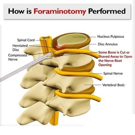
Foraminotomy
Partial Discectomy
Nucleoplasty
Radiofrequency Ablation (RFA) Or Radiofrequency Neurotomy For The Chronic Or Acute Spine Pain
Radiofrequency ablation (RFA) or radiofrequency neurotomy is a minimally invasive spine surgery. The primary goal of radiofrequency ablation or neurotomy is to destroy the nerves that senses pain. The term Neurotomy is descriptive of surgical interventions that cut or damage nerves that trigger symptoms of pain. Ablation is a general term meaning taking away or removal. In surgical scenarios, ablation applies to the surgical removal of tissue.
In short, Radiofrequency ablation and radiofrequency neurotomy are interchangeable terms used in the destruction of the innate or inherent functions of nerves. It is carried out through radiofrequency, which generates heat by radio waves. RF ablation has the most conflicting research when it comes to clinical success. Some call it an absolute failure, while others cite mild respite of pain temporarily.
All About Radiofrequency Ablation (RFA Or RF Ablation) Of The Spine:
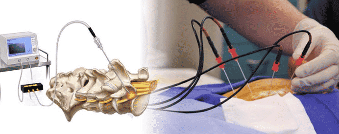
Radiofrequency ablation or RF ablation is a minimally invasive spine surgical procedure that takes about one to two hours. The process requires using several specialized needles inserted in the vicinity of the pain-sensing nerve (medial branch nerve) in your lower back to destroy the nerve with heat. RFA or radiofrequency ablation for back patients is performed in an operating room under live X-ray. You will be positioned on your stomach, sedated through local anesthetics, and conscious during the procedure.
The surgeon will insert three to four needles after carefully assessing you, making sure that you are a candidate for the procedure. Once the needles are in place, the radiofrequency machine connected to the needles will start to send strong radio waves to the tip of the needles. Radio waves will heat the tip of these needles and burn the nearby tissues and nerves.
Most patients may experience increased pain, and some may report an intense burning-like sensation similar to a severe sunburn in the area cared for. According to published reports, you should notice an improvement in symptoms two to three weeks following the procedure. However, RFA is not a cure. In fact, it may harm the nearby tissues, including the muscle that stabilizes your spine. To understand this better, we have outlined the functions of the medial branch nerve, which is a target of radiofrequency ablation surgery, below:
Sense pain in your spinal joints
Controls the ligaments of your spinal joints (medially)
The only nerve the innervates and controls the multifidus muscles
RFA for back pain: what it targets and why the details matter
What RFA aims to do: Radiofrequency ablation (RFA) uses heat at the tip of a small probe to create a tiny lesion next to the medial branch nerve. That nerve carries pain signals from the small joints of the spine (facet joints). By disrupting that signal, RFA can reduce joint-related back pain for some people often for a number of months.
What else the medial branch does: Besides pain signaling from the facet joints, the medial branch also provides a small motor supply to the multifidus, a deep stabilizing muscle that helps you stand upright, extend, side-bend, and rotate through the trunk. Because of that connection, RFA may also affect the multifidus in the treated area.
Why this matters: After medial branch RFA, fatty/atrophic change in the multifidus can occur on imaging for some patients and may be difficult to reverse. That’s why careful patient selection and a protect-the-muscle rehabilitation plan (spine-neutral motor control, graded loading, no provocative twisting early on) are important. Pain relief can also fade over time as nerves regrow, and some people consider repeat procedures another reason to plan strengthening around the stabilizers from the start.
Good questions to ask before RFA
Is my pain clearly coming from the facet joints (how was this confirmed)?
What result should I expect (pain, function, meds, return to work), and for how long?
What’s the plan to protect and retrain the multifidus after the procedure?
If relief is partial or short-lived, what are my next non-invasive options?
Therefore, many people first consider conservative, whole-spine care delivered non-invasively before procedures like RFA. For a plain-language overview, see this article on radiofrequency ablation (RFA) and conservative alternatives. (External educational resource; not medical advice. Discuss options with a registered clinician.)
Considering Conservative Care Before or After RFA
RFA can provide symptom relief for appropriately selected patients, but the duration and degree of benefit vary. Because the procedure changes pain signaling rather than directly restoring the underlying joint or surrounding muscle function, rehabilitation and movement management remain important.
For a non-emergency presentation, patients may also wish to discuss conservative options before proceeding with an interventional procedure. These may include appropriate physiotherapy, chiropractic care, graded rehabilitation, movement retraining, and other non-invasive approaches selected after assessment.
The appropriate choice depends on the suspected pain source, clinical findings, previous care, neurological status, imaging when relevant, and the patient’s goals.
To ask about an assessment or appointment availability, contact CSC in KL.
Spinal Decompression Through Laminectomy/Laminotomy
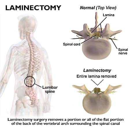
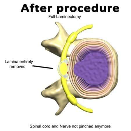
The lamina is the bony structure at the rear of the spine. They protect the spinal canal and enable the attachment of critical ligaments for spinal stability. Your spinal canal starts at the base of the skull and runs down to the tailbone. Laminectomy is a minimally invasive spine surgery that removes the lamina. Earlier, we mentioned that the lamina has the attachment point for a critical ligament. Ligamentum flavum is a highly specialized ligament that connects spinal segments through their laminal attachment points.
The ligamentum flavum limits excessive motion and has the contractile ability to reposition the spine into proper alignment following a flexion or forward bending. Repetitive traumatic events, poor posture, and prolonged sitting can lead to hypertrophy of the ligamentum flavum. Hypertrophy of the ligamentum flavum can increase the chance of developing spinal canal stenosis.
A laminectomy removes part or all of the lamina to create additional space within the spinal canal. It may be performed for spinal stenosis or other conditions in which neural structures are compressed. Depending on the case, associated ligament, bone, or other tissue may also be addressed.
The amount of bone removed and the surgical approach vary. Some procedures use smaller, minimally invasive approaches, while others require wider decompression.
As with other spinal procedures, outcomes depend on the underlying diagnosis, the amount of decompression required, spinal stability, alignment, patient health, and other factors. Persistent symptoms, recurrent narrowing, instability, and further surgery can occur in some patients and should be discussed as part of informed consent.
Decompression Of Spine Performed Through A Foraminotomy
Spinal nerves originate from the spinal cord. As they leave the cord, they pass through a passageway. The passageway provides protection and an exit point for the spinal nerves. These passageways are termed intervertebral foramina or foramen. But, the foramina are not a passage just for the nerves.
Other structures also pass through this opening, including the spinal artery, veins, and sinuvertebral nerve (the major pain-sensing nerve for spinal discs, spinal joints, and joint ligaments). The spinal discs' borders are formed by the spinal discs, spinal bones (vertebral bodies) anteriorly, and spinal joints (facets) posteriorly. Arthritic or degenerative changes of spinal discs, vertebral bodies, and facet joints will result in the foramina's narrowing.
Narrowed spinal foramina (vertebral foramina) are classified as stenotic. The term used to describe such conditions is called foraminal stenosis. The narrowed foramina (foraminal stenosis) compress the spinal nerves and vessels within the passageway (vertebral foramen).
Foraminotomy is the process of removing portions of the bony walls (pedicels of the top of facets or parts of vertebral bones), spinal discs, or both. The amount of bone, disc material, or other tissue removed during foraminotomy depends on the source and location of the narrowing. Because these structures contribute to spinal mechanics, the surgeon must balance adequate nerve decompression with preservation of stability.
Persistent symptoms, recurrent narrowing, instability, or the need for additional surgery may occur in some patients, but the likelihood varies by diagnosis, surgical technique, number of levels, and individual factors.
Nucleoplasty: What Recent Studies Show About Benefits and Risks
Some studies of intradiscal procedures have reported limited or inconsistent improvement in carefully selected patients. Changes in an average pain score should be interpreted according to the study’s predefined outcome measures rather than reclassified as either “success” or “failure” using a different definition after the fact.
The authors reported the average patient pain level before nucleoplasty at 6.7 (on a Visual Analog Scale or VAS of 1-10, with 10 being excruciatingly severe). Nine months after nucleoplasty, pain levels were documented at 5.6, equating to a 10% decrease in pain.
The important questions are whether the change was clinically meaningful, whether function improved, how long any improvement lasted, how the intervention compared with a control or sham procedure, and what adverse events occurred.
Are There Different Types Of Nucleoplasty For The Spine?
Two approaches are described in the literature: nucleoplasty alone and nucleoplasty combined with IDET. Evidence of benefit is limited and inconsistent; selection and conservative optimization first are recommended.
Nucleoplasty without intradiscal electrothermal therapy (IDET)
Nucleoplasty with intradiscal electrothermal therapy (IDET)
The process of inserting a catheter (introducer) at the center of the spinal disc is damaging, but cooking it through electrothermal means blows the mind. However, most troubling is the motive behind this rootless procedure's continued use in light of its documented failures!
A randomized, double-blind study from the Department of Orthopedics at the Royal Adelaide Hospital in South Australia concluded no differences between the nucleoplasty with IDET and the sham IDET procedure. The study was published in the Spine Journal.
Intradiscal procedures differ in how they act on disc tissue, and evidence should be considered separately for each technique rather than treating IDET and nucleoplasty as identical procedures.
One randomized, double-blind, placebo-controlled trial of IDET enrolled 57 participants: 38 received IDET and 19 received a sham procedure. At six months, IDET did not show a meaningful advantage over the sham procedure using the study’s predefined outcome measures.
The study therefore raises an important question about expected benefit in appropriately selected patients, while also demonstrating why results from one intradiscal technique should not automatically be applied to every disc procedure.
IDET vs Placebo: What a Randomized Controlled Trial Found
IDET heats the outer disc (annulus) to try to shrink/“seal” tears; nucleoplasty is different but related it removes/disrupts inner disc material (nucleus). In a rigorous randomized, double-blind, placebo-controlled trial of IDET for chronic discogenic low back pain (patients had 1-2 symptomatic levels with annular tears and had already tried conservative care), 57 people were randomized (38 IDET; 19 sham). At 6 months, no patient in either group met the study’s predefined “successful outcome”, and standard measures (Oswestry Disability Index, SF-36, etc.) did not improve meaningfully with IDET compared with placebo. There were no permanent neurologic complications reported.
What this means for you: if you’re being offered IDET or a nucleus-targeting procedure like nucleoplasty, ask how “success” will be defined for you (pain, function, meds, return to work), what benefit is realistically expected, and what non-invasive options remain. This trial suggests IDET did not outperform placebo at 6 months in a carefully selected group, so it’s reasonable to seek a second opinion and review conservative, image-informed, low-force care before choosing a disc procedure.
Citation: Freeman BJC, Fraser RD, Cain CMJ, Hall DJ, Chapple DCL. A randomized, double-blind, controlled trial: intradiscal electrothermal therapy versus placebo for the treatment of chronic discogenic low back pain. Spine (Phila Pa 1976). 2005;30(21):2369-2377; discussion 2378. doi:10.1097/01.brs.0000186587.43373.f2.
Discectomy Is The Type Of Operation That Removes The Spinal Disc In Part Or Totally.
The spinal discs are the separators, connectors, shock absorbers, and stabilizers of the spine that provide mobility and weight-bearing. Poor posture, prolonged sitting, and repetitive traumatic events lead to degenerative changes that impact the health of spinal discs.
A degenerated spinal disc is a weak structure that is unable to provide efficient loadbearing and mobility. In time, a degenerated disc can succumb to stresses of daily life, causing the spinal disc to tear, bulge, herniate, or rupture. A bulging disc or a herniated disc is better known by its layperson term, slipped disc or slip disc.
As the spinal disc fibers protrude, bulge, or herniate beyond their normal boundary, they can push or pinch the spinal nerve or spinal cord.
Discectomy removes part, and less commonly all, of an intervertebral disc when disc material is believed to be contributing to clinically significant nerve compression or other surgical indications.
Some disc herniations can decrease in size over time without surgery, while others produce persistent or progressive neurological symptoms for which surgical assessment may be appropriate. The decision depends on neurological findings, symptom severity and duration, imaging correlation, and response to appropriate conservative care.
Removing disc material changes the structure of the treated segment and may influence spinal mechanics. The clinical significance of those changes varies between patients and procedures.
Spinal Fusion: An intervention For Severe Cases Or For Failed laminectomy (laminotomy), Facetectomy, Foraminotomy, Nucleoplasty & Partial Discectomy.
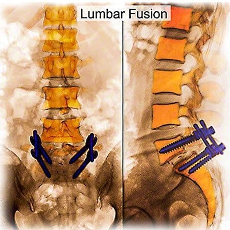
Spinal fusion is a surgical intervention that has been around for decades. It was the go-to method of spine surgery for most surgeons. In spinal fusion, the surgeon attempts to fuse spinal bones through implants, a bone graft, or a combination of the two. Fusion aims to stop mobility or motion in hypermobile, unstable, or excessively damaged segments. There are various methods of spinal fusion, including minimally invasive spine surgery.
Each type of method has its merits and disadvantages. In some patients, the surgical method is performed from the front, the back, or the front and back. It is often harvested from the patients’ hip or a synthetic source if the bone graft is used. In spinal fusion, a surgeon removes the facet joints and the spinal disc.
They may pack the void with spacers or a bone graft. Also, screws may be used to hold the implant and spacer (hardware) in place. The bone graft will join one or more spinal segments into a single solid mass. With fusion, there can be a loss of mobility. Restricted motion is a common cause of additional stress on segments adjacent to the fused vertebrae.
Spinal fusion may increase mechanical stress on nearby segments (adjacent-segment changes), which can contribute to problems over time in some patients. Not everyone develops this; risk varies with factors like number of levels fused, alignment, and activity.
If symptoms remain after previous spine surgery, a fresh assessment can help clarify whether the current symptoms arise from the original condition, postoperative changes, another spinal level, or a different source.
If the first surgery didn’t help, pause before the second
Before another operation is considered, it is reasonable to establish what is now producing the symptoms and whether the proposed procedure addresses that finding.
When the situation is not urgent, allowing time for reassessment and consideration of appropriate non-surgical options can help patients make a more informed decision.
Smarter next steps (before surgery #2 or #3)
Reassess the cause: correlate symptoms with a fresh clinical exam and available imaging; confirm the actual pain generator(s).
Try a time-boxed conservative plan: gentle, non-rotatory care with clear checkpoints (e.g., visits 5/10/15) and measurable goals.
Get two opinions: one surgical and one non-surgical. If both agree surgery is necessary or there’s a true emergency proceed informed.
(Educational guidance, not medical advice. Decisions should follow a thorough assessment by a registered clinician.)
How Long Does It Take To Recuperate From The Operation?
Recovery time after spine surgery varies substantially according to the procedure, number of levels treated, diagnosis, age, general health, neurological status, postoperative rehabilitation, and the physical demands of work and daily life.
Some procedures allow relatively early return to selected activities, while recovery from more extensive surgery can take considerably longer. The operating surgeon should provide procedure-specific restrictions, rehabilitation milestones, and expected timeframes.
Some patients achieve substantial symptom and functional improvement, while others continue to experience residual symptoms or activity limitations. Expectations should therefore be discussed in terms of both symptom improvement and meaningful function.
For a non-emergency decision, obtaining another qualified opinion, surgical or non-surgical, can help clarify the available options before proceeding.
How Successful Is The Back Operation? Let's See What Research On The Spine Journal Reported.
According to a published research article in the peer-reviewed Spine Journal, fusion causes spinal stenosis, disc herniations, and instabilities next or adjacent to the surgically fused segment. In other words, patients who were operated on through spine fusion surgery developed spinal disc problems in healthy segments before surgery.
Spine surgery (laminectomy, foraminotomy, discectomy, nucleoplasty, or spinal fusion) can reduce pain if successful, but it will also hamper your spine’s ability to function normally. In time, the operated-on segment or those near the surgical site will decay or degenerate faster. Spinal fusions have the highest rate of degenerative changes in the good ones. We have provided the recap of that research below:
All fifty-eight patients acquired spinal stenosis, disc herniation, or disturbance in a section next to previously asymptomatic or healthy spinal segments. In short, spinal fusions could result in damage and symptoms that arise from segments that are adjacent to the operated site.
Impact Of Cervical Laminectomy
Another published research on the clinical outcomes of 64 cervical laminectomy noted abnormal changes in spinal curvatures in 36% of patients. Also, 14% of the patient cases studied resulted in reversed cervical curvatures or kyphotic curves two years post-surgery. Furthermore, two of the 64 patients needed additional surgeries. Additional surgeries were needed two years following the first surgery to address the instabilities caused by laminectomy.
Unfortunately, this study did not elaborate on the level of pain, function, symptoms, or quality of life following surgery. Also, the study only reviewed cases 2-years post-surgical intervention. We are confident that had they examined patient cases at five years and seven years (post-surgical study), the reported statistics would undoubtedly be much higher among those needing a second or even a third operative procedure. Nonetheless, from the limited information gathered, one could persuasively argue that laminectomy has a success rate of 50%.
The most astonishing report on the long-term success or outcomes of a lumbar fusion surgery comes from research conducted by the Ohio Bureau of Workers’ Compensation records. We have provided a link to your review's abstract of a historical cohort on surgical spinal fusion.
Why Spine Surgery Sometimes Falls Short
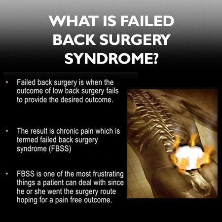
Clinicians sometimes use the term failed back surgery syndrome (FBSS) when pain returns or never fully settles after an operation. That doesn’t mean surgery never helps many people do well. It means there are biomechanical and diagnostic reasons why relief can be incomplete.
What can go wrong and why:
Load shifts to other levels. Fusing or reshaping one segment can increase stress on the discs and joints above/below (often called adjacent-segment changes), which may become irritated over time.
Scar and sensitivity. Healing can create scar tissue around nerves (epidural fibrosis) or leave tissues more reactive, keeping symptoms going even after the original compression is treated.
Muscle shutdown and deconditioning. Pain and incision protectiveness can inhibit deep stabilizers (e.g., the multifidus). Without a plan to rebuild them, support stays weak and movement stays guarded.
Target mismatch. Imaging may show several findings, but not all findings cause pain. If the true “pain generator” isn’t addressed, symptoms can persist.
Movement/posture drivers remain. Habitual flexed sitting, repeated end-range bending, or poor hip mechanics can keep loading the same segment, regardless of what was done surgically.
More surgery ≠ better odds. Re-operations are typically more complex (more scar tissue, altered mechanics). That’s why a careful re-assessment is vital before “surgery #2 or #3.”
Minimally invasive ≠ consequence-free. Smaller incisions still alter mechanics and tissues; they can help selected cases, but they are not immune to the issues above.
Practical takeaway: If you’re not facing an emergency, exhaust a measured, non-invasive plan first calm-first care, image-informed decisions, and staged rehab that rebuilds stabilizers and movement control. If surgery is needed (or already done), pair it with a targeted rehabilitation plan and clear checkpoints so progress is tracked and gaps are addressed early.
(Educational information only. Decisions should follow a thorough assessment by a registered clinician.)
European Spine Journal on Spine Surgery
An article in the European Spine Journal reported on the eliminating of a patient pain or symptoms through invasive spinal decompression methods by experienced surgeons at 90% two months after the operation. The failure rate drops to 75% after five years with medication.
A study in the European Spine Journal reported that after lumbar decompression (laminectomy, foraminotomy, facetectomy, or partial discectomy), 90% of patients still had leg symptoms at 2 months. At 5 years, 75% continued to report symptoms, often requiring medication; 25% reported no leg pain. In the same cohort, 24% later had a second surgery (fusion), and a smaller subset underwent a third (5-year lumbar decompression outcomes (Eur Spine J, 2010).
The paper nevertheless described the operations as “successful” highlighting a key gap between research success (any measurable improvement) and patient success (low pain, good function, fewer meds, return to normal life). In other words, symptom reduction ≠ symptom resolution.
Any decrease in patients’ symptoms backed the author’s claims of surgical successes. In other words, any evidence of improvement was deemed as the success achieved. You can download and read this research below:
Spine Fusion Failure Was Related To Permanent Disabilities In 74% Of Workers In The U.S. State Of Ohio.
Researchers examined the clinical outcomes of 1,450 patients using the Ohio Bureau of Workers’ Compensation database. The researchers specifically targeted patients with a diagnosis of disc degeneration, disc herniation, or radiculopathy. Let’s define what radiculopathy is first.
Radiculopathy is pain that runs down one or both limbs due to nerve compression or nerve damage. Patients complaining of radiculopathy may experience pain, tingling (pins and needles), burning sensations, and weakness in one or both limbs.
Those with neck-related radiculopathy may experience symptoms in the upper and lower extremities. Radiculopathy involving the lower back mainly impacts the lower buttocks and legs.
Now, let’s get back to the research article. Researchers divided the database into two equal groups, surgical and non-surgical. It is important to note that both groups had patients who suffered from the same diagnosis and severity. In short, of the 1,450 cases, 750 were operative patients, and the remaining 750 received non-invasive care. The researcher compared the clinical outcomes in each group after two years. Keep in mind that patients in both groups had identical complaints. Although they were similar, the recovery and overall success were not the same.
Only 26% of the injured workers that were operated on returned to work. The remaining 74% of fusion patients were considered medically disabled and as such, unfit to continue their work-related activities.
The published statistics equate to a resounding failure rate of 74% for surgical patients. Had the surgery been successful, surely the patients would have returned to work! Now, let’s see how the non-surgically cared for patients did. The non-surgical group got targeted physical therapy (physiotherapy) methods.
Of the 750 physiotherapy patients, 67% returned to work within two years. The study offers clear evidence that spine surgery may not allow the patient to return to an active life post-surgery. Additionally, the research establishes the impact of clinically targeted physiotherapy methods as opposed to surgery.
Why Should You Seek A Second Opinion From A Non-Surgical Provider When Your Surgeon Recommends A Procedure?
We live in a world where the opinions and recommendations of neurosurgeons and orthopedic surgeons are not questioned. Simply put, when spine surgeons recommend surgery, patients follow their advice.
There is nothing wrong with putting your trust in the doctors and surgeons who care for you. After all, you willingly went seeking their professional opinion.
But, as with any medical recommendation, you may want to seek a second and even third option in non-life-threatening situations. Orthopedic surgeons and neurosurgeons spend years learning surgical methods for the spine.
They are physicians who specialize in spine surgery, and their research, training and continuing education center on surgical skills and knowledge. A non-surgical opinion looks at the same problem from the side of conservative care.
A surgical second opinion and a non-surgical second opinion can both be useful. A non-surgical second opinion comes from chiropractors and physiotherapists who assess the spine and explain the conservative options, including for people whose earlier surgery did not settle their symptoms.
Why Should You Seek Out Second Opinions From Chiropractors When The Surgeon Has Recommended An Operation For The Spine?
A spine surgeon’s clinical focus is on surgical interventions. A chiropractor's focus is entirely dedicated to non-surgical methods. Chiropractors help with focused non-surgical spine care, helping with spine-related conditions daily, weekly, monthly, and yearly. In short, a highly experienced clinical chiropractor is the recommended individual to visit for non-surgical second opinions.
Chiropractors and physical therapists, or physiotherapists, have been caring for the spine for over a century. For obvious reasons, a surgeon’s education and training are focused on surgical methods of care. Likewise, a chiropractor or physiotherapist (physical therapist) spends years learning non-surgical methods. However, it must be said that surgeons, chiropractors, and physiotherapists may hold degrees but differ. Some surgeons excel more than others!
The same holds for chiropractors and physiotherapists. So, care must be taken when choosing one. Please don't shy away or be afraid to ask your neurosurgeon, orthopedic surgeon, chiropractor, or physiotherapist about their education, training, expertise, and experience.
Ask them about their personal experiences in similar conditions and results achieved. Question them on surgical and non-surgical methods. Ask them about the time they spend researching surgical and non-surgical techniques of spine care. Don’t be afraid; it is your spine, health, and long-term well-being at stake.
So, speak up and get as much information from each specialty about your options. Doing so will enable you to make a better-informed decision.
Non-Surgical Spinal Decompression Therapy Conservative Care Options for the Neck & Back
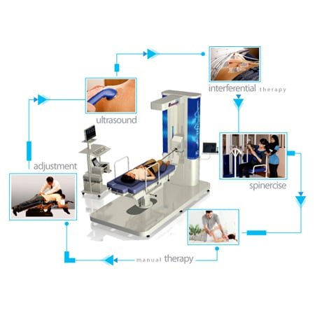
NSD Therapy® is an alternative, non-invasive method of spine care.. It is a multi-faceted spine care system rendered by research-based chiropractors, and clinical physiotherapy is provided through manual and integrated spine-specific technologies. If your surgeon is recommending spine surgery, NSD Therapy® can help. Visit us today to discover the recommended alternative to spine surgery.
Our clinical teams of Chiropractors and Physiotherapists use spine-specific methods and technologies such as the decompression therapy, Laser, and flexion-distraction to help your recovery for neck and back issues. Best of all, the care you get from us is non-invasive. We have cared for many across our center who sought our help with their slipped disc, neck pain, and back pain.
NSD Therapy®: a gentle, customizable, non-surgical plan
NSD Therapy® is one of our non-surgical options for selected spine conditions. It’s a gentle, non-rotatory, team-delivered protocol that aims to calm irritated tissues and improve movement and, for some people, may reduce the likelihood of needing injections or surgery. We first introduced NSD Therapy® at our Bukit Damansara (Kuala Lumpur) center and now offer it at several CSC locations. To ask whether it suits your case, call 03-2093 1000.
How we tailor NSD Therapy®
Stepwise assessment: history, movement and posture screen, segment-by-segment palpation, and a focused neurological check.
Image-informed planning: our registered clinicians review your MRI images (T1/T2) in addition to the radiologist’s report. We look for the exact level(s) involved, annular tears, Modic/end-plate changes, facet irritation, soft-tissue signal suggesting inflammation, and muscle quality (e.g., fatty infiltration). We also consider tissue depth (how many cm below the skin) to choose modalities that can actually reach the target safely.
Calibrated, comfort-first care: forces, angles, and session length are adjusted to your tolerance; no forceful twisting or end-range “cracking.”
Measured progress: clear goals and checkpoints; we modify the plan based on your response.
What we may use (case-by-case)
Angle-controlled spinal decompression to reduce load at the involved level(s).
Flexion-distraction (brief, non-rotatory).
SOT blocking + Activator for precise, low-force alignment cues.
High-intensity laser (Class IV) when deeper penetration is needed (dose-controlled).
Therapeutic ultrasound applied with a smooth, gel-cushioned glide (not pressure massage).
Focused shockwave at low settings for superficial tender points when appropriate.
Electrotherapy and spine-neutral motor control to rebuild support without flaring symptoms.
What NSD Therapy® is not
Not a single machine or one-size-fits-all protocol.
Not aggressive stretching, deep paraspinal pressure, or rotatory thrusts.
Not planned from a report alone we correlate your exam findings with the actual MRI images to decide what’s safe and likely to help.
Educational information only; not a diagnosis or medical advice. Care decisions should follow a thorough assessment by a registered clinician. Individual results vary.
What Conservative Care Should Have Included Before Surgery Is Reasonable
Most guidelines assume a real trial of non-surgical care before elective spine surgery. The trouble is that what counts as a trial is rarely defined, and many people arrive at a surgical consultation having had far less than they think. A reasonable trial includes all of this:
A program of graded exercise, supervised, over eight to twelve weeks. Not a handout. Not three sessions. Exercise therapy for chronic low back pain has been studied at that scale (Hayden JA, Ellis J, Ogilvie R, Malmivaara A, van Tulder MW. Cochrane Database Syst Rev. 2021;(9):CD009790).
Load that was actually increased. If week ten looked like week one, the program was not tested.
An explanation you understood. What is thought to be causing the problem, and why the plan follows from that.
Attention to sleep, activity and the things that keep pain going. These are not extras. They change how a spine responds to everything else.
A reassessment with measurements, not impressions. Walking distance, sitting tolerance, what you have and have not got back.
If your course of care missed several of these, the useful question before consenting to surgery is whether conservative care failed or was never properly delivered.
Questions Worth Asking Your Surgeon
These are ordinary questions and a good surgeon will welcome them.
What exactly are you operating on, and how confident are you that it explains my symptoms?
Which of my symptoms do you expect to change, and which do you not? Leg pain and back pain often respond differently.
What happens to people like me who do not have this operation?
How many of these do you do in a year, and what does your own follow-up look like?
What is the plan if this does not work, and what would the next operation be?
What does recovery actually involve, week by week, and what rehabilitation is arranged?
When Spine Surgery Should Not Wait
Nothing on this page applies to these. They are urgent:
Go to an emergency department today.
Weakness that is getting worse, a foot that drags, or wasting of muscle.
Loss of hand coordination, unsteadiness when walking, or symptoms in both arms or both legs.
Fever with back pain, especially with a recent infection, injection or procedure.
Spine pain after significant trauma, or in anyone with osteoporosis, cancer or long-term steroid use.
What does “successful” mean in studies and how is that different from what patients expect?
In clinical papers, any measurable improvement can count as success (e.g., a small drop in a pain or disability score). For most people, success means low enough pain to live normally, fewer meds, better sleep/walking tolerance, and a return to work or sport. Your page explains this gap and shows why procedures labeled “successful” in journals may still leave some with persistent symptoms. Agree on patient-meaningful targets with your clinician (pain ≤3/10 most days, walk/sit 30-60 minutes, med step-down, no new deficits) and review them at set checkpoints (e.g., weeks 2/4/8).
When is spine surgery usually considered?
What are the main risks and limitations of common procedures (laminectomy, foraminotomy, discectomy, fusion)?
All surgery is invasive “minimally invasive” still cuts tissue. Reported issues include dural tears, nerve irritation/injury, infection, and adjacent-segment problems. Your page summarizes research where decompression procedures often left residual leg symptoms, and where fusion cohorts showed higher reoperation/complication and opioid-use rates and lower return-to-work in specific populations. Outcomes vary by person, procedure and surgeon; that’s why shared definitions of success and clear follow-up plans matter.
Are “minimally invasive” procedures more successful?
Smaller incisions don’t guarantee better long-term results. A keyhole is still an incision, and to see the target the surgeon has to move a probe and a camera through several angles, which can disturb the tissue around the entry point. Keyhole approaches can still change spinal mechanics, create scar tissue, and in some people still lead to further surgery within a few years. Decisions should weigh imaging-symptom match, response to conservative care, and personal goals, not incision size alone.
If my first surgery didn’t help, what should I do before considering a second or third?
Reassess the cause: fresh clinical exam + available imaging to confirm the pain generator(s). Time-boxed conservative plan: gentle, non-rotatory care with checkpoints (e.g., visits 5/10/15) and measurable goals. Two opinions: one surgical and one non-surgical. If both point to surgery or there’s an emergency proceed informed. Repeating the same approach without a new diagnosis rarely improves the odds; risks typically rise with each operation.
What does a good non-invasive plan look like and when might I feel a change?
Start calm and precise: no forceful twisting or end-range “cracking” during inflammatory phases; correlate exam with MRI images (not just the report); use dose-controlled modalities (e.g., angle-controlled decompression, brief flexion-distraction, high-intensity laser, ultrasound, focused shockwave at low settings when indicated). Add movement re-education and core control only when tissues tolerate loading. Many notice steadier days in a few weeks when flare drivers are reduced, with progress reviewed at planned checkpoints. Individual results vary.
What is NSD Therapy® and who might it suit?
NSD Therapy® is a gentle, team-delivered protocol not a single machine. It starts with stepwise assessment (history, movement, segment palpation, focused neuro check) and image-informed planning (your clinicians review T1/T2 MRI images alongside the report to identify levels, tears, Modic/end-plate and facet changes, soft-tissue signals, and muscle quality). Care is calibrated to tissue depth and tolerance (no rotatory thrusts), and may include angle-controlled decompression, brief flexion-distraction, SOT/Activator alignment cues, high-intensity laser, therapeutic ultrasound, focused shockwave for superficial tender points, and electrotherapy with spine-neutral motor control. It’s considered for selected spine conditions after individual evaluation.
Peer-Reviewed Medical References:
Deyo RA, Mirza SK, Martin BI. Back pain prevalence and visit rates: estimates from U.S. national surveys, 2002. Spine. 2006;31(23):2724-2727.
Fritz JM, Childs JD, Wainner RS, Flynn TW. Primary care referral of patients with low back pain to physical therapy: impact on future health care utilization and costs. Spine. 2012;37(25):2114-2121.
Bronfort G, Haas M, Evans R, Leininger B, Triano J. Effectiveness of manual therapies: the UK evidence report. Chiropr Osteopat. 2010;18:3.
Cherkin DC, Deyo RA, Sherman KJ, et al. A randomized trial comparing acupuncture, simulated acupuncture, and usual care for chronic low back pain. Arch Intern Med. 2009;169(9):858-866.
The Author of Spine Surgery Risks: Complications & Safer Options
Spine Surgery Risks: Complications & Safer Options was written by Yama Zafer, D.C., who has 30+ years in chiropractic and physiotherapy and founded Chiropractic Specialty Center in Kuala Lumpur in 2006.
Last Updated: Spine Surgery Risks & Non-Invasive Alternatives
Spine Surgery Risks & Non-Invasive Alternatives was last reviewed and updated on October 4, 2026.
Five years with compression on both sides at L4-L5 raises two questions that are worth settling, because bilateral involvement behaves differently from one-sided nerve compression.
The first is what pattern your symptoms actually follow. If both legs feel heavy or ache after walking a certain distance, and that eases specifically when you sit down or lean forward rather than merely when you stop, the canal is narrowed and this is neurogenic claudication. The confirming sign is being able to walk much further pushing a trolley or going uphill than on level ground. That version is managed by using forward-leaning positions deliberately to build walking tolerance, and the measure that matters is walking distance rather than pain score. If instead your symptoms follow a defined line down each leg with numbness in specific areas, that is bilateral root involvement and the emphasis differs.
The second is whether your imaging is current. Five-year-old imaging no longer describes your spine, and if a decision of any kind is approaching, a current scan is the starting point rather than a re-reading of the old one.
Now the part that most often explains why five years have passed without resolution. Care for disc problems commonly settles symptoms and stops there. Pain eases well before the muscles supporting the segment have recovered, so the underlying capacity is never rebuilt, and the problem returns each time demand rises. If nothing in your five years has included progressive strengthening, continued for months past the point where symptoms settled, then the main lever has never been pulled. That is more likely than the idea that your case is untreatable.
A program that would address it: restoring the movement lost above and below L4-L5 and at the hips, since stiff hips push their work into this level; progressive loading of the deep trunk muscles and the gluteals, increased in small steps every week or two rather than repeated at the same level; and rebuilding tolerance for sitting, standing, and walking as measured quantities rather than by feel. Bending technique matters too, since L4-L5 tolerates rounded loaded bending least of all.
Some have found spinal decompression therapy by devices like the RxDecom or similar to be helpful for spinal disc related issues, including those that at times cause nerve issues.
If your symptoms steadily get worse, or the care you are receiving is not helping, please arrange a medical review. New numbness around the genitals or anus, new difficulty passing urine, or loss of bladder or bowel control needs urgent medical attention: a thorough assessment by a competent clinician the same day.
Straight answer first: spinal manipulation is not appropriate for you. Instrumented segments are not manipulated, and neither is a spine four years after a cord injury. Anyone offering that should be declined.
That said, there is a great deal in chronic spinal cord injury that is genuinely treatable, and most of it concerns everything other than the injured segments.
Shoulders are the first priority and the most commonly neglected. In manual wheelchair users the shoulders take over the work of the legs, and rotator cuff problems become extremely common over the years, particularly with transfers and with propelling on inclines. Losing shoulder function after a cord injury costs independence directly. Preventive work matters here: strengthening the muscles that hold and rotate the shoulder blade, balancing the front-loaded work of pushing with pulling exercise, keeping the chest and front shoulder tissue from shortening, and having transfer technique and wheelchair setup reviewed. If either shoulder already hurts, that is worth assessing now rather than after it limits you.
Second, any new or changing symptom above your level of injury deserves prompt medical assessment rather than being attributed to the original injury. A recognized late complication after traumatic cord injury is the development of a fluid cavity within the cord, which can appear years afterward and presents as new pain, a rising level of sensory loss, increasing weakness, or increasing spasticity. It is diagnosed by MRI and it is treatable, but it needs to be looked for. Given your injury four years ago, that is worth knowing about specifically.
Third, the ongoing management that has the largest effect on long-term health: skin and pressure care, bladder and bowel management, spasticity and contracture prevention, and cardiovascular conditioning, which is harder to maintain after a cord injury and matters as much as it does for anyone else.
Physiotherapy has a real role in all of that, and registered physiotherapy is provided here. Whether travel is worthwhile for it is a different question, and specialist spinal rehabilitation services closer to home are usually the better route for ongoing care.
Seek urgent medical assessment if you notice a rising level of numbness, new weakness in the arms, sudden increases in spasticity, or symptoms of autonomic dysreflexia such as a pounding headache with sweating and flushing above your injury level.







PIVD L4 L5 nerve compression on both sides for the last 5 years.
Five years with compression on both sides at L4-L5 raises two questions that are worth settling, because bilateral involvement behaves differently from one-sided nerve compression.
The first is what pattern your symptoms actually follow. If both legs feel heavy or ache after walking a certain distance, and that eases specifically when you sit down or lean forward rather than merely when you stop, the canal is narrowed and this is neurogenic claudication. The confirming sign is being able to walk much further pushing a trolley or going uphill than on level ground. That version is managed by using forward-leaning positions deliberately to build walking tolerance, and the measure that matters is walking distance rather than pain score. If instead your symptoms follow a defined line down each leg with numbness in specific areas, that is bilateral root involvement and the emphasis differs.
The second is whether your imaging is current. Five-year-old imaging no longer describes your spine, and if a decision of any kind is approaching, a current scan is the starting point rather than a re-reading of the old one.
Now the part that most often explains why five years have passed without resolution. Care for disc problems commonly settles symptoms and stops there. Pain eases well before the muscles supporting the segment have recovered, so the underlying capacity is never rebuilt, and the problem returns each time demand rises. If nothing in your five years has included progressive strengthening, continued for months past the point where symptoms settled, then the main lever has never been pulled. That is more likely than the idea that your case is untreatable.
A program that would address it: restoring the movement lost above and below L4-L5 and at the hips, since stiff hips push their work into this level; progressive loading of the deep trunk muscles and the gluteals, increased in small steps every week or two rather than repeated at the same level; and rebuilding tolerance for sitting, standing, and walking as measured quantities rather than by feel. Bending technique matters too, since L4-L5 tolerates rounded loaded bending least of all.
Some have found spinal decompression therapy by devices like the RxDecom or similar to be helpful for spinal disc related issues, including those that at times cause nerve issues.
If your symptoms steadily get worse, or the care you are receiving is not helping, please arrange a medical review. New numbness around the genitals or anus, new difficulty passing urine, or loss of bladder or bowel control needs urgent medical attention: a thorough assessment by a competent clinician the same day.
I'm a paraplegic. I injured my spine 4 years ago. I have surgical instruments in L4 and T6.
Straight answer first: spinal manipulation is not appropriate for you. Instrumented segments are not manipulated, and neither is a spine four years after a cord injury. Anyone offering that should be declined.
That said, there is a great deal in chronic spinal cord injury that is genuinely treatable, and most of it concerns everything other than the injured segments.
Shoulders are the first priority and the most commonly neglected. In manual wheelchair users the shoulders take over the work of the legs, and rotator cuff problems become extremely common over the years, particularly with transfers and with propelling on inclines. Losing shoulder function after a cord injury costs independence directly. Preventive work matters here: strengthening the muscles that hold and rotate the shoulder blade, balancing the front-loaded work of pushing with pulling exercise, keeping the chest and front shoulder tissue from shortening, and having transfer technique and wheelchair setup reviewed. If either shoulder already hurts, that is worth assessing now rather than after it limits you.
Second, any new or changing symptom above your level of injury deserves prompt medical assessment rather than being attributed to the original injury. A recognized late complication after traumatic cord injury is the development of a fluid cavity within the cord, which can appear years afterward and presents as new pain, a rising level of sensory loss, increasing weakness, or increasing spasticity. It is diagnosed by MRI and it is treatable, but it needs to be looked for. Given your injury four years ago, that is worth knowing about specifically.
Third, the ongoing management that has the largest effect on long-term health: skin and pressure care, bladder and bowel management, spasticity and contracture prevention, and cardiovascular conditioning, which is harder to maintain after a cord injury and matters as much as it does for anyone else.
Physiotherapy has a real role in all of that, and registered physiotherapy is provided here. Whether travel is worthwhile for it is a different question, and specialist spinal rehabilitation services closer to home are usually the better route for ongoing care.
Seek urgent medical assessment if you notice a rising level of numbness, new weakness in the arms, sudden increases in spasticity, or symptoms of autonomic dysreflexia such as a pounding headache with sweating and flushing above your injury level.