


Meniscus Tear: Symptoms, Causes and Non-Invasive Care
A meniscus tear usually causes pain on the inner or outer side of the knee, swelling that builds over a day, and catching or locking. A torn meniscus hurts most with twisting, squatting, kneeling and stairs, and the knee may not straighten fully.
Every tear comes from injury. It can be one sudden twist on a planted foot, as in football or a slip on a wet floor. Or it can be many small strains from squatting, kneeling, stairs and sport that wear the meniscus thin over the years. Tight thigh muscles and a knee that sits slightly out of line add to the strain.
Non-invasive care starts with a full assessment, using X-rays, MRI or, in most cases, both. It then combines gentle, non-rotatory chiropractic adjustment matched to the knee, physiotherapy, hands-on soft tissue work and precise exercise. Two major trials support starting this way. Trimming a worn meniscus did no better than a sham (pretend) operation, and physiotherapy did not fall short of keyhole surgery. Even a locked knee can start with this care, and surgery is the next option if it does not help. The sections below explain these trials, the tear types, complex tears, diagnosis, the care plan and recovery.
For more information about a meniscus tear, contact the Bukit Damansara (KL) main center: WhatsApp+60 17 269 1873 Call+603 2093 1000
Meniscus Tear: Key Takeaways
| Point | What to know |
|---|---|
| Ten years after trimming | Knees that had a worn meniscus trimmed scored worse than knees given a sham operation. More of them showed arthritis getting worse on X-ray (Kalske and colleagues, 2026). |
| Knee replacement later | In England, 22% of women and 10% of men who had a meniscus trimmed had a knee replacement within 15 years (Abram and colleagues, 2019). |
| Serious complications | About 3 in 1,000 meniscus trimmings in England were followed within 90 days by a serious complication, such as a joint infection or a blood clot in the lung (Abram and colleagues, 2018). |
| Scans and pain | Among adults aged 50 to 90 with a torn meniscus on MRI, 61% had no knee pain, aching or stiffness in the previous month (Englund and colleagues, 2008). |
| Which tears heal | Only the outer 10 to 25% of an adult meniscus has blood vessels, so tears at the outer edge are the most likely to heal (Makris and colleagues, 2011). |
| Alignment | In my experience, a torn meniscus will not settle until the shinbone and thighbone are back in line. I check alignment from the first visit. |
| Diagnosis | MRI is the scan of choice for finding a torn meniscus and the pattern of the tear (Nguyen and colleagues, 2014). An X-ray adds the bones and the knee's alignment. |
Meniscus Tear: Page Guide & Key Sections
- Torn Meniscus Surgery vs Conservative Care: What the Major Trials Found
- Knee and Meniscus Assessment at Two KL Centers
- Meniscus Tear Symptoms: Pain, Swelling, Catching and Locking
- What Causes a Torn Meniscus? The Mechanism of Injury
- Meniscus Tear Types: Radial, Horizontal, Bucket-Handle, Flap and Complex
- Medial vs Lateral Menisci: Why Each Tears Differently
- Complex Meniscus Tears: What They Are and How They Are Treated
- How a Torn Meniscus Is Diagnosed: Examination, X-Ray and MRI
- Meniscus Tear Treatment Without Surgery
- Can a Torn Meniscus Heal on Its Own?
- When Surgery or an Injection Is Considered
- What the Meniscus Is and Why It Matters for Knee Stability
- Who Non-Surgical Care Suits, and When Referral Makes Sense
- Torn Meniscus: Frequently Asked Questions
- References
Torn Meniscus Surgery vs Conservative Care: What the Major Trials Found
Keyhole surgery to trim a torn meniscus has been tested against a sham (pretend) operation and against physiotherapy. These trials, and national records of what happens after the operation, support conservative care first for a worn meniscus.
The Sham-Surgery Trial: Trimming Against a Pretend Operation
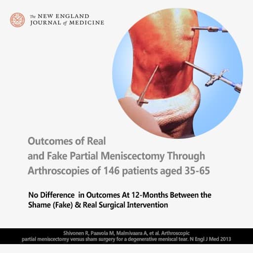
A Finnish trial took 146 adults aged 35 to 65 with a worn tear of the inner (medial) meniscus and no knee arthritis. Their pain had lasted more than three months despite conservative treatment. People whose tear began with one obvious injury were not included.
After a camera check inside the knee, 70 had the tear trimmed and 76 had a sham procedure in which nothing was removed. No one knew which they had received. At 12 months, the two groups did equally well on both knee scores and on pain after exercise (Sihvonen and colleagues, N Engl J Med. 2013;369(26):2515-2524). The journal makes the full article free as a PDF.
Five and Ten Years After Trimming
At five years, the trimmed knees showed no benefit, more mechanical symptoms and a slightly higher risk of arthritis on X-ray (Sihvonen and colleagues, 2020).
At ten years, with 91% of the patients followed up, the trimmed knees scored worse on a meniscus questionnaire than the sham knees. X-rays showed arthritis getting worse in 81% of trimmed knees and 70% of sham knees.
Eight trimmed patients and three sham patients had gone on to a knee replacement or an operation to realign the shinbone. The authors found no benefit from trimming and a suggestion of worse outcomes (Kalske and colleagues, N Engl J Med. 2026;394(17):1757-1759). The authors' accepted version is free to read.
Physical Therapy Against Keyhole Surgery: The ESCAPE Trial
The ESCAPE trial, run in nine hospitals in the Netherlands, compared early keyhole surgery with a structured physical therapy (physiotherapy) program. It enrolled 321 people with a torn meniscus that did not lock the knee. At two years, physical therapy did not fall short of surgery for knee function (van de Graaf and colleagues, 2018).
At five years it still did not. The authors concluded that physical therapy should be the preferred treatment over surgery for degenerative meniscal tears (Noorduyn and colleagues, 2022). These are the tears that build up from many small injuries.
Knee Replacement and Complications After a Meniscus Is Trimmed
Records from outside the trials point the same way:
- National hospital records from England followed 834,393 people who had a meniscus trimmed. Within 15 years, 22% of the women and 10% of the men had a knee replacement. The trimmed knee was replaced three times as often as the other knee (Abram and colleagues, 2019).
- The Osteoarthritis Initiative is a large US study of knee arthritis. In it, people who had a meniscus trimmed were about three times as likely to have a knee replacement later as closely matched people who did not (Rongen and colleagues, 2017).
- An open-access review of 20 studies of keyhole surgery for an arthritic knee put the rate of a later knee replacement at about 2.5% a year (Winter and colleagues, 2017). The full review can be downloaded as a PDF.
- Among almost 700,000 meniscus trimmings in England, about 3 in 1,000 were followed within 90 days by a serious complication. The most common were a joint infection needing more surgery and a blood clot in the lung. Both were more common than in the general population (Abram and colleagues, 2018).
Before agreeing to have a meniscus trimmed, three questions are worth asking. Is the tear the source of the pain? What did the sham-controlled trials find for this operation? And what would a proper course of conservative care from an experienced knee provider involve first?
Knee and Meniscus Assessment at Two KL Centers
A first visit for a torn meniscus at Chiropractic Specialty Center checks the meniscus, the ligaments and the knee's alignment. It also looks at how the hip, knee, ankle and foot work together during walking and squatting. Existing X-ray or MRI reports are worth bringing. Either Kuala Lumpur center can arrange it:
- Bukit Damansara (KL, main center): Call +603 2093 1000 or WhatsApp +60 17 269 1873 to see the Bukit Damansara / KL center.
- Bandar Sri Damansara (KL): Call +603 6262 5777 or WhatsApp +60 12 455 6939 to see the Bandar Sri Damansara center.
Meniscus Tear Symptoms: Pain, Swelling, Catching and Locking
Meniscus tear symptoms depend on where the tear sits, how large it is and how it happened. Most people notice several of these:
- Pain along the joint line, on the inner side of the knee for a medial tear or on the outer side for a lateral tear.
- Swelling and stiffness that usually build slowly, over a day or so after the injury.
- A deep ache inside the knee, often worse at the end of the day.
- Catching, clicking or a popping feeling when the knee bends or turns.
- Locking: the knee gets stuck and will not straighten fully.
- The knee giving way, or feeling as if it might.
- Pain when twisting or turning the body with the feet planted.
- Pain when squatting, kneeling or getting up after sitting.
- Pain on stairs, especially going down.
- Trouble bending the knee fully or straightening it fully.
- A sharp pain when turning over in bed, in a knee that is already worn.
Sudden and Gradual Tears Feel Different
A tear from one sudden injury usually follows a twist in sport or a fall. Many people feel a pop, and the knee swells by the next day. A gradual tear builds up from many small injuries, such as squatting, kneeling, stairs or a knee that is loaded unevenly. There may be no single moment to remember, and the pain creeps in over weeks, often after a long day on the feet.
Why the Knee Can Lock, Catch or Stop Straightening
A torn piece of meniscus can get in the way as the thighbone and shinbone glide over each other. In flap tears and bucket-handle tears, a loose piece can shift when the knee bends. It can catch, or stop the knee from straightening fully. Twisting and turning on a planted foot often bring this on.
Catching that comes and goes is different from true locking. A knee that is stuck and will not straighten points to a displaced tear. The section on when surgery is considered explains how such a knee is cared for.
Why a Torn Meniscus Hurts
Only the outer edge of the meniscus has nerves, so a tear there can hurt directly (Makris and colleagues, 2011). Cartilage itself has no nerves, but the bone just beneath it does. When a thinned or torn meniscus lets the cartilage of the thighbone press on the cartilage of the shinbone, the wear can irritate that bone. This is one source of the deep ache many people describe.
What Causes a Torn Meniscus? The Mechanism of Injury
Every meniscus tear comes from injury, either one sudden injury or many small ones that add up over months or years. Many knees have both. These are the common causes.
A Twist on a Planted Foot
The classic mechanism of injury is a twist on a planted foot, often described as a twisted knee. The foot stays planted while the body turns. The thighbone is forced to rotate inward while the foot holds the shinbone in place. It happens when turning sharply in football, futsal, badminton or basketball, getting out of a car, or slipping on a wet floor.
In my experience, this twist does two kinds of harm. It strains the knee's ligaments, which can weaken them, stretch them beyond their limit and sometimes tear them. It also puts a pulling and shearing load on the meniscus itself.
Where the meniscus is already worn thin, the twist can make the cartilage of the thighbone rub sharply against the cartilage of the shinbone. This happens in the open center of the meniscus, where no meniscus covers the shinbone.
A review of 11 studies with 7,358 people found that playing football (soccer) and rugby are strong risk factors for sudden meniscus tears (Snoeker and colleagues, 2013).
Many Small Injuries and a Thinning Meniscus
Each deep squat, kneel, flight of stairs or awkward landing puts a small strain on the meniscus. Over months and years those strains add up, and the meniscus wears and its fibers begin to fray. A tear can then appear after an ordinary movement, such as rising from a deep squat. Research calls these degenerative tears.
In the same review, being over 60, being male, kneeling and squatting at work, and climbing many flights of stairs were strong risk factors for them (Snoeker and colleagues, 2013).
As the meniscus thins, it cushions less. The thighbone then sits closer to the shinbone, and its cartilage is pressed onto the cartilage of the shinbone that the meniscus no longer covers. That raises the load on both surfaces and adds to their wear (Englund and colleagues, 2009).
Tight Thigh Muscles and Pressure From the Kneecap
In my experience, tight quadriceps muscles on the front of the thigh press the kneecap hard against the thighbone. The kneecap glides in a groove at the front of the thighbone, between its two rounded ends. Over time, the cartilage there can crack. Those cracks can lead to tears and wear within the meniscus as the thighbone rolls forward and back on it with every bend of the knee.
A Thighbone Sitting Out of Line on the Shinbone
The meniscus is attached to the top of the shinbone, and the rounded ends of the thighbone, the femoral condyles, rest on it. I find that when these rounded ends sit slightly angled or rotated on the shinbone, the meniscus is loaded unevenly and tears can follow.
Sleeping and Resting Positions
Habits that hold the knee twisted add strain over time. A person with a worn knee may sleep on one side with the leg turned, then feel a sudden sharp pain when turning over. In my experience, that pain comes from ligaments that have weakened. Long periods of kneeling or squatting also load the meniscus.
Other Things That Raise the Risk
- An earlier ACL tear, or a knee that gives way.
- Weak or tight muscles around the hip and thigh, which change how load reaches the knee.
- Earlier meniscus surgery, because the meniscus left behind carries more load.
- A discoid meniscus, a rounder and thicker lateral meniscus that some people are born with.
Meniscus Tear Types: Radial, Horizontal, Bucket-Handle, Flap and Complex
Meniscus tears are classified by where they sit, how deep they go and the pattern of the tear (Makris and colleagues, 2011). The type changes the symptoms, how the tear behaves and which care suits it.
| Type | What it looks like | What it tends to do |
|---|---|---|
| Partial (incomplete) | A tear that does not cross the full thickness of the meniscus. | Often follows many small injuries. It may cause an ache and little else. |
| Radial | A split that runs from the thin inner edge toward the outer rim, across the C. | Weakens the meniscus in its job of spreading load. |
| Horizontal | A split that divides the meniscus into a top layer and a bottom layer. | Common in middle age, after many small injuries. |
| Longitudinal | A split that runs along the length of the C. | Often follows a sudden injury in a younger knee. |
| Bucket-handle | A long split along the C whose inner piece flips into the middle of the joint, like the handle of a bucket. | A common cause of a locked knee. |
| Flap | A loose flap of meniscus, sometimes called a parrot-beak tear. | Can catch or lock the knee. |
| Complex | Two or more tear patterns together. | Common in a worn meniscus. |
| Root | A tear where the meniscus attaches to the bone at its front or back end. | These anchor points let the meniscus turn body weight into tension around its C shape, so a root tear can stop it sharing the load. |
An MRI report also names where the tear sits. It may be in the front end of the meniscus (anterior horn), its middle (body) or its back end (posterior horn). The report also says whether the tear reaches the outer edge or stays in the inner part.
A report may also mention intrasubstance degeneration or signal. That describes wear inside the meniscus that has not reached its surface. It is not a tear, but it is linked with a higher risk of a tear in the same part of the meniscus (Jarraya and colleagues, 2017).
Medial vs Lateral Menisci: Why Each Tears Differently
A medial meniscus tear sits on the inner side of the knee, and a lateral meniscus tear on the outer side. The two menisci are built differently, so they tear in different ways, hurt in different places and need slightly different care.
Medial Meniscus Tear
The medial meniscus is C-shaped and firmly attached, so it moves less than the lateral one. It is also the one torn more often (Makris and colleagues, 2011). A medial meniscus tear usually hurts along the inner joint line, where the thighbone meets the shinbone. The pain is worst when squatting, turning on a planted foot or going down stairs.
Many of these tears sit in the back part of the meniscus, called the posterior horn. An MRI report may call them horizontal, radial or complex.
A sudden medial tear follows a twist on a bent knee with the foot planted. It happens in football, futsal and badminton, or in a slip on a wet floor. A gradual tear builds up over years of squatting, kneeling and climbing stairs. That is the kind of tear the sham-surgery trial studied (Sihvonen and colleagues, 2013).
In a medial tear, I check how the thighbone sits on the shinbone. In Is Chiropractic Safe? 13 Questions Answered (Video), I explain why this matters. A knee out of alignment tilts the thighbone, and the tilted bone irritates the meniscus and wears it down.
Medial Meniscus Tear Treatment
Medial meniscus tear treatment at CSC follows the same order as care for any torn meniscus. It starts with an in-depth assessment, then care stabilizes the knee and restores its alignment. I start strengthening only once the knee is stable. It covers the hips, knees and ankles, with the aim of taking stress off the knee joint.
For a complex tear of the medial meniscus, the plan comes from the imaging and the examination together. The examination shows whether the knee swells, catches, locks or gives way.
Lateral Meniscus Tear
The lateral meniscus is rounder, close to a circle, and moves more freely. It is torn less often than the medial meniscus (Makris and colleagues, 2011). A lateral meniscus tear hurts along the outer joint line.
In sport, it often comes with a sudden injury to the ACL, a ligament inside the knee that joins the thighbone to the shinbone. Some people are born with a thicker, disc-shaped lateral meniscus, called a discoid meniscus, which tears more easily.
Lateral meniscus tear treatment follows the same order, and so does care for a complex tear of the lateral meniscus. The outer side of the knee and its ligaments are checked too. So is the iliotibial band, the long band of tissue down the outside of the thigh.
With any knee problem, my assessment also takes in the hip, the ankle and the lower back. A knee problem often comes with trouble in those places as well.
Complex Meniscus Tears: What They Are and How They Are Treated
A complex meniscus tear has two or more tear patterns in the same meniscus, such as a horizontal split and a radial split together (Nguyen and colleagues, 2014). It can occur in the medial or the lateral meniscus. Complex tears are grouped with the tears that build up with wear, and these are common from middle age on (Makris and colleagues, 2011).
How serious a complex tear is depends on how the knee behaves, not on the label: whether it swells, catches, locks or gives way. Many tears seen on MRI cause no pain at all, as the section on diagnosis shows.
Surgical repair works mainly for tears in the outer part of the meniscus, where there is a blood supply (Makris and colleagues, 2011). Care for a complex tear follows the same plan as for any other tear, set out under Meniscus Tear Treatment Without Surgery.
How a Torn Meniscus Is Diagnosed: Examination, X-Ray and MRI
A torn meniscus is diagnosed from the story of the injury, a hands-on examination and imaging.
The examination checks the joint line for tenderness, any swelling, how far the knee bends and straightens, and any locking. Meniscus tests, such as the McMurray test, bend and turn the knee to see whether this brings on pain or a click along the joint line. The ligaments are tested at the same time.
An X-ray shows the bones, the joint space and the knee's alignment, but not the meniscus itself. MRI is the scan of choice for finding the tear and describing its pattern. Normal parts of the knee can look like a tear on MRI, so careful reading helps prevent unnecessary surgery (Nguyen and colleagues, 2014). Radial tears are missed on MRI more often than other types (Jarraya and colleagues, 2017).
A tear on a scan is not always the source of pain. In a study of 991 adults aged 50 to 90 who were not chosen for knee problems, many had a tear or other meniscus damage on MRI. It was found in 19% of women aged 50 to 59 and in 56% of men aged 70 to 90.
Of the people with a tear, 61% had had no knee pain, aching or stiffness in the previous month (Englund and colleagues, 2008). So the examination and the symptoms decide what a tear on a scan means.
Meniscus Tear Treatment Without Surgery
Treatment for a meniscus tear without surgery follows an order I have used through my 30+ years in chiropractic and physiotherapy. First comes an in-depth assessment. Next, the knee is stabilized and its alignment restored. Then come precise strengthening and stretching.
Care is hands-on, with superficial or deeper soft tissue work as the knee allows, combined with technologies such as high-intensity laser and shockwave therapy. The plan includes the hip, ankle, foot and walking pattern, because they change how much load reaches the knee. The aim is full range of motion and a return to the activities the person values, from a walk in the park to golf, football or tennis.
Depending on the findings, non-surgical care for a torn meniscus may include:
- Registered physiotherapy with guided exercise that builds strength, balance and control through the hip, knee and ankle.
- Rehabilitation that moves from motion and control to heavier strengthening as the knee allows.
- High-intensity laser therapy when deeper joint tissues are part of the plan.
- Therapeutic ultrasound for more superficial tissues, such as the collateral ligaments and the patellar tendon.
- Shockwave therapy in selected cases with tendon changes or long-standing irritation around the knee.
- Electrotherapy as part of symptom management and movement re-education.
- Manual soft tissue and trigger point work on the quadriceps, hamstrings, calf muscles and iliotibial band.
- Gentle, non-rotatory knee mobilization when joint mechanics need attention.
Newer Treatments for a Torn Meniscus
Newer treatments for a torn meniscus fall into three groups: injections, operations that repair the meniscus, and technology used alongside hands-on care. Surgery has changed as well. Surgeons now try to save a torn meniscus instead of removing it (see When Surgery or an Injection Is Considered).
At CSC, two of these technologies can be added to hands-on care, and neither needs a needle or an incision. High-intensity laser therapy is used when the deeper tissues of the joint are part of the plan. Shockwave therapy is used for long-standing irritation around the knee.
Can a Chiropractor Help a Torn Meniscus?
A chiropractor experienced with knee injuries can be part of the care. The chiropractic part checks how the kneecap, the thighbone on the shinbone and the top of the fibula sit. What is out of line is corrected with gentle Activator adjustment, never forceful, aggressive or twisting manipulation.
In my experience, a torn meniscus will not settle until the alignment of the shinbone matches that of the thighbone. In my view, if the alignment is not restored, the rest of the work cannot hold. Exercise on its own does not bring a joint back into alignment, so adjustment is coordinated with registered physiotherapy and guided exercise.
Chiropractic care for a torn meniscus is condition-based. Each adjustment is chosen for the tear, the alignment found and the stage of healing. The Activator method delivers a quick, light impulse with no twisting, which suits a knee whose meniscus or ligaments are already strained.
Physiotherapy and Exercises, Step by Step
Exercise starts once the knee is settled and stable, and each stage begins when the knee is ready for it.
- Settle the knee: ease the swelling and protect it from twisting and deep bending.
- Restore movement: regain full bending and straightening with gentle range-of-motion work.
- Build strength: the quadriceps, especially the inner part just above the kneecap, the hamstrings at the back of the thigh, the calf and the hip muscles that keep the knee in line.
- Train balance and control: single-leg balance and controlled steps that teach the knee to resist twisting.
- Return to activity: walking distance, stairs, then the movements of the person's sport or work, added one at a time.
Stretching is added where muscles are tight, especially the quadriceps, because a tight quadriceps raises the pressure on the kneecap. The video on this page shows knee exercises, from the warm-up at 07:55 to leg-press tips at 16:51.
Home Care: What to Do and What to Avoid
For the first days, the NHS advice on knee pain is to put as little weight as possible on the knee. That means, for example, not standing for a long time. An ice pack wrapped in a towel can be used for up to 20 minutes every 2 to 3 hours. After that, these steps ease repeated strain on the meniscus:
- Avoid twisting or pivoting on the affected knee.
- Limit deep squatting and kneeling.
- Go up stairs one step at a time with the stronger leg first, and come down with the sore leg first, holding the rail.
- Keep walking distances manageable, and build them up as the knee allows.
- Avoid sudden jumps in exercise or sport.
- Avoid resting or sleeping with the leg turned to one side.
- Note when the knee locks, swells, clicks or gives way, and what brings it on.
Can a Torn Meniscus Heal on Its Own?
Some meniscus tears heal, and many settle without causing trouble. Where the tear sits decides much of it.
Only the outer 10 to 25% of an adult meniscus has blood vessels and nerves. This outer part is called the red-red zone, and tears there are the most likely to heal. The inner part, the white-white zone, has neither and relies on joint fluid, so tears there heal poorly. Between them lies the red-white zone, which shares features of both. The blood supply also declines with age (Makris and colleagues, 2011).
Meniscus Tear Recovery Time
For a mild to moderate meniscus tear, I plan for the knee to settle within four to six weeks, with chiropractic and physiotherapy combined. For a complex tear, or a tear in the inner white zone with no blood supply of its own, I plan for about two months. The assessment sets the grade, and a ligament injury beside the tear adds to the time.
No single timeline fits every tear. Recovery depends on the tear's type, size and location, whether ligaments are injured too, the knee's alignment and how the knee responds to care. Progress is measured by range of motion, strength compared with the other leg, and how the knee handles stairs and walking.
What Happens If a Torn Meniscus Is Left Untreated
A tear that keeps catching, swelling or giving way can, over time:
- grow, or turn into a flap that catches or locks the knee;
- wear the cartilage on the thighbone and shinbone, because the meniscus no longer spreads the load;
- irritate the bone beneath that cartilage;
- lead to a Baker's cyst, a fluid swelling behind the knee;
- raise the long-term risk of knee osteoarthritis, the wear-related form of arthritis, which a review of meniscus tear risk factors puts at about four times higher (Snoeker and colleagues, 2013).
These changes build slowly, which leaves time to have the knee assessed and start the right care.
When Surgery or an Injection Is Considered
Surgery for a torn meniscus is considered sooner after major trauma. For every other tear, my position is that a thorough assessment and a proper course of focused conservative care come first. By that I mean care planned from the imaging and examination findings and aimed at the causes they document, taking in the hip, the ankle and the lower back as well as the knee, and doing no harm. Routine knee treatment is not that, and many forms of it are useless. That includes a knee that truly locks and a bucket-handle tear that has displaced. The care comes from a provider thoroughly familiar with knee physiotherapy, knee chiropractic care, knee rehabilitation and knee exercise. Surgery is the next option if it has not helped.
The choice also depends on age, activity goals, the tear's type and location, tissue quality, and whether the ACL, PCL or cartilage is injured too.
Meniscectomy, Repair and Reconstruction
A partial meniscectomy removes the torn part of the meniscus. A meniscus repair stitches the tear instead. Because removing meniscus tissue is linked with later wear of the joint (Englund and colleagues, 2003), current practice favors saving the meniscus where possible. Meniscal reconstruction, with a scaffold or a donor meniscus (allograft transplant), may be considered after a large part of the meniscus has been removed.
Steroid Injections: Why I Advise Against Them
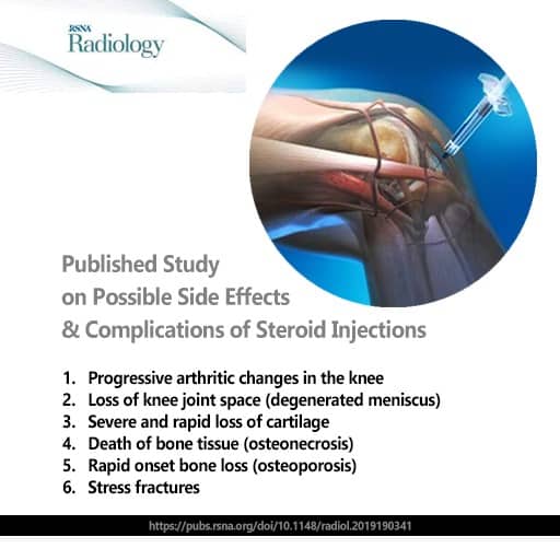
I advise against steroid injections for a torn meniscus or any other knee problem. In my view they are invasive and painful, they repair nothing, and they can harm soft tissue and cartilage. If focused conservative care does not help, my advice is to avoid steroids and go straight to a surgical opinion.
Steroid injections are sometimes offered when a knee is swollen or inflamed. In a two-year trial in people with knee osteoarthritis, steroid injections every three months led to more cartilage loss than salt-water injections. Knee pain was no different (McAlindon and colleagues, 2017).
What the Meniscus Is and Why It Matters for Knee Stability
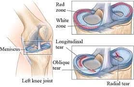
The meniscus is a firm, rubbery cartilage pad inside the knee. Each knee has two. The medial meniscus, on the inner side, is C-shaped, and the lateral meniscus, on the outer side, is rounder, close to a circle. Both sit between the thighbone (femur) and the shinbone (tibia) and spread body weight during standing, walking, stairs and squatting.
Each meniscus is wedge-shaped and thicker at its outer rim. Together they form a shallow cup that helps the rounded end of the femur sit securely on the flat top of the tibia.
Beyond cushioning, the meniscus steadies the joint and protects the cartilage that covers the ends of the bones.
Video: How Knee Load and Movement Affect the Meniscus
Knee load depends on how force passes through the hips, pelvis, ankle, foot and surrounding muscles. In this 18-minute video, I explain how movement, cartilage load and the meniscus fit together. I also show why knee symptoms can return after rest, and which exercises strengthen the knee.
Key moments and an edited transcript of this video are on our Knee Pain Causes and Exercises video page.
Key moments from the video:
- 00:00 Why knee symptoms develop and what may drive repeated strain
- 00:40 How the knee works as a weight-transfer joint
- 01:52 Meniscus and cartilage load during movement
- 03:05 How alignment changes may affect knee-wear patterns
- 05:40 When imaging such as X-ray or MRI may be discussed
- 06:45 Conservative care and movement-based options
- 07:55 Knee warm-up and controlled-movement exercises
- 08:58 Tubing and range-of-motion drills
- 10:12 Wall-squat mechanics and form
- 11:49 Inner- and outer-thigh muscle strengthening
- 14:09 Gym-based knee rehabilitation movements
- 15:40 Quadriceps and hamstring balance
- 16:51 Leg-press and adductor-machine tips
Who Non-Surgical Care Suits, and When Referral Makes Sense
Non-surgical care suits many people with a torn meniscus, but not every knee. A medical opinion comes first for a suspected fracture or major trauma, signs of joint infection or a suspected blood clot. The same applies to an inflammatory joint condition and to a knee that swells quickly after an injury. Care is also adjusted for people with osteoporosis, those taking blood-thinning medication, those with heart or circulation problems, and those recovering from recent knee surgery.
At CSC, knee care brings together clinical physiotherapists and chiropractors in Kuala Lumpur, led by Yama Zafer, D.C.
Fees are listed on the chiropractic price in Kuala Lumpur and physiotherapy fees in Kuala Lumpur pages.
Torn Meniscus: Frequently Asked Questions
Will walking on a torn meniscus make it worse?
Walking is part of recovery, but distance and timing matter. For the first days, the NHS advises putting as little weight as possible on a painful knee, for example by not standing for a long time. After that, walking distance is built up as the knee allows. Walking that brings on swelling, locking or giving way is a sign to cut back and have the knee assessed. Turning the whole body with the feet, rather than pivoting on a planted foot, keeps twisting load off the meniscus.
Is a torn meniscus the same as torn knee cartilage?
In everyday speech, often yes. The knee has two kinds of cartilage. Each meniscus is a rubbery pad of fibrocartilage between the thighbone and the shinbone, and a tear in it is often called torn cartilage. The thin, smooth cartilage that covers the ends of these bones is a different tissue, called articular cartilage. Wear of that layer is the hallmark of knee osteoarthritis, and it can develop alongside a meniscus tear.
Can a meniscus tear happen together with ACL or PCL injuries?
Yes. Meniscus tears often occur with ACL injuries and sometimes with PCL injuries, especially after sports trauma, twisting or a sudden change of direction. A meniscus tear on its own is also common. The ACL (anterior cruciate ligament) and the PCL (posterior cruciate ligament) are the two ligaments inside the knee that stop the shinbone sliding forward and backward. When one of them is torn, the thighbone can slide on the shinbone, which adds strain to the meniscus. That is why the assessment checks the ligaments together with the meniscus.
Can a teenager with a torn meniscus have chiropractic care?
Yes, with care matched to a growing knee. Knee injuries in children and teenagers are assessed with the growth plates in mind. For teenagers, a low-force method such as the Activator may be used, while very young children receive non-force care only. A parent or guardian can stay throughout every visit. The guide to chiropractic care for children explains the first visit and a safe return to sport.
When should a painful knee be assessed?
An assessment makes sense when the knee keeps swelling, catches or locks, or gives way. It also makes sense when the knee has not settled after a few weeks of sensible changes to activity. A knee that is locked and will not straighten should be seen sooner, by a provider experienced with knee injuries. The assessment looks at the tear, the ligaments and the knee's alignment, so that care is planned for that knee, not from the scan alone.
What is the ICD-10 code for a meniscus tear?
A current meniscus tear is coded under the ICD-10-CM family S83.2- (tear of meniscus, current injury). More specific codes show the medial or lateral meniscus, the tear type, the side and the encounter. The code supports records and billing and does not decide the treatment. It is chosen by the clinician who examines the knee and reads the imaging.
How to Read This Guide
This guide is general education and cannot assess a particular knee. The exact tear type, and whether nearby ligaments or cartilage are involved, come from a history, an examination and, when needed, imaging read alongside the symptoms.
Peer-Reviewed References
- Makris EA, Hadidi P, Athanasiou KA. The knee meniscus: structure-function, pathophysiology, current repair techniques, and prospects for regeneration. Biomaterials. 2011;32(30):7411-7431.
- Snoeker BA, Bakker EW, Kegel CA, Lucas C. Risk factors for meniscal tears: a systematic review including meta-analysis. J Orthop Sports Phys Ther. 2013;43(6):352-367.
- Sihvonen R, Paavola M, Malmivaara A, et al. Arthroscopic partial meniscectomy versus sham surgery for a degenerative meniscal tear. N Engl J Med. 2013;369(26):2515-2524.
- Kalske R, Sihvonen R, Paavola M, et al. Arthroscopic partial meniscectomy for degenerative tear: 10-year outcomes. N Engl J Med. 2026;394(17):1757-1759.
- Abram SGF, Judge A, Beard DJ, Carr AJ, Price AJ. Long-term rates of knee arthroplasty in a cohort of 834 393 patients with a history of arthroscopic partial meniscectomy. Bone Joint J. 2019;101-B(9):1071-1080.
- Noorduyn JCA, van de Graaf VA, Willigenburg NW, et al. Effect of physical therapy vs arthroscopic partial meniscectomy in people with degenerative meniscal tears: five-year follow-up of the ESCAPE randomized clinical trial. JAMA Netw Open. 2022;5(7):e2220394.
- Winter AR, Collins JE, Katz JN. The likelihood of total knee arthroplasty following arthroscopic surgery for osteoarthritis: a systematic review. BMC Musculoskelet Disord. 2017;18(1):408.
- Englund M, Guermazi A, Gale D, et al. Incidental meniscal findings on knee MRI in middle-aged and elderly persons. N Engl J Med. 2008;359(11):1108-1115.
- Englund M, Guermazi A, Lohmander SL. The role of the meniscus in knee osteoarthritis: a cause or consequence? Radiol Clin North Am. 2009;47(4):703-712.
- van de Graaf VA, Noorduyn JCA, Willigenburg NW, et al. Effect of early surgery vs physical therapy on knee function among patients with nonobstructive meniscal tears: the ESCAPE randomized clinical trial. JAMA. 2018;320(13):1328-1337.
- Rongen JJ, Rovers MM, van Tienen TG, Buma P, Hannink G. Increased risk for knee replacement surgery after arthroscopic surgery for degenerative meniscal tears: a multi-center longitudinal observational study using data from the osteoarthritis initiative. Osteoarthritis Cartilage. 2017;25(1):23-29.
- Abram SGF, Judge A, Beard DJ, Price AJ. Adverse outcomes after arthroscopic partial meniscectomy: a study of 700 000 procedures in the national Hospital Episode Statistics database for England. Lancet. 2018;392(10160):2194-2202.
- Sihvonen R, Paavola M, Malmivaara A, et al. Arthroscopic partial meniscectomy for a degenerative meniscus tear: a 5 year follow-up of the placebo-surgery controlled FIDELITY (Finnish Degenerative Meniscus Lesion Study) trial. Br J Sports Med. 2020;54(22):1332-1339.
- Englund M, Roos EM, Lohmander LS. Impact of type of meniscal tear on radiographic and symptomatic knee osteoarthritis: a sixteen-year followup of meniscectomy with matched controls. Arthritis Rheum. 2003;48(8):2178-2187.
- McAlindon TE, LaValley MP, Harvey WF, et al. Effect of intra-articular triamcinolone vs saline on knee cartilage volume and pain in patients with knee osteoarthritis: a randomized clinical trial. JAMA. 2017;317(19):1967-1975.
- Nguyen JC, De Smet AA, Graf BK, Rosas HG. MR imaging-based diagnosis and classification of meniscal tears. Radiographics. 2014;34(4):981-999.
- Jarraya M, Roemer FW, Englund M, et al. Meniscus morphology: Does tear type matter? A narrative review with focus on relevance for osteoarthritis research. Semin Arthritis Rheum. 2017;46(5):552-561.
These references describe the meniscus, meniscus tears, their risk factors and diagnosis, meniscus surgery and knee replacement after it in general. None of them evaluates care at Chiropractic Specialty Center.
The Author of Meniscus Tear: Symptoms, Causes & Non-Invasive Care
Meniscus Tear: Symptoms, Causes & Non-Invasive Care was written by Yama Zafer, D.C., who has 30+ years in chiropractic and physiotherapy and founded Chiropractic Specialty Center in Kuala Lumpur in 2006.
Last Updated: Meniscus Tear: Symptoms, Causes and Non-Invasive Care
Meniscus Tear: Symptoms, Causes and Non-Invasive Care was last reviewed and updated on October 23, 2026.