

L1–L2 Spine: Joint, Disc & Nerve Care in Kuala Lumpur
At CSC, our L1–L2 spine care in Kuala Lumpur focuses on the upper-lumbar link between the thoracic and lumbar regions. You’ll see MRI terms like disc bulge/protrusion, annular tear, facet hypertrophy, or ligamentum flavum thickening. This page explains what they mean for the exiting L1 and traversing L2 nerve paths, why direction matters, and how to match positions and exercises to this segment.
Care is non-invasive and gentle & focused—no twisting. Plans pair chiropractic mobilization with physiotherapy, calibrated spinal decompression, and segment-specific exercise to improve motion and day-to-day function.
Takeaway: Safe, Integrated Care for L1–L2 Spine
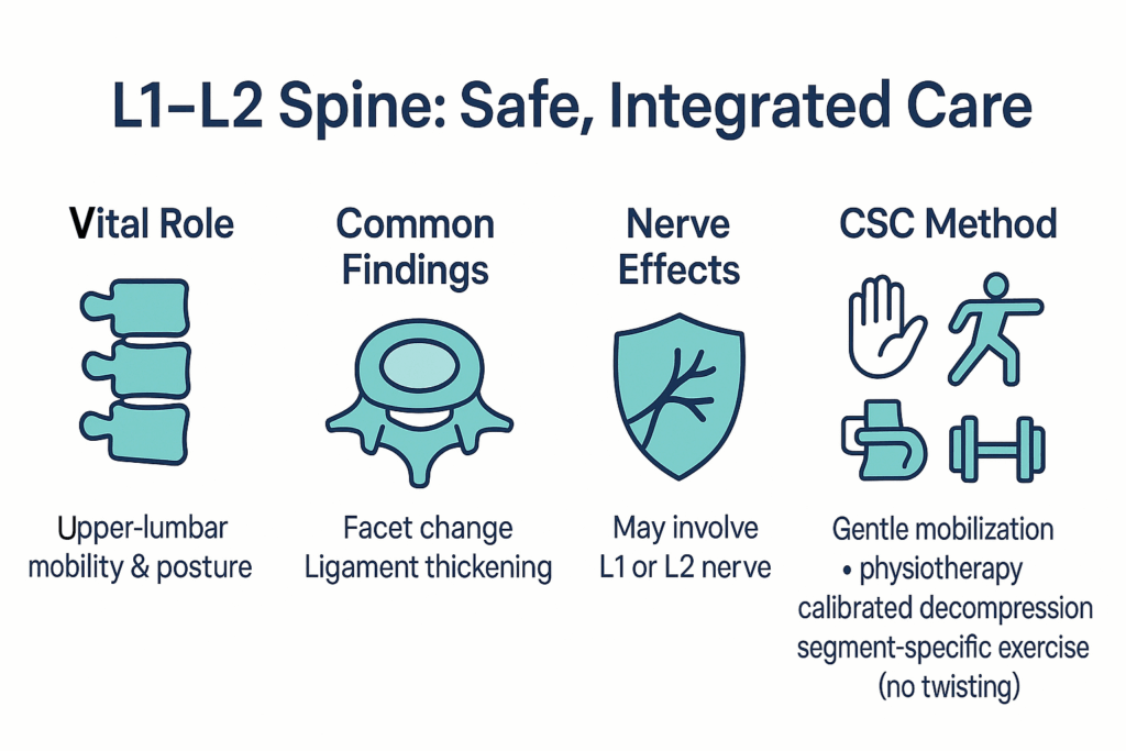
- Vital Role: L1–L2 links upper-lumbar stability with posture control.
- Common Issues: Disc bulge/protrusion, facet change, ligament thickening.
- Nerve Impingment: May leads to a pinched nerve involving th L2 or the L1 nerve.
- CSC Method: Gentle mobilization + physiotherapy + calibrated decompression + segment-specific exercise (no twisting).
Understanding the L1–L2 Segment
L1–L2 stabilizes the trunk and allows controlled bending/rotation. The disc cushions load; facet joints guide motion; ligamentum flavum and deep muscles add stability. Two nerve corridors sit nearby: the foramen where the L1 nerve exits, and the lateral recess under the disc where the L2 nerve traverses. Height loss, facet thickening, or a posterior disc change can narrow these spaces—often felt more in upright standing or arching back.
Nerve Supply:
L1–L2 contributes to lumbar nerve roots involved in sensation and muscle control in the lower abdomen, hips, and upper thighs.
Evidence: Bogduk N. 2005; Adams & Dolan 2012.
Common Conditions
- Ligamentum flavum hypertrophy: inward buckle reduces canal/lateral recess.
(Avoid the 40% stat unless you’ll cite a specific, high-quality source.)
Evidence: Fardon et al. 2014; Sairyo et al. 2005; Fujiwara et al. 1999. - Disc bulge/protrusion at L1–L2: broad vs focal outpouching; direction decides corridor.
- Disc dehydration (degeneration): less height; more load to facets.
- Facet hypertrophy: thicker joint margins can narrow corridors.
Non-Invasive Solutions for L1–L2 Problems
- Segment-specific exercise: trunk endurance, hip strategy, posture resets, graded loading.
Evidence: Qaseem et al. 2017; Chou et al. 2016. - Gentle mobilization (no twisting) to restore segment motion.
- Calibrated spinal decompression for disc mechanics & hydration.
- Physiotherapy modalities (laser, ultrasound, electrotherapy) for tissue loading.
This team-based approach ensures registered physiotherapists and chiropractors work together to reach deeper structures while protecting the disc and nerves.
L1–L2: What Happens & What May Help?
L1–L2 links your mid-back to your lower back. It shares everyday load and helps you stand, sit, and turn. Two nerve spaces sit right beside it: a side opening where the L1 nerve exits, and a space under the disc where the L2 nerve runs. Standing very upright or bending back can shrink these spaces; a slight forward lean often feels easier.
MRI words, in simple terms:
- Degenerative / desiccated disc: the disc has less water and height; it stiffens and shares load less evenly.
- Disc bulge: a broad rim swell; the outer wall is still intact.
- Protrusion / prolapse: a focal bump that pushes out more than a bulge; usually still partly contained.
- Herniation / disc rupture: the wall is torn and inner material pushes outward.
- Extrusion: a larger piece extends out with a narrow “neck” at its base.
- Fragment / sequestration: a piece breaks off and may move a little from the disc.
What usually helps (non-surgical care): gentle, segment-focused hands-on care without twisting, calibrated spinal decompression/traction, targeted physiotherapy, simple exercise for trunk and hips, posture resets, short walk breaks, and hip-hinge lifting. Avoid high-force twisting, deep back bends with load, and heavy rounded lifts.
Imaging? MRI Imaging is useful if things persist or if strength, reflex, or feeling changes—get checked sooner if groin or upper-thigh numbness/weakness is getting worse or night symptoms continue.
How Nearby Levels (L2–S1 & Tailbone) Shape L1–L2
Many reports list changes at more than one level. Use this quick map to see how each nearby segment can influence L1–L2 and day-to-day movement.
- L2–L3 (upper-lumbar link): When this level stiffens or the disc loses height, extra load often shifts down to L1–L2. Posterolateral change here involves the traversing L3 tunnel; far-lateral/foraminal change can involve the exiting L2 doorway—often noticed with upright standing or arching back.
- L3–L4 (mid-lumbar bridge): Shares load and guides bending. Posterolateral direction tends to narrow the lateral recess for the traversing L4path; far-lateral/foraminal direction can involve the exiting L3 path. People often describe front-thigh or knee tingling in long standing, eased by a slight forward lean.
- L4–L5 (high-motion level): Common site for broad bulges and focal changes. Narrowing here frequently relates to the L5 corridor (lateral recess/foramen), which can affect foot control and steady walking—especially with loaded bending or long sitting.
- L5–S1 (lumbosacral junction): Transfers load into the pelvis. Foraminal change can involve the exiting L5 root; posterolateral change can involve the traversing S1 path. Positions near full extension can feel tighter; hip-hinge and keeping loads close usually help.
- Sacrum & Tailbone (coccyx): Form the base for lumbar load-sharing and sitting posture. Ligaments and pelvic-floor attachments here influence comfort in prolonged sitting; neutral posture and micro-breaks often make a clear difference.
L1–L2 Disc Changes: a plain-English guide to “slipped disc” types
L1–L2 is the upper-lumbar link between the mid-back and lower back. The disc here works like a cushion. When it changes, direction and size decide which corridor feels tight: the exiting L1 doorway (foramen) at the side, or the traversing L2 tunnel (lateral recess) under the disc.
- Degenerative / Desiccated disc (DDD): The disc loses water and a little height; the outer wall stiffens and more load shifts to the small joints. This raises the chance of small wall splits and later bulge-type changes at L1–L2.
- Disc bulge (broad-based): A wide swell of the disc rim with the outer wall intact. It can reduce space in the foramen or lateral recess, especially if the disc has already lost some height.
- Protrusion & prolapse (contained → early non-contained): A protrusion is a focal bump still held by the outer wall. Some reports use prolapse for a more pronounced bump or early step toward a non-contained change. Posterolateral direction tends to involve the L2 tunnel; far-lateral/foraminal direction can involve the L1 doorway.
- Herniation & disc rupture: A tear in the wall lets inner material push outward. “Rupture” is often used for a more severe disruption. Effects depend on direction, size, and remaining space.
- Extruded disc (non-contained herniation): Material extends beyond the disc space with a narrow “neck” at its origin. This has a higher chance of contacting nearby nerve paths at L1–L2.
- Fragmented & sequestered disc: A piece breaks off (fragmented); if fully detached, it’s sequestered. Fragments can move slightly; impact depends on where they sit.
Watch Video Series on Common Types of Spinal Disc Damage
Summary
L1–L2 sits where the mid-back meets the lower back. When the disc loses height or the small joints/ligament thicken, space can narrow in two nerve corridors—the exiting L1 doorway (foramen) and the traversing L2 tunnel (lateral recess). Direction matters: changes toward the back-and-side often relate to the L2 path; far-lateral changes can involve L1.
What people commonly notice: lower-back stiffness after standing, groin or upper-thigh tingling, and early leg tiredness on hills or stairs. A slight forward lean often feels easier because it opens space.
Care at CSC is non-invasive and gentle & focused—no twisting. Plans are segment-specific and may include chiropractic care and mobilization, physiotherapy modalities, calibrated spinal decompression, and exercise for trunk endurance, hips, and posture. Ergonomics (hip-hinge, posture resets, short walk cycles) helps protect this level day to day.
Meet Yama Zafer, D.C: the Author of L1–L2 Spinal Joint & Disc Care in Kuala Lumpur
Written by Y. Zafer [D.C.], this article on L1–L2 Spine: Joint, Disc & Nerve Care in Kuala Lumpur reflects nearly three decades of clinical experience combining chiropractic and physiotherapy for targeted spine recovery.
Last Updated: L1–L2 Spine: Joint, Disc & Nerve Care in Kuala Lumpur
This page was last clinically reviewed and updated on August 11, 2025, to ensure accuracy and relevance in L1–L2 care.
Peer-Reviewed Evidence for L1–L2 & L3–L4 (Disc, Joint & Nerve Care)
- Adams MA, Dolan P. (2012). Intervertebral disc degeneration: evidence for two distinct phenotypes. Journal of Anatomy, 221(6), 497–506.
- Bogduk N. (2005). Clinical Anatomy of the Lumbar Spine and Sacrum (4th ed.). Elsevier Churchill Livingstone.
- Brinjikji W, Luetmer PH, Comstock B, et al. (2015). Imaging features of spinal degeneration in asymptomatic populations: systematic review. AJNR American Journal of Neuroradiology, 36(4), 811–816.
- Chou R, Deyo R, Friedly J, et al. (2016). Noninvasive treatments for low back conditions: a systematic review. Annals of Internal Medicine, 165(7), 493–505.
- Fardon DF, Williams AL, Dohring EJ, Murtagh FR, Rothman SLG, Sze GK. (2014). Lumbar disc nomenclature: version 2.0. Spine Journal, 14(11), 2525–2545.
- Fujiwara A, Tamai K, An HS, et al. (1999). The relationship between disc degeneration and facet joint osteoarthritis of the lumbar spine. Spine, 24(14), 1476–1480.
- Genevay S, Atlas SJ. (2010). Lumbar spinal stenosis. Best Practice & Research Clinical Rheumatology, 24(2), 253–265.
- Ishimoto Y, Yoshimura N, Muraki S, et al. (2013). Prevalence of symptomatic lumbar spinal stenosis and association with physical performance. Osteoarthritis and Cartilage, 21(6), 783–788.
- Jenis LG, An HS. (2000). Lumbar foraminal stenosis. Spine, 25(3), 389–394.
- Kalichman L, Hunter DJ. (2008). Diagnosis and conservative management of degenerative lumbar spondylolisthesis. European Spine Journal, 17(3), 327–335.
- Khan AM, Girardi FP. (2005). Spinal lumbar synovial cysts: diagnosis and management. Spine, 30(12), 144–149.
- Komori H, Shinomiya K, Nakai O, et al. (1996). The natural history of lumbar disc herniation with radiculopathy. Spine, 21(2), 225–229.
- Lucantoni C, Than KD, Wang AC, et al. (2011). Tarlov cysts: pathophysiology, clinical features, and outcomes (review). Neurosurgical Focus, 31(6), E14.
- Macki M, Hernandez-Enriquez M, Bydon M, et al. (2014). Spontaneous regression of lumbar disc herniation: meta-analysis. World Neurosurgery, 82(6), 1357–1366.
- McGill SM. (2016). Low Back Disorders (3rd ed.). Human Kinetics.
- Qaseem A, Wilt TJ, McLean RM, Forciea MA. (2017). Noninvasive treatments for acute, subacute, and chronic low back conditions: ACP clinical practice guideline. Annals of Internal Medicine, 166(7), 514–530.
- Sairyo K, Biyani A, Goel V, et al. (2005). Pathomechanism of ligamentum flavum hypertrophy: a multidisciplinary investigation. Spine, 30(23), 2649–2656.
FAQ – L1–L2 Spinal Joint, Disc & Nerve Care
What does L1–L2 do?
It links the mid-back and lower back, shares load, and protects two nerve corridors (exiting L1 doorway; traversing L2 tunnel).
Which areas can L1–L2 relate to?
Groin and upper-thigh feeling, hip-flexor control, and steadiness with stairs or uphill walking.
Why do upright standing and arching back feel worse?
Those positions reduce space in the foramen/lateral recess at L1–L2. A slight forward lean usually opens space.
What do “bulge,” “protrusion,” “prolapse,” and “herniation” mean?
Bulge = broad swell; protrusion = focal bump contained by the wall; prolapse/herniation = material pushed beyond the border to varying degrees. Direction (posterolateral vs far-lateral) signals which corridor is involved.
Do all L1–L2 findings need imaging first?
Not always. Many start with an assessment and a position-matched plan. Imaging helps if symptoms persist or if strength/reflex/sensation changes.
What usually helps (non-invasive)?
Gentle segment-aware mobilization (no twisting), calibrated spinal decompression, physiotherapy modalities, segment-specific exercise (trunk/hip endurance), and ergonomics (hip-hinge, posture resets, short walk bouts).
What should I avoid for now?
If groin/upper-thigh numbness or weakness progresses, your knee/hip feels less steady, or night symptoms continue.
Summary Table – L1–L2 Spine
| Feature | Details |
|---|---|
| Segment | L1–L2 (upper-lumbar transition between mid-back and lower back). |
| Role | Shares upper-body load; adds controlled motion for posture and turning. |
| Nerve corridors | Exiting L1 via the foramen; traversing L2 in the lateral recess. |
| What these nerves influence | Groin and upper-thigh sensation; hip-flexor control; contributes to steady walking and stairs. |
| Typical MRI terms | Disc degeneration, bulge, protrusion/prolapse, herniation/extrusion, fragment/sequestration; facet hypertrophy; ligamentum flavum hypertrophy; foraminal or lateral recess stenosis. |
| Direction → involvement | Posterolateral change → often the L2 corridor; far-lateral/foraminal change → often the L1corridor. |
| Functional clues (non-diagnostic) | Lower-back stiffness after standing, groin/upper-thigh tingling, early leg tiredness on hills or stairs; slight forward lean often feels easier. |
| Helpful positions & cues | Short walk bouts, posture resets, slight forward lean when needed, hip-hinge for lifts, keep loads close. |
| Non-invasive care (CSC) | Gentle, segment-aware mobilization (no twisting), calibrated spinal decompression, physiotherapy modalities, segment-specific exercise (trunk/hip endurance), ergonomics coaching. |
| Avoid (for now) | High-force twisting/long-lever moves, rounded heavy lifts, one-size-fits-all routines that ignore your findings. |
| When to get assessed | Ongoing groin/upper-thigh numbness or tingling, new weakness with hip flexion, or symptoms that persist/worsen despite basic adjustments to posture and activity. |
| Contact | Main center: KL (Bukit Damansara) for service info and routing. Also: Sri Petaling & Bukit Jalil; Bandar Sri Damansara; Shah Alam (Setia Alam & Kota Kemuning); Bangi. |
L1–L2 Spine Care in Kuala Lumpur — Book a Segment-Specific Review
Prefer a simple, step-by-step plan matched to your MRI? Our KL main center (Bukit Damansara) will review your L1–L2 wording, note corridor direction (exiting L1 vs traversing L2), and outline non-invasive options—no twisting.
Call Us: +603 2093 1000 or SMS / WhatsApp: +60 17 269 1873 and we’ll route you to the nearest CSC (Sri Petaling & Bukit Jalil • Bandar Sri Damansara • Shah Alam: Setia Alam & Kota Kemuning • Bangi) or see you at our main center in Kuala Lumpur, and don’t forget to bring your MRI if you have one