

C1-C2 (Atlas & Axis): Symptoms, Nerves and Adjustment
C1-C2 symptoms can start with a neck that turns less easily to one side, first noticed when reversing a car or checking a blind spot. The atlas (C1) and the axis (C2) form the most mobile joint of the neck, and turning is its main work. When that joint is strained or misaligned, pain can gather high in the neck.
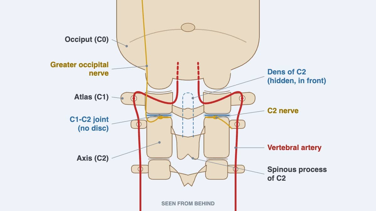
The C2 nerve runs close to the joint. Pressure on it, or on the greater occipital nerve it feeds, can bring brief, sharp, shooting pain over the back of the scalp. Common contributors to C1-C2 strain include a neck injury, long hours looking down at a screen and tight muscles at the back of the neck. A jaw problem on the same side can add to it.
At C1, I use a non-force contact on the side of the atlas, with no twisting, kept within what you can comfortably tolerate. Physiotherapy, light soft tissue work and exercises suited to the neck join it in one plan. Below are the hospital warning signs, how this joint turns the head, my method at C1, and exercises to check with your provider first.
For more information about the C1-C2 level, contact the Bukit Damansara (KL) main center: WhatsApp+60 17 269 1873 Call+603 2093 1000
C1-C2 Symptoms and Care: Key Takeaways
| Question | Short answer |
|---|---|
| How much of the neck's turning happens at C1-C2? | About half (Kim and colleagues, 2024). The joint above it, between the skull and the atlas, does more of the nodding. Measured ranges vary between people (Lopez and colleagues, 2015). |
| How is a jaw problem linked to C1? | In my experience, the jaw problem is often at its worst in parallel with C1 trouble on the same side. Upper cervical care is therefore the most important part of my jaw care, with chiropractic, physiotherapy and exercises for the jaw alongside it. Jaw Joint (TMJ) Problems and C1-C2: A Two-Way Link covers both directions. |
| Is there a disc between C1 and C2? | No. The atlas has no vertebral body, so the two bones meet at three small joints. One sits at the front around the dens, a peg rising from C2, and one on each side (Kaiser and colleagues, 2023). |
| Which nerves pass close to C1? | Four cranial nerves, the vagus among them, pass through a narrow gap between the atlas and the styloid process (Ozveren and colleagues, 2003). The facial nerve runs close by, and the brainstem sits just above. |
| Which nerve brings sharp pain at the back of the head? | Most often the greater occipital nerve, which grows from the C2 nerve. Attacks last seconds to minutes and often feel stabbing, with a tender scalp over the nerve (Djavaherian and Guthmiller, 2023). |
| What do I mean by C1-C2 misalignment? | In my experience, the atlas commonly misaligns in two ways: it rotates, or slides slightly to one side, a lateral listhesis. An examination shows whether either is present; a symptom alone cannot. |
| How do I adjust C1? | With a non-force contact on its lateral mass, the solid side block of the atlas, at an angle suited to you. On a stable neck, I may add the Activator at level one, very gently, on the muscles beside it. |
| Which signs need a hospital first? | Sudden stroke signs need 999 at once, without driving. Signs of a possible spine injury need 999 too, with the person kept still. Both lists appear in full under Warning Signs That Need a Hospital. |
| What makes C1-C2 unstable? | In people with no risk factor, instability here is extremely rare. Known causes include injury, Down syndrome, rheumatoid arthritis and, in adults, degenerative disease. Most people with it have no symptoms (Lacy and colleagues, 2023). |
| Is an X-ray needed before care? | Not always. The rule I give in Is Chiropractic Safe? 13 Questions Answered: imaging comes only once the examination shows a reason. A neck X-ray shows the bones; an MRI also shows the ligaments and the space the nerves occupy. |
C1-C2 Guide: Sections at a Glance
- C1-C2 Nerve Symptoms: Compression, Irritation or Damage
- Having the Upper Neck Assessed in Kuala Lumpur
- Atlantoaxial Joints (C1-C2): How the Atlas Turns on the Axis
- C1-C2 Key Statistics From Research
- Suboccipital Muscles, the Dura and Blood Flow
- Cranial Nerves, Vagus and Brainstem Near C1
- Jaw Joint (TMJ) Problems and C1-C2: A Two-Way Link
- What Causes C1-C2 Misalignment or Strain?
- C1-C2 Adjustment: How I Work at the Atlas Without Twisting
- Exercises With Daily Habits for the Upper Neck
- Related Cervical Segments and Their Roles
- C1-C2 Segment Overview: Key Facts at a Glance
- Frequently Asked Questions
C1-C2 Nerve Symptoms: Compression, Irritation or Damage
Eight pairs of nerves leave the neck. The first exits above the atlas and the second between C1 and C2 (Kaiser and colleagues, 2023). The rear branch of C1, the suboccipital nerve, supplies only the small muscles under the skull and has no skin branch (Graefe and Jozsa, 2024).
For symptoms at the back of the head, the C2 nerve matters most. Its rear branch becomes the greater occipital nerve, which climbs through the neck muscles to the back of the scalp. A front branch forms the lesser occipital nerve, which runs up behind the ear (Djavaherian and Guthmiller, 2023).
When either nerve is pinched or irritated, the pain it brings is called occipital neuralgia. These are its features (Djavaherian and Guthmiller, 2023):
- Sudden attacks of sharp, stabbing or shooting pain over the back of the head, each lasting seconds to minutes.
- Tenderness, altered feeling, or pain from a light touch over the scalp in the same area.
- Nearly always one side at first; about a third have it on both sides.
- A steady, dull ache is less typical and likely points to another cause.
The greater occipital nerve is behind about 90 percent of cases. It can be pressed where the C2 nerve root leaves the spine, inside the muscles it passes through, or where an artery crosses it. Tense or enlarged neck muscles are thought to add pressure, and past injury or bony change can too (Djavaherian and Guthmiller, 2023).
Not every pain at the top of the neck comes from a nerve. The joints and the muscles can be the source too, and the examination sorts one from the other.
Misalignment Symptoms and Segment Dysfunction
By misalignment, I mean the atlas sitting slightly rotated, or shifted a little to one side, which I call a lateral listhesis. Both are common in my practice. C1-C2 segment dysfunction is the wider term for a joint here that is not moving or sitting as it should. Across the top of the neck, the same idea is called upper cervical dysfunction. No single symptom proves either; an examination decides.
In Neck Trigger Points and Exercises, I show two quick checks. Turning the head right and left should be “equal in both directions.” When it is not, the cause “could be muscular or joint involvements.”
Neck Trigger Points and Exercises gives a second clue. When turning brings discomfort on the side you turn toward, I say “it could be a joint involvement.” A joint problem can also grow out of a muscle problem left alone too long.
Warning Signs That Need a Hospital
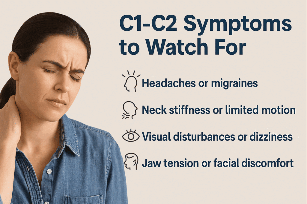
A few signs point past the joint and need a hospital first, whatever the neck feels like. The US Centers for Disease Control and Prevention gives these signs of stroke:
- Numbness or weakness that starts suddenly in an arm, a leg or the face, especially on one side of the body.
- Confusion that comes on suddenly, or new difficulty speaking or understanding speech.
- A sudden change in sight, in one eye or both.
- Sudden dizziness, a loss of balance or coordination, or new trouble walking.
- A sudden, severe headache without a known cause.
If one appears, call 999 for an ambulance right away instead of driving.
Mayo Clinic's first-aid guidance treats some injuries as possible spine injuries. Heavy force on the head or back counts. The same goes for severe neck or back pain, a head injury with changing alertness, and weakness, numbness or paralysis. Poor control of the limbs, bladder or bowels counts too, as does a neck or body twisted out of shape. Keep the person still, without moving the head or neck, and call 999.
Instability between C1 and C2 is a separate matter, and most people with it have no symptoms. When it does cause symptoms, the listed effects include neck pain, restricted movement and signs of pressure on the spinal cord. Others are breathing failure, a tear in the vertebral artery and paralysis of the limbs. The lower cranial nerves, which serve swallowing and speech, can also weaken (Lacy and colleagues, 2023). Apart from neck pain or restricted movement on their own, signs like these call for an urgent hospital assessment.
Weakness in an arm or hand that keeps getting worse should not be ignored. It calls for a careful assessment by a competent provider, and conservative care can continue only while the weakness itself is improving. When I speak of conservative care, I mean focused care aimed at what the scans and examination show. Routine neck treatment is not the same, and much of it is of no use.
In my Back Pain to Leg Pain talk, I describe how cord pressure high in the neck can reach the arms, the legs and breathing. As I put it in that talk: “The higher up, the more important.”
Having the Upper Neck Assessed in Kuala Lumpur
CSC has two centers in Kuala Lumpur, at Bukit Damansara and Bandar Sri Damansara. It has looked after upper neck problems with non-invasive care since 2006, without high-force or invasive techniques. At both centers, chiropractors and physiotherapists plan care together, under the clinical lead of Yama Zafer, D.C.
The assessment covers the head, the neck and the jaw joints, with particular care for the jaw and for C1-C2 down to C2-C3. Its aim is to find subtle neck stiffness or misalignment. The neck pain assessment guide explains what the wider neck examination covers. A first visit lasts from 30 minutes to two hours. If you have neck X-rays or an MRI, bring the images as well as the reports.
The contact page gives directions and opening hours. Each center also has its own page: contact details for the Kuala Lumpur headquarters and contact details for Bandar Sri Damansara.
- Bukit Damansara (KL, main center): Call +603 2093 1000 or WhatsApp +60 17 269 1873 to see the Bukit Damansara / KL center.
- Bandar Sri Damansara (KL): Call +603 6262 5777 or WhatsApp +60 12 455 6939 to see the Bandar Sri Damansara center.

Atlantoaxial Joints (C1-C2): How the Atlas Turns on the Axis
The atlas has no vertebral body; it is a ring. Rising into the front of that ring is the dens, also called the odontoid process, a bony peg on top of the axis. The atlas pivots around it when the head turns (Kaiser and colleagues, 2023).
The joint is built more for turning than for bending forward or back (Kaiser and colleagues, 2023). About half of all the neck's turning happens here (Kim and colleagues, 2024).
The two bones meet at three joints. One sits at the front, between the dens and the front arch of the atlas. The other two, the lateral atlantoaxial joints, sit one on each side. They are fairly shallow, which allows a large range of motion (Kaiser and colleagues, 2023). On each side the C2 nerve leaves between the two bones, and its branch climbs to the scalp.
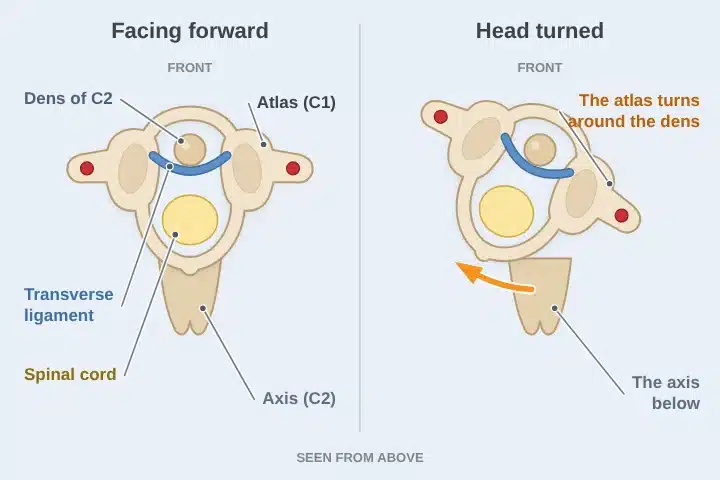
Ligaments hold the arrangement steady. The transverse ligament runs behind the dens, from one side of the atlas to the other. It is considered the strongest ligament attachment in the neck. The alar ligaments tie the tip of the dens to the skull (Lacy and colleagues, 2023).
Measured ranges show how the work is divided between the top two joints:
| Movement | C1-C2 (atlas on axis) | C0-C1 (skull on atlas) |
|---|---|---|
| Turning to one side | About 23.3 to 38.9 degrees | About 2.4 to 7.2 degrees |
| Nodding forward and back | About 10.1 to 22.4 degrees | About 23 to 24.5 degrees |
| Tilting to the side | About 6.7 degrees | About 3.4 to 5.5 degrees |
The figures come from several studies gathered in one review (Lopez and colleagues, 2015), and they vary between people. Even within one healthy neck, the range is not constant; it changes over time and with the direction of movement (Bogduk and Mercer, 2000). The joint above, between the skull and the atlas, is covered in the occiput and atlas guide.
On each side, the vertebral artery leaves the opening in C2 and bends outward into the opening in C1. From there it turns back over the rear arch of the atlas to enter the skull (Kaiser and colleagues, 2023).
My video Neck Cracks Are Dangerous explains how aggressive handling of the neck can disturb this artery. That is one reason I keep forceful twisting away from the top of the neck.
C1-C2 Key Statistics From Research
Each point below comes from the research listed at the end of this page:
- About half of the neck's turning happens at C1-C2 (Kim and colleagues, 2024).
- The joint between the skull and the atlas does more of the nodding than C1-C2 (Lopez and colleagues, 2015).
- C1 and C2 have no disc between them; they meet at three small joints (Kaiser and colleagues, 2023).
- About 70 percent of cervicogenic headaches, headaches that start in the neck, involve the C2-C3 joint, their most frequent source (Al Khalili and colleagues, 2022).
- An international study group set out the diagnostic criteria for cervicogenic headache (Sjaastad and colleagues, 1998).
- In 120 healthy adults aged 20 to 30, turning the head fully reduced blood flow in the vertebral arteries (Mitchell, 2003). The drop was greater on the side opposite the turn.
- The two vertebral arteries carry roughly a fifth of the blood that reaches the brain; the carotid arteries carry the rest (Benjamin and Lui, 2025).
- The transverse ligament limits forward bending and forward sliding of the atlas on the axis; the alar ligaments limit turning (Dvorak and colleagues, 1988).
- About 4 percent of people have an elongated styloid process, and only about 4 percent of those have symptoms from it (Dua and Mohseni, 2026).
Suboccipital Muscles, the Dura and Blood Flow
The deep muscles under the skull number four. Two of them start on C2 and act across C1-C2 (George and Tadi, 2023):
- Obliquus capitis inferior: from the spinous process of C2 out to the transverse process of C1. It turns the head toward its own side, working directly across C1-C2.
- Rectus capitis posterior major: from the spinous process of C2 up to the inferior nuchal line of the skull. It extends the head and turns it toward its own side.
The other two, the rectus capitis posterior minor and the obliquus capitis superior, start on C1 and act at the skull. The occiput and atlas page covers them. The rear branch of the C1 nerve, the suboccipital nerve, supplies all four (George and Tadi, 2023).
Both C2 muscles are tied to the dura, the covering of the spinal cord. The obliquus capitis inferior sent visible strands of tissue to the side of the dura in all 12 specimens studied. Under the microscope, the link was confirmed in 75 percent; the rest could not be assessed. In the one sample stained for nerve tissue, nerve fibers ran through the bridge (Pontell and colleagues, 2013).
The rectus capitis posterior major has a bridge too. Because it is larger, a review suggests it may pull on the dura more strongly than the minor muscle does. In a whiplash-type movement, the deep muscles may pass that pull through the bridge to the dura (Enix and colleagues, 2014).
Researchers call these links myodural bridges. A recent review groups them with the nuchal ligament as a single complex at the top of the spine. Its proposed roles are keeping the dura from folding as the head moves, a part in the flow of spinal fluid and reporting head position. The review also connects changes in it with headache and dizziness that start in the neck (Zhang and colleagues, 2026).
These muscles are dense with muscle spindles, the stretch sensors behind the sense of head position. The obliquus capitis inferior had the most in one fetal study, about 242 per gram (Kulkarni and colleagues, 2001). In a small study, people with chronic neck pain stood less steadily than controls. MRI also showed marked wasting of both rectus muscles, with fat infiltration (McPartland and colleagues, 1997).
In my experience, a disorder of the upper neck can heighten Eagle-like symptoms or sensations as well. People with a C1-C2 problem may also have headaches or migraines of varying degree, mental fog or dizziness. It is not the usual picture, but in my experience it can happen.
The research allows for these. In one population study, 85.7 percent of people with headache disorders had neck pain in the past year (Ashina and colleagues, 2015). Among people without them, the figure was 56.7 percent. Dizziness from the neck is diagnosed only after other causes are excluded (Reiley and colleagues, 2017). A review of 22 whiplash studies found a consistent pattern of poorer memory and attention (Kessels and colleagues, 2000).
The blood supply for the back of the brain runs through this level. The vertebral artery leaves C2, passes through C1 and crosses the suboccipital triangle, where it feeds these muscles (George and Tadi, 2023). In people with marked hardening of the arteries, StatPearls adds, contraction of these muscles may compress the artery a little. The reduced flow can bring light-headedness and dizziness (George and Tadi, 2023). Nerve branches from C1 to C3 travel beside the artery above C3. Yet in monkeys, stimulating the sympathetic nerves that serve the artery had only a minimal effect on its flow (Bogduk and colleagues, 1981).
Cranial Nerves, Vagus and Brainstem Near C1
The upper cervical spine runs from the base of the skull to C3. Its atlantoaxial joint, C1-C2, is the most mobile part of the neck (Kaprive and Das, 2023). Within a small space, the upper neck holds or borders:
- The occipital nerves, which carry feeling from the back of the head and the scalp (Djavaherian and Guthmiller, 2023).
- The vertebral arteries, which feed the brainstem, cerebellum, thalamus and occipital lobes (Benjamin and Lui, 2025).
- The C1 to C3 spinal nerves, which relay pain from the upper neck toward the head and face (Al Khalili and colleagues, 2022).
- Cranial nerves that serve swallowing and speech, among them the vagus, which reaches the heart, lungs and gut (Finsterer and Grisold, 2015).
- The styloid process and its ligament. When too long or hardened, they can press on nearby nerves and vessels, as in Eagle syndrome (Dua and Mohseni, 2026).
Because these structures share a narrow space, joint misalignment and muscle tension here may act together. In my view, the possible result is a mix of symptoms, such as dizziness, headache, neck stiffness, visual strain, swallowing difficulty or facial tingling. This is why a problem from the occiput to C3 can be felt well beyond the neck.
The brainstem, which controls breathing and regulates the heart rate, sits just above the atlas. Serious injuries here are dangerous partly because of that closeness, and because of the blood vessels nearby (Kaprive and Das, 2023). The two vertebral arteries join inside the skull to form the basilar artery, which supplies the brainstem (Kaprive and Das, 2023).
Four cranial nerves run down past the atlas: the glossopharyngeal (IX), the vagus (X), the accessory (XI) and the hypoglossal (XII). With the internal jugular vein, they share a narrow space between the transverse process of the atlas and the styloid process (Ozveren and colleagues, 2003). The styloid process is a thin spike of bone pointing down from the skull, below and in front of the ear (Abuhaimed and colleagues, 2023).
In 40 dissected bodies, the accessory nerve lay in front of the atlas's transverse process in 77.5 percent of dissections (Durazzo and colleagues, 2009). The facial nerve (VII) leaves the skull just behind the styloid process and passes along its outer side (Abuhaimed and colleagues, 2023). That is why I say five cranial nerves pass just a short distance from the lateral mass of C1.
These lower cranial nerves serve swallowing, speech, taste and parts of the body's automatic control. The vagus reaches the heart, the lungs and the gut. When they are affected, the listed signs include disturbed speech or swallowing, altered taste, and pain in the head, throat or neck. Heart or digestive problems are listed too, with weakness of the trapezius, sternocleidomastoid or tongue muscles (Finsterer and Grisold, 2015).
The position of C1 can matter to these nerves. One reported patient with fractures of the atlas had palsy of all four, a pattern called Collet-Sicard syndrome (Hsu and colleagues, 2004). That patient also had a birth difference where the skull meets the spine. In another case, C1 dislocated in rotation on C2, with palsy of the nerve to the tongue (Chien and colleagues, 2005). That person had ankylosing spondylitis.
The atlas can press on the jugular vein too. One study looked at 69 narrowed internal jugular veins (Ding and colleagues, 2020). The transverse process of C1 pressed on 35 of them by itself, and on 31 together with the styloid process. A review of 149 patients with this kind of narrowing found the symptoms non-specific (Scerrati and colleagues, 2021). The most frequent were headache (46.3 percent), tinnitus (43.6 percent) and insomnia (39.6 percent).
One 2025 paper proposes that slowly stretched neck ligaments can strain the vagus nerves, which lie just in front of the neck bones. Its authors link that stretch to a forward, face-down posture from excessive phone and computer use (Hauser and colleagues, 2025). They call it a hypothesis that still has to be proven. The symptoms they looked at included anxiety, dizziness, fatigue, insomnia and neck pain.
Not every effect needs direct pressure on a nerve. The neck's sensory receptors connect with the balance and visual systems. When a neck problem alters their signals, measurable changes can follow in joint position sense, eye movement control and balance. People with such changes report dizziness and unsteadiness (Treleaven, 2008).
Pain can travel the same way. The C1 to C3 nerves send pain signals to the same relay as the trigeminal nerve, which serves the face. A neck problem can therefore be felt in the head or face (Al Khalili and colleagues, 2022). In my view, an atlas shifted to one side may also crowd the upper neck nerves where they leave the spine.
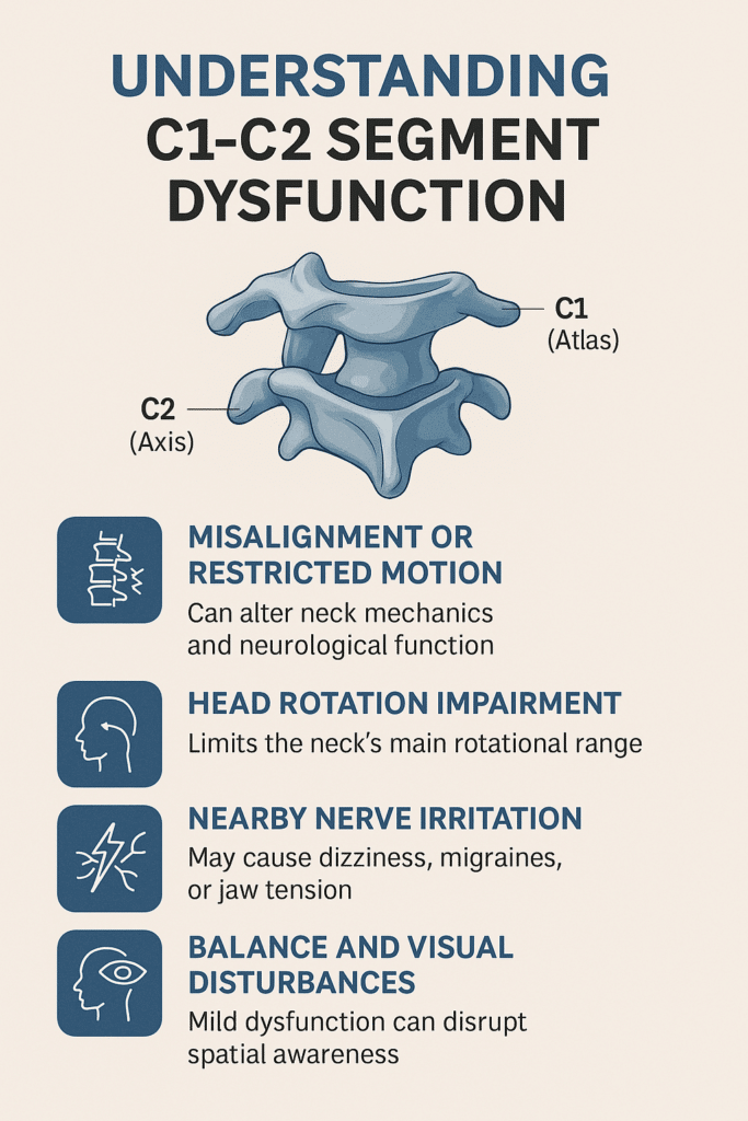
Muscles matter here as well. As the rear branches of the C1 to C3 nerves leave the spine, tight suboccipital muscles can compress them. So can the trapezius and splenius capitis (Kaprive and Das, 2023). In one study, people with episodic tension-type headache had more active trigger points in the upper trapezius, sternocleidomastoid and temporalis muscles. They also held the head further forward and had less neck movement (Fernández-de-las-Peñas and colleagues, 2007).
The cord itself is a concern too. Where the spinal canal in the neck is narrowed, abnormal or excessive movement adds stretch and shear to the cord. These forces seem to play a part in cord injury (Henderson and colleagues, 2005).
Eagle Syndrome: How the Upper Neck Can Mimic It
Eagle syndrome comes from a styloid process that is too long or abnormally angled, or from a hardened ligament on it (Dua and Mohseni, 2026). Eagle described two such patients in 1937 (Eagle, 1937). About 4 percent of people have an elongated styloid, and only about 4 percent of those have symptoms from it. Symptoms are more common in women (Dua and Mohseni, 2026).
The pressure falls most often on the glossopharyngeal nerve (IX). The trigeminal (V), facial (VII), vestibulocochlear (VIII) and vagus (X) nerves can be involved as well (Dua and Mohseni, 2026). Raina and colleagues (2009) add the hypoglossal nerve (XII) to the structures an elongated styloid can press on. In my experience, it is often five of these nerves, and it can be six:
- Glossopharyngeal (IX), the most often affected: a dull, constant throat pain and a feeling of something stuck in the throat. Swallowing can be difficult, with sharp pain shooting to the ear or the back of the tongue.
- Trigeminal (V): facial pain, jaw discomfort or nerve-type pain.
- Facial (VII): it runs along the outer side of the styloid, so stretching or pressure may add facial discomfort.
- Vestibulocochlear (VIII): less often involved, but nearby irritation or vascular change can occasionally add tinnitus or dizziness.
- Vagus (X): it lies on the inner side of the styloid. Pressure there can bring throat or voice box symptoms, such as a change in the voice.
- Hypoglossal (XII): it runs beside the styloid, and pressure here can, rarely, weaken the tongue.
Symptoms may worsen with turning the head or swallowing. When the styloid presses on the carotid artery instead, brief stroke-like episodes, vertigo and fainting can follow. Brief stroke-like episodes, fainting or a new loss of function in part of the body need an urgent medical assessment (Dua and Mohseni, 2026). Sudden stroke signs need 999, as listed under Warning Signs That Need a Hospital.
In my view, the upper neck from the occiput to C3 can mimic the symptoms often linked with Eagle syndrome. A moderately or severely malpositioned C1 above all can mimic them, because the same nerves pass there. StatPearls makes a related point: pain from the cervical spine may mimic Eagle syndrome. It does so particularly when neck movement is limited and imaging shows bony hardening (Dua and Mohseni, 2026).
Some people have a styloid that is slightly long but still within normal length. On its own, it often causes no symptoms. In my experience, when that styloid meets an upper neck malalignment, Eagle-like symptoms of varying degree become possible, even probable. Sorting them out takes a clinician who is alert to these findings. That clinician must judge whether the upper neck is involved, complicated by the styloid and perhaps worsened by a jaw joint problem.
CT with 3D reconstruction is the preferred way to measure the styloid's length and angle (Dua and Mohseni, 2026). The Eagle syndrome guide covers the condition itself.
One Kinematic Chain: Styloid, Jaw and Mid to Lower Neck
In my experience, none of these problems stands alone. The upper neck, the styloid and the jaw joint work like a kinematic chain: when one goes, the other goes.
The upper neck also depends on the stability of the middle and lower neck. As I see it, instability, nerve compression or cord compression lower in the neck can lead to an upper neck problem as well. The levels below C2 are outlined under Related Cervical Segments and Their Roles.
Jaw Joint (TMJ) Problems and C1-C2: A Two-Way Link
Problems in the temporomandibular joints (TMJ), the jaw joints just in front of the ears, can affect how the upper neck moves, C1-C2 in particular. The jaw and the neck share nerves and muscles. A restricted jaw or an uneven bite may add tension to the suboccipital muscles and the upper neck ligaments.
In one study, people with jaw joint pain had less upper neck rotation in a flexion-rotation test than people without symptoms (Grondin and colleagues, 2015).
I see the link run both ways. A chronic upper neck problem can lead to jaw problems, and the reverse is also true. When the disc in the jaw joint has slipped, C1 often needs close attention as well. On the side of the jaw problem, C1 may be involved too. Such a case then needs either chiropractors and physiotherapists working together, or a chiropractic adjustment at the upper neck that is non-force and non-rotatory.
This is why my care for the jaw does not stop at the jaw. In my experience, a jaw problem is often most intense in parallel with C1 trouble on the same side. Chiropractic, physiotherapy and exercises for the jaw are one part of the plan, and the most important part is upper cervical care at C1.
Where C1-C2 or C2-C3 shows restricted movement or misalignment, assessing the jaw joints can reveal contributing factors. Jaw posture, the range of jaw opening and the symmetry of the bite belong in a full upper neck assessment. The jaw pages on this site go further:
- TMJ Disorders: Jaw Clicking, Stiffness and Assessment
- Slipped Disc in the Jaw: TMJ Displacement, Causes & Care Options
- Jaw Chiropractor in KL: TMJ Care Guide
- TMJ Treatment in Malaysia: How Options Are Decided
- Jaw and TMD in Malaysia: Non-Invasive Care Options
- Temporomandibular Joint Dysfunction Care By Jaw Experts
- Causes of TMJ Pain: Jaw Concerns You Should Know
- Jaw Pain and TMD: What the Symptoms Can Mean
What Causes C1-C2 Misalignment or Strain?
These are among the common contributors to C1-C2 strain or dysfunction:
- A neck injury, such as a road crash, a fall or a knock to the head. Injuries that tear the ligaments here often come with a head injury (Lacy and colleagues, 2023).
- Long hours with the head bent over a phone or laptop. In my Back Pain to Leg Pain talk, I call it tech neck.
- Sleeping with the head turned hard to one side for hours.
- Muscle tension that is left long enough to affect the joint. I describe that progression in Neck Trigger Points and Exercises.
- A jaw problem on the same side. When the disc in the jaw joint has slipped, I find C1 often needs attention as well.
- Forceful neck cracking or pulling, the subject of my video Neck Cracks Are Dangerous.
True instability, as distinct from strain, has these known causes (Lacy and colleagues, 2023):
- Injury, including a broken C1 or C2 or a torn ligament.
- Conditions present from birth, such as Down syndrome. Instability shows on X-rays in up to 30 percent of people with Down syndrome, but only about 1 percent have symptoms.
- Inflammatory disease, most often rheumatoid arthritis, which can loosen the transverse ligament and wear the bone.
- Degenerative disease, in adults.
In my experience, age is a factor in neck wear but never the only one. Habits, posture, past injury and genetics all play a part.
C1-C2 Adjustment: How I Work at the Atlas Without Twisting
Depending on my findings, I may adjust C1 and C2 together, or only one of them. In most instances, the adjustment is at C1.
My work at C1 goes through the lateral mass, the solid block on each side of the atlas. The contact is non-force: the lightest pressure, at an angle chosen for that person, along the atlas, with no turn of the neck. This is the non-rotatory adjustment I refer to throughout.
When the neck is stable, chiropractic manipulation that is non-rotatory using the Activator instrument is an option alongside the contact. I keep it on level one, its lowest force, cushioned, with you seated. I generally apply it very gently to the neck's soft tissues beside the lateral mass, not to the lateral mass itself.
Now and then I let the level one impulse pass through my contact finger first, which softens it again. Once a neck proves unstable, the non-force contact alone is my choice.
Clinics that call their care upper cervical work in very different ways at C1 and C2. My own view is that this joint may not suit anything stronger than a non-force touch on the atlas. For the muscles beside it, more than a very low force may not be suitable either. Before agreeing to care at this level, ask any clinic what will be used and how much force it carries.
During the same visit, I may also adjust the jaw, the rib joints at the spine or breastbone, or lower parts of the neck. The examination decides which. Light soft tissue work on the trapezius and rhomboids may accompany it. In Neck Trigger Points and Exercises, I hold each point a few seconds and never press deep.
Gentle mobilization, light fascia work and muscle activation for the suboccipital group and the trapezius may sit in the same plan. Physiotherapy covers posture, jaw position and neck movement, without high-force adjustments.
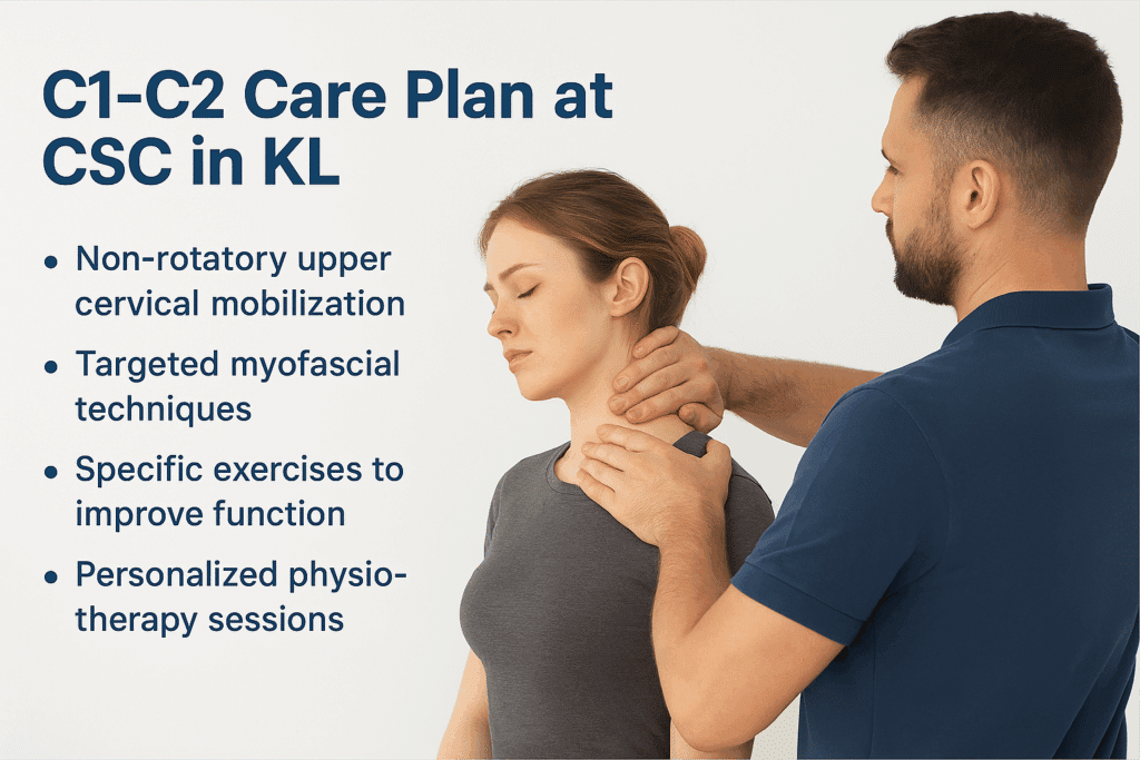
Each step is kept within what you can comfortably tolerate. Any discomfort means the care is too aggressive, a point I make in Is Chiropractic Safe? 13 Questions Answered. Tell the clinician at once, however slight it feels. As my motto puts it: “No pain, more gain.”
Is Chiropractic Safe? 13 Questions Answered also carries my advice on combining care: “Don't choose between one or the other. Choose both.” Physiotherapy sits beside the adjustment in the same plan. I also set two checkpoints there. With no improvement by the fifth session, I look again at the reasons. With none by the tenth, we decide together whether to go on.
Very young children receive non-force care only. For teenagers, I may use chiropractic manipulation that is non-rotatory using the Activator instrument. A parent or guardian can stay with the child throughout every visit. The guide to neck manipulation in children sets out what studies have found in children.
Exercises With Daily Habits for the Upper Neck
Before trying any exercise, show it to your doctor or healthcare provider; I make that request in Neck Trigger Points and Exercises. With that check done, these are the ones I teach for the top of the neck and the upper back:
- Isometrics: rest a hand against your head and press gently into it, to the front, back and each side, keeping the neck still. Each hold lasts about five seconds, or three when five feels too long.
- My instruction in Neck Trigger Points and Exercises is plain: “Do not get someone else to push on you.”
- Stop if any of it brings numbness, tingling or discomfort. That means the neck is not ready for it yet.
- The W, T and L arm positions, with the shoulders drawn down and never shrugged, to work the upper back.
Self-Support Tips: Gentle Home Guidance
These gentle self-care steps suit the C1-C2 segment, once your provider has seen them:
- Slow turns: turn the head gently from side to side, five to eight times each way, staying within a comfortable range.
- Support under the skull: lie on your back with a small rolled towel under the skull's base, and rest for up to five minutes.
- Breathing with light massage: breathe slowly into the belly, and as you breathe out, lightly massage the top of the neck and upper trapezius.
- Posture: keep the shoulders relaxed and the head balanced over the spine.
- Reading and writing: use an angled surface, so the head does not bend down for long.
Do not continue any of these if you feel pain, dizziness or any other symptom, and stop if symptoms worsen. Check with your chiropractor or another healthcare professional before starting any home program.
In my view, yoga does not fit care at this level. Its neck turns and inversions load a joint built for turning, and with a spinal disc problem in the neck, yoga must be avoided.
The daily side matters as much as the exercises. I ask patients to do more of what supports the neck and less of what strains it:
- Set the screen so your eyes sit slightly below its top edge; my video Desk Posture Mistakes shows how.
- With a painful neck or back, get up every 15 to 20 minutes and move for half a minute to a minute; Desk Posture Mistakes sets every 15 minutes as the least. With no neck or back problem, take a break every 30 to 40 minutes.
- Sleep on your side with the neck level, or on your back with its curve supported, rather than with the head twisted for hours. That advice comes from my Back Pain to Leg Pain talk.
- At the desk, place the phone opposite your writing hand or use a headset, a habit I cover in Desk Posture Mistakes.
My Back Pain to Leg Pain talk puts it most simply: “The most important thing is to make lifestyle changes.”
Fees for each service are on the chiropractic price list and the physiotherapy fee list. The fee for a plan is explained and agreed before care begins.
Related Cervical Segments and Their Roles
Each neck level shares movement, load and nerve pathways with its neighbors:
- Occiput and atlas (C0-C1): nodding, and the balance of the head on the neck.
- C2-C3: the bridge between the upper and middle neck, and the first disc. The third occipital nerve, from C3, supplies its joint, the most frequent source of headache from the neck (Al Khalili and colleagues, 2022).
- C3-C4: side bending and head posture. The C4 nerve leaves here; with C3 and C5, it forms the phrenic nerve to the diaphragm.
- C4-C5: neck and shoulder movement together. The C5 nerve leaves here, a key nerve for the shoulder.
- C5-C6: one of the most mobile levels, busy in bending, reaching and turning. The C6 nerve leaves here, toward the thumb side of the forearm.
- C6-C7: close to where the neck meets the upper back. The C7 nerve leaves here, toward the middle finger.
- C7-T1: the base of the neck, steadying shoulder and arm movement. The C8 nerve leaves here, below C7 (Kaprive and Das, 2023).
Because each level carries part of the load, a problem lower down can reach the top of the neck, as I note above.
C1-C2 Segment Overview: Key Facts at a Glance
C1-C2 does about half of the neck's turning and sits just below the brainstem, near the vagus and other cranial nerves. Strain, muscle imbalance or misalignment here can change how the head moves and how nearby nerves behave.
| Category | Details |
|---|---|
| Anatomy | The atlas (C1) on the axis (C2), joined by three small joints and strong ligaments, with no disc |
| Main function | About half of the neck's turning, and a steady base for the skull |
| Common problems in my practice | The atlas rotated or shifted slightly to one side, restricted turning and muscle tension |
| Nearby structures | The brainstem above; the C2 and greater occipital nerves; four cranial nerves, the vagus among them, in front of the atlas |
| Symptoms that may be linked, in my experience | Pain at the back of the head, restricted turning, headaches including migraine, dizziness, jaw tension, changes in vision or hearing, and palpitations |
| Care at CSC | A non-force, non-rotatory contact on the atlas, the Activator at level one on the muscles beside it, physiotherapy and light soft tissue work |
| Self-guided care | Slow turns within comfort, towel support under the skull, slow breathing with light massage, and posture habits, once your provider has seen them |
| Safety | Stop anything that brings pain, dizziness or other symptoms, and ask your practitioner for guided care |
Frequently Asked Questions
Can I Adjust C1 and C2 Myself?
No. An adjustment is chiropractic care, and in Is Chiropractic Safe? 13 Questions Answered, I say that chiropractic care must only be done by a chiropractor. In Neck Cracks Are Dangerous, I add: “Aggressive neck care is never good and should always be avoided.” If someone offers to yank or pull your neck, I say in that video that you should “ask them to stop.” The vertebral artery runs through small openings beside these bones. At home, keep to gentle isometric holds against your own hand, once your provider has seen them.
Can C1 and C2 Cause Dizziness?
Dizziness has many possible causes, so C1 and C2 are never assumed to be the cause. They can play a part, though. Neck problems can alter the signals from the neck's sensory receptors, and people with such changes report dizziness and unsteadiness (Treleaven, 2008). Sudden dizziness is one of the stroke signs the CDC lists. Like every sign on that list, it needs 999 at once rather than a drive to the hospital. The full list is under Warning Signs That Need a Hospital above. When dizziness comes and goes with neck movement, the neck deserves an examination, along with the other possible causes.
Does a C1 Adjustment Have Side Effects?
The possible effects and risks of a C1 adjustment are explained before care begins. In Is Chiropractic Safe? 13 Questions Answered, I describe chiropractic as safe when a competent, registered chiropractor delivers it for the condition found. In that video I also say that “safety of chiropractic depends on the chiropractor and their experience and the technique that they use.” At C1, the contact I use is non-force and the neck is not twisted. If anything during care feels uncomfortable, say so at once, so that the approach can be changed.
What Do C1 and C2 Control?
Movement first: C1-C2 does more of the head's turning than the joint above, which does more of the nodding. The rear branch of the C1 nerve supplies the small muscles under the skull. The C2 nerve carries feeling from the back of the scalp through the greater occipital nerve. Its lesser occipital branch covers the area behind the ear. The spinal cord passes through both bones and carries signals for the whole body below. Severe pressure on it here can affect the limbs and even breathing (Lacy and colleagues, 2023).
Can a C1-C2 Misalignment Affect the Nervous System?
It can, though it may cause no clear symptoms at first. The C2 nerve, the brainstem and four cranial nerves, the vagus among them, sit close to this joint. Reported cases show a broken or dislocated C1 paralyzing some of those cranial nerves (Hsu and colleagues, 2004; Chien and colleagues, 2005). One 2025 paper proposes, as an unproven hypothesis, that neck instability can strain the vagus nerve. It studied dizziness, fatigue and insomnia, among other symptoms (Hauser and colleagues, 2025). Heart or digestive problems are listed for lower cranial nerve disorders (Finsterer and Grisold, 2015). In my view, a malpositioned C1 can bring symptoms like those of Eagle syndrome, because the same nerves pass there. An examination decides.
Are Self-Guided Techniques Safe for C1-C2?
Gentle ones can be, once your provider has seen them. Slow breathing, a rolled towel under the base of the skull, light massage and slow turns within comfort are the kind I mean. Stop at once if any of them brings pain, dizziness, numbness or tingling, or if symptoms worsen. Self-cracking, pulling or twisting the neck is a different matter. In Neck Cracks Are Dangerous, I explain why aggressive neck care should always be avoided. The isometric holds I teach in Neck Trigger Points and Exercises are another gentle start, with your own hand as the only resistance.
Why Avoid Twisting the Upper Neck?
The vertebral artery winds through C2 and C1 and over the rear arch of the atlas into the skull (Kaiser and colleagues, 2023). In 120 healthy young adults, turning the head fully reduced blood flow in these arteries, more on the side opposite the turn (Mitchell, 2003). Forceful twisting drives the neck to that end position with speed. That is why my contact at C1 is non-force and non-rotatory. My video Neck Cracks Are Dangerous covers the risk to this artery in more detail. The ligaments that hold C1 on C2 are spared a sudden pull as well.
Which Neck Exercises Should Be Avoided?
Avoid anything that yanks or forces the upper neck, such as towel pulls or a Y-Strap. In Neck Cracks Are Dangerous, I explain that the neck's small joints make these dangerous. In my Back Pain to Leg Pain talk, I show what tilting the head to one side to stretch it does. It stretches the muscles on one side but squeezes the joints on the other. With a disc problem lower in the neck, leave general neck stretching out. In your fifties, skip that side stretch even without disc damage, and keep any stretch gentle. In the same talk, I advise against yoga in your forties and fifties, even without pain.
How Does C1-C2 Differ From C2-C3?
C1-C2 has no disc, so its problems involve the joints, ligaments, muscles and the C2 nerve. The first disc of the neck sits between C2 and C3, so disc problems can only begin there or lower. The third occipital nerve comes from C3 and curves around the C2-C3 joint. It supplies a small patch of skin just below the base of the skull (Djavaherian and Guthmiller, 2023). The C2-C3 guide covers that level.
How Does CSC Approach C1-C2 Problems?
CSC has provided non-invasive, non-surgical care in Kuala Lumpur since 2006, with chiropractors and physiotherapists working in the same plan. The assessment covers the head, the neck and the jaw joints, from C1-C2 down to C2-C3. At C1, I use a non-force, non-rotatory contact on the lateral mass. On a stable neck, I may add chiropractic manipulation that is non-rotatory using the Activator instrument, at level one, on the muscles beside it. Gentle mobilization, muscle activation and myofascial work around the upper neck may join it, with exercises and daily habits. The possible effects and risks are explained before care begins.
How to Read This Guide
Statements written as “I” give my own position, drawn from my videos, talks or written answers. The video or talk is named wherever it is the source. Each study appears by author and year, and the reference list links to it. Where the research is a single case or a hypothesis, the text says so. This page cannot examine your neck or say what causes your symptoms; an assessment in person does that.
References
- Abuhaimed AK, Alvarez R, Menezes RG. Anatomy, head and neck, styloid process. StatPearls. Treasure Island (FL): StatPearls Publishing; 2023.
- Al Khalili Y, Ly NK, Murphy PB. Cervicogenic headache. StatPearls. Treasure Island (FL): StatPearls Publishing; 2022.
- Ashina S, Bendtsen L, Lyngberg AC, et al. Prevalence of neck pain in migraine and tension-type headache: a population study. Cephalalgia. 2015;35(3):211-219.
- Benjamin R, Lui F. Vertebrobasilar insufficiency. StatPearls. Treasure Island (FL): StatPearls Publishing; 2025.
- Bogduk N, Lambert GA, Duckworth JW. The anatomy and physiology of the vertebral nerve in relation to cervical migraine. Cephalalgia. 1981;1(1):11-24.
- Bogduk N, Mercer S. Biomechanics of the cervical spine. I: Normal kinematics. Clin Biomech. 2000;15(9):633-648.
- Centers for Disease Control and Prevention. Signs and symptoms of stroke. CDC; 2026.
- Chien JT, Chen IH, Lin KH. Atlantoaxial rotatory dislocation with hypoglossal nerve palsy in a patient with ankylosing spondylitis: a case report. J Bone Joint Surg Am. 2005;87(7):1587-1590.
- Ding JY, Zhou D, Pan LQ, et al. Cervical spondylotic internal jugular venous compression syndrome. CNS Neurosci Ther. 2020;26(1):47-54.
- Djavaherian DM, Guthmiller KB. Occipital neuralgia. StatPearls. Treasure Island (FL): StatPearls Publishing; 2023.
- Dua A, Mohseni M. Eagle syndrome. StatPearls. Treasure Island (FL): StatPearls Publishing; 2026.
- Durazzo MD, Furlan JC, Teixeira GV, et al. Anatomic landmarks for localization of the spinal accessory nerve. Clin Anat. 2009;22(4):471-475.
- Dvorak J, Schneider E, Saldinger P, Rahn B. Biomechanics of the craniocervical region: the alar and transverse ligaments. J Orthop Res. 1988;6(3):452-461.
- Eagle WW. Elongated styloid process: report of two cases. Arch Otolaryngol. 1937;25(5):584-587.
- Enix DE, Scali F, Pontell ME. The cervical myodural bridge, a review of literature and clinical implications. J Can Chiropr Assoc. 2014;58(2):184-192.
- Fernández-de-las-Peñas C, Cuadrado ML, Pareja JA. Myofascial trigger points, neck mobility, and forward head posture in episodic tension-type headache. Headache. 2007;47(5):662-672.
- Finsterer J, Grisold W. Disorders of the lower cranial nerves. J Neurosci Rural Pract. 2015;6(3):377-391.
- George T, Tadi P. Anatomy, head and neck, suboccipital muscles. StatPearls. Treasure Island (FL): StatPearls Publishing; 2023.
- Graefe SB, Jozsa F. Neuroanatomy, suboccipital nerve. StatPearls. Treasure Island (FL): StatPearls Publishing; 2024.
- Grondin F, Hall T, Laurentjoye M, Ella B. Upper cervical range of motion is impaired in patients with temporomandibular disorders. Cranio. 2015;33(2):91-99.
- Hauser RA, Matias D, Rawlings BR. Cervicovagopathy: ligamentous cervical instability and dysstructure as a potential etiology for vagus nerve dysfunction in the cause of human symptoms and diseases. Front Neurol. 2025;16:1572863.
- Henderson FC, Geddes JF, Vaccaro AR, Woodard E, Berry KJ, Benzel EC. Stretch-associated injury in cervical spondylotic myelopathy: new concept and review. Neurosurgery. 2005;56(5):1101-1113.
- Hsu HP, Chen ST, Chen CJ, Ro LS. A case of Collet-Sicard syndrome associated with traumatic atlas fractures and congenital basilar invagination. J Neurol Neurosurg Psychiatry. 2004;75(5):782-784.
- Kaiser JT, Reddy V, Launico MV, Lugo-Pico JG. Anatomy, head and neck: cervical vertebrae. StatPearls. Treasure Island (FL): StatPearls Publishing; 2023.
- Kaprive JF, Das JM. Anatomy, head and neck: atlantoaxial joint. StatPearls. Treasure Island (FL): StatPearls Publishing; 2023.
- Kessels RP, Aleman A, Verhagen WI, van Luijtelaar EL. Cognitive functioning after whiplash injury: a meta-analysis. J Int Neuropsychol Soc. 2000;6(3):271-278.
- Kim D, Viswanathan VK, Munakomi S, Menger RP. C1 fractures. StatPearls. Treasure Island (FL): StatPearls Publishing; 2024.
- Kulkarni V, Chandy MJ, Babu KS. Quantitative study of muscle spindles in suboccipital muscles of human foetuses. Neurol India. 2001;49(4):355-359.
- Lacy J, Bajaj J, Gillis CC. Atlantoaxial instability. StatPearls. Treasure Island (FL): StatPearls Publishing; 2023.
- Lopez AJ, Scheer JK, Leibl KE, Smith ZA, Dlouhy BJ, Dahdaleh NS. Anatomy and biomechanics of the craniovertebral junction. Neurosurg Focus. 2015;38(4):E2.
- Mayo Clinic Staff. Spinal injury: first aid. Mayo Clinic; 2023.
- McPartland JM, Brodeur RR, Hallgren RC. Chronic neck pain, standing balance, and suboccipital muscle atrophy: a pilot study. J Manipulative Physiol Ther. 1997;20(1):24-29.
- Mitchell JA. Changes in vertebral artery blood flow following normal rotation of the cervical spine. J Manipulative Physiol Ther. 2003;26(6):347-351.
- Ozveren MF, Türe U, Ozek MM, Pamir MN. Anatomic landmarks of the glossopharyngeal nerve: a microsurgical anatomic study. Neurosurgery. 2003;52(6):1400-1410.
- Pontell ME, Scali F, Enix DE, Battaglia PJ, Marshall E. Histological examination of the human obliquus capitis inferior myodural bridge. Ann Anat. 2013;195(6):522-526.
- Raina D, Gothi R, Rajan S. Eagle syndrome. Indian J Radiol Imaging. 2009;19(2):107-108.
- Reiley AS, Vickory FM, Funderburg SE, Cesario RA, Clendaniel RA. How to diagnose cervicogenic dizziness. Arch Physiother. 2017;7:12.
- Scerrati A, Norri N, Mongardi L, et al. Styloidogenic-cervical spondylotic internal jugular venous compression, a vascular disease related to several clinical neurological manifestations: diagnosis and treatment, a comprehensive literature review. Ann Transl Med. 2021;9(8):718.
- Sjaastad O, Fredriksen TA, Pfaffenrath V. Cervicogenic headache: diagnostic criteria. Headache. 1998;38(6):442-445.
- Treleaven J. Sensorimotor disturbances in neck disorders affecting postural stability, head and eye movement control. Man Ther. 2008;13(1):2-11.
- Zhang L, Song X, Chen C, et al. The myodural bridge complex: a comprehensive review of morphology, physiology, developmental biology and pathology. Front Med (Lausanne). 2026;13:1790220.
None of these sources evaluated care at Chiropractic Specialty Center. They describe the upper neck, its nerves and blood vessels, Eagle syndrome, headache from the neck, stroke and first aid in general.
The Author of C1-C2 Nerve & Misalignment Symptoms: Causes and Care
C1-C2 Nerve & Misalignment Symptoms: Causes and Care was written by Yama Zafer, D.C., who has 30+ years in chiropractic and physiotherapy and founded Chiropractic Specialty Center in Kuala Lumpur in 2006.
Last Updated: C1-C2 (Atlas & Axis): Symptoms, Nerves and Adjustment
C1-C2 (Atlas & Axis): Symptoms, Nerves and Adjustment was last reviewed and updated on October 21, 2026.
Baca halaman ini dalam Bahasa Malaysia: Disfungsi C1-C2 (Atlas dan Aksis): Gejala, Saraf dan Pelarasan