

Ligamentum Flavum Hypertrophy: MRI Findings and Narrowing
Ligamentum flavum hypertrophy means thickening of the elastic ligament that lines part of the back wall of the spinal canal. It is usually associated with age-related change and can occur alongside facet-joint enlargement, disc-height loss and other features of spinal stenosis. The thickness measured on MRI does not by itself tell how much trouble the finding is causing, because similar changes can appear in people with very different symptoms.
Position can matter. The ligamentum flavum folds inward more during spinal extension and becomes more taut in flexion, which is one reason symptoms from lumbar canal narrowing may be worse with prolonged standing or walking and easier when sitting or leaning forward. That pattern is a clue, not a diagnosis, and circulation, hip problems and other neurological causes may produce similar complaints.
This Clear Guide explains ligamentum flavum hypertrophy, MRI findings, and narrowing so readers can understand what the ligament is, how thickness varies by spinal region, why measurements and symptoms may differ, and what balanced next steps look like.
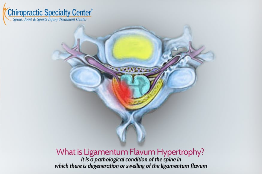
Ligamentum flavum hypertrophy means thickening of a paired elastic ligament that forms part of the back wall of the spinal canal. On MRI it is commonly reported as thickening or hypertrophy. The finding is frequent with age-related change. Thickness alone does not decide symptoms. A full clinical picture that includes history, examination, and the complete imaging context remains essential. This page gives general educational information only. It is not a diagnosis or a personal care plan.
For more information about ligamentum flavum hypertrophy, contact the Bukit Damansara (KL) main center: WhatsApp+60 17 269 1873 Call+603 2093 1000
Why the Ligamentum Flavum Is Frequently Misunderstood
The ligamentum flavum lies deep inside the spinal canal, behind the spinal cord and nerve roots. It cannot be seen or felt from outside the body.
Because it is hidden, many clinicians, including some chiropractors and physiotherapists, focus first on the disc or the facet joints. Some assume that once thickening appears on an MRI report, little can be done with conservative care. That view is incomplete.
The disc, the facet joints, and the ligamentum flavum belong to the same spinal segment. They function as one connected system. Change in one structure affects the others. Understanding the unique properties of this ligament helps explain why some people have symptoms that do not match a single number on an MRI report.
What Makes the Ligamentum Flavum Unique
The ligamentum flavum is not ordinary soft tissue. It is a highly specialized structure.
It contains a very high proportion of elastin: the protein that allows tissues to stretch and then return to their original shape. No other ligament in the body has quite the same combination of elasticity and location.
It has no true muscle belly and no conventional tendon in the usual sense. Its structure and function are distinct. This is one reason it behaves differently from ordinary ligaments or muscles when it thickens or loses elasticity.
When healthy, the high elastin content lets it lengthen during forward bending and then spring back, helping control motion and maintain space inside the spinal canal. When it becomes thickened and less elastic, that protective ability is reduced.
Normal Thickness by Spinal Region
Thickness is not the same at every level of the spine. Approximate average ranges reported in the literature are:
- Cervical spine (neck): about 1.5 mm to 1.7 mm
- Thoracic spine (mid-back): about 2.0 mm to 3.0 mm
- Lumbar spine (lower back): about 3.0 mm to 4.0 mm
These are average figures, not fixed rules. Actual values vary by person, age, exact spinal level, and the method used to measure.
In the lumbar spine, many radiology reports use a threshold near 4 mm when describing hypertrophy. In the cervical spine, values above roughly 1.7 mm are often noted as thickened. No single number applies to every patient or every study.
Breathing, posture, and changes in spinal fluid pressure can also alter the apparent thickness or shape of the ligament from one moment to the next.
How the Ligament Changes with Breathing, Posture, and Load
The ligamentum flavum is not rigid. It responds to pressure inside the spinal canal and to movement of the spine and rib cage.
During inhalation, pressure in the chest and abdomen rises. Blood is pushed into the epidural veins that surround the spinal canal. This can temporarily increase pressure and change the tension or position of the ligament.
During exhalation the pressure falls and the veins drain. A healthy ligament with good elastin content adapts smoothly to these shifts.
When the ligament becomes thickened and loses elasticity, it can no longer adapt as well. Under load or during spinal extension it may buckle inward into the canal. This can reduce space for the nerves even when a resting MRI measurement looked only moderately increased.
This difference between a lying-down MRI and real-world movement is sometimes called dynamic stenosis. It helps explain why symptoms can fluctuate with posture, walking, or breathing.
A Clinical Pattern That Often Points to Ligament Involvement
One pattern that raises the possibility of significant ligamentum flavum involvement is this:
A person has noticeable stiffness or difficulty rising from a chair or after prolonged sitting. After taking a few steps the stiffness eases and movement becomes easier.
This pattern can occur when a thickened, less elastic ligament reduces canal space in certain positions. Once the person begins to move, small changes in posture and load may temporarily improve the available space. The pattern is not diagnostic on its own, but it is clinically useful and is often overlooked.
How the Ligament Works with the Disc and Facet Joints
The disc, the facet joints, and the ligamentum flavum share the same spinal segment. They are not separate structures.
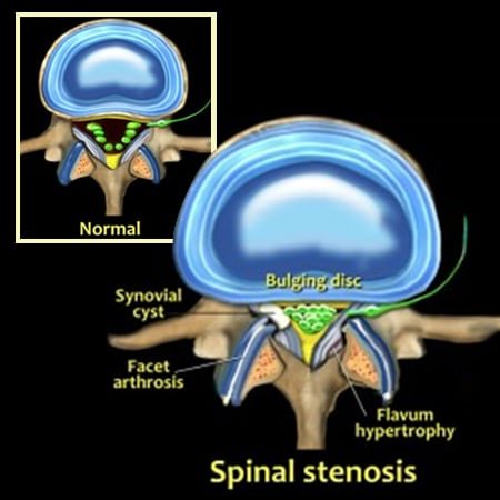
When disc height decreases, the space between the vertebral arches narrows. The ligament can then fold or thicken more easily. Enlargement of the facet joints can further reduce available space.
Because these structures often change together, an MRI report frequently lists ligamentum flavum hypertrophy alongside disc changes and facet arthropathy. The relative contribution of each structure differs from person to person.
Related educational pages: slipped disc and disc changes and facet joint hypertrophy.
Why Thickness Measurements and Symptoms May Not Match
A thicker ligament on MRI does not automatically produce pain, numbness, or weakness. Some people with clear thickening have few symptoms. Others with more modest measurements have significant limitations.
Reasons for the mismatch include:
- Thickness is only one dimension. Shape, laterality, and relationship to the nerve roots also matter.
- Symptoms depend on the space available during standing and walking, which a lying MRI may not fully capture.
- Coexisting disc or facet changes can increase or decrease the clinical effect of the ligament.
- Individual nerve sensitivity and daily activity demands differ.
Matching the imaging to the person’s history and examination remains essential.
The Risk of Aggressive Rotatory Force on a Thickened Ligament
When the ligamentum flavum is already thickened and under tension, high-velocity rotatory (twisting) manipulation can place sudden shear stress on the tissue.
In some cases this force may produce microscopic tears within the ligament. Repeated micro-trauma of this kind can contribute to further scarring and additional thickening over time.
For this reason, aggressive rotatory techniques are generally avoided when imaging shows significant canal narrowing or clear ligamentum flavum hypertrophy. Gentle, non-rotatory methods that respect the mechanical limits of the segment are preferred when care is considered.
Mechanical Nature of the Problem
Ligamentum flavum hypertrophy is primarily a mechanical issue. The thickened tissue physically occupies space inside the spinal canal or the neural foramina.
Oral medication and epidural steroid injections do not change the thickness or elasticity of the ligament. They may reduce chemical inflammation around the nerves for a period of time and thereby ease symptoms temporarily. They do not reverse the structural encroachment. Repeated injections also carry documented risks to local tissues.
Further thickening is often driven by ongoing segmental load and micro-instability. Approaches that improve load distribution, movement quality, and soft-tissue condition are therefore more relevant to the underlying mechanics than chemical approaches alone.
Approaches That Focus on Load, Movement, and Soft-Tissue Quality
After individual assessment, a care plan may include gentle methods aimed at reducing mechanical stress and supporting function. Examples that are sometimes considered include:
- Flexion-distraction techniques that gently open the canal and reduce load on the posterior structures
- Controlled, non-rotatory spinal decompression when suitable
- Specific soft-tissue and stretching approaches directed at the posterior spinal tissues
- Selected physical modalities such as shockwave therapy, high-intensity laser, or therapeutic ultrasound applied with appropriate parameters and positioning
- Movement and stability work that improves the muscular support around the segment
No method is claimed to shrink the ligament or reverse established hypertrophy. The aim is to support function, reduce avoidable mechanical stress, and help the person move with greater comfort and confidence. Suitability is always decided by examination and the full clinical picture.
At Chiropractic Specialty Center, care is planned after assessment and may coordinate chiropractic and registered physiotherapy approaches. See the main center page: Chiropractor in Kuala Lumpur
Balanced Pathways After Assessment
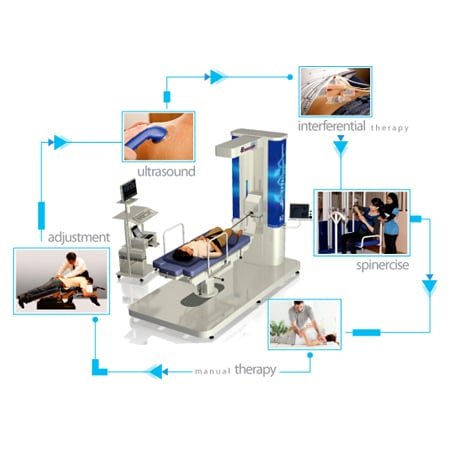
Management decisions are individualized. Options commonly discussed after clinical and imaging review include:
Conservative and supportive approaches
Activity modification, postural awareness, and carefully selected exercise under professional guidance are often considered first when neurological deficits are absent or mild.
Rehabilitative and non-invasive care
Structured rehabilitation aimed at improving mobility, muscle support, and movement patterns may be appropriate. High-velocity rotatory techniques are generally avoided when significant stenosis and ligament thickening are present.
Interventional and surgical pathways
When symptoms are progressive, function is significantly limited, or neurological deficits are present, medical specialists may discuss image-guided procedures or surgical decompression. The decision rests on the full clinical picture and the person’s values. No single pathway is appropriate for every individual.
Ligamentum Flavum Buckling or True Thickening?
On an MRI report, a thick ligamentum flavum can mean two different things. The ligament may have truly grown thicker, with scar-like tissue building up over time. Or it may have folded inward, or buckled, because the disc in front of it has lost height and the space has shortened.
Think of a rubber band between two fingers. Move the fingers closer, and the band sags and bunches up without growing any bigger.
A 2024 study compared the ligament’s thickness on MRI with the real thickness of tissue removed during surgery in 70 people with spinal stenosis. The MRI made the ligament look thicker than it really was. Buckling added to that look, and buckled ligaments had less true thickening but more worn discs.
This matters because buckling depends partly on posture and disc height. Standing tall and leaning back tend to fold the ligament inward, while bending forward stretches it out. That is why many people with this finding feel better sitting or leaning on a shopping cart, and why care at CSC often uses flexion-based methods, spinal decompression and exercise to keep the canal as open as possible.
What “Indenting the Thecal Sac” and “Mild Hypertrophy” Mean
Reports often pair a thick ligament with words like “mild,” “indenting the thecal sac” or “narrowing the canal.” The thecal sac is the fluid-filled tube around the nerve roots. “Indenting” means the ligament touches the back of that tube and pushes its outline in a little.
“Mild” usually means there is still room around the nerves. On its own, mild thickening is common with age and often causes no symptoms. What counts is the total space left in the canal, which depends on the ligament, the disc in front and the facet joints at the side, all together. Studies that measure the canal most often describe central stenosis as a front-to-back width under 10 millimeters or an area under 70 square millimeters.
The pattern of symptoms matters as much as the numbers. Leg pain, heaviness or tiredness that builds with walking and eases with sitting or bending forward fits this kind of narrowing. When the exam and the scan agree, conservative care focuses on movement, strength and positions that open the canal, with regular checks of walking distance to track progress.
Related Educational Pages
These pages provide additional detail on closely related topics:
- Slipped disc and disc changes: slip disc care in Kuala Lumpur without surgery
- Sciatica and nerve-related leg symptoms: sciatica treatment in Kuala Lumpur
- Facet joint hypertrophy: facet hypertrophy and non-invasive care
- Neck care: neck pain assessment and care
- Back care: back pain care in KL, PJ and Selangor
- Main CSC Center in Bukit Damansara: our chiropractic center in Kuala Lumpur
CSC Centers in Kuala Lumpur
Chiropractic Specialty Center has two active centers:
Chiropractic Specialty Center Bukit Damansara (Flagship Center)
Plaza Damansara, No. 71, Jalan Medan Setia 1, 50490 Kuala Lumpur. Operating Hours: Mon, Fri: 8:00 AM, 8:00 PM Sat, Sun: 8:00 AM, 6:00 PM. Call: +603 2093 1000 WhatsApp / SMS: +60 17 269 1873
Chiropractic Specialty Center Bandar Sri Damansara
Operating Hours: Mon, Fri: 8:00 AM, 8:00 PM Sat, Sun: 8:00 AM, 6:00 PM. Call: +603 6262 5777 WhatsApp / SMS: +60 12 455 6939
- Main Bandar Sri Damansara center pagePage last updated on Thursday, April 19,, 2026, with the H1 title: “Ligamentum Flavum Hypertrophy: Safe Care Options.”.
- Full contact details & map
Ligamentum Flavum Hypertrophy, Disc Changes & Nerve Effects Explained (2-Hour Guide Video)
In this in-depth session, Yama Zafer explains how spinal disc changes, ligamentum flavum thickening, and joint stress can influence nerve function. The video breaks down how these structures work together, why symptoms may travel into the arms or legs, and how posture, sitting, and movement patterns affect the spine over time.
This discussion is especially useful for understanding how ligamentum flavum hypertrophy may develop alongside disc bulges, herniations, and facet joint changes.
Video’s Key Moments:
- 1:33:12 Non-Invasive vs Procedural Considerations
- 00:00 Understanding Back & Nerve-Related Symptoms
- 04:06 Key Functions of the Spine & Load Handling
- 06:22 Spinal Disc Structure & Support System
- 18:30 Disc Changes & Degeneration Explained
- 28:49 Stages of Disc Bulge, Protrusion & Herniation
- 38:00 Sitting & Spinal Pressure Effects
- 45:12 Load Changes: Sitting vs Standing vs Leaning
- 51:05 Sleep & Spinal Positioning
- 57:13 Movements to Be Careful With in Disc Conditions
- 1:12:46 Nerve Involvement & Sciatic Patterns
- 1:21:40 Movement & Muscle Balance Around the Spine
- 1:33:12 Non-Invasive vs Procedural Considerations
Planning a visit? You can review the current chiropractic price in Kuala Lumpur and physiotherapy fees in Kuala Lumpur before you come in.
Frequently Asked Questions on Ligamentum Flavum Hypertrophy
These answers are educational. They do not replace individual assessment of ligamentum flavum hypertrophy, spinal joints, spinal discs, or nerves. .
What does ligamentum flavum hypertrophy mean on my MRI report?
It means the radiologist observed thickening of the elastic ligament that forms part of the back wall of the spinal canal. The finding is common with age-related change. It must be interpreted together with your symptoms and the rest of the imaging.
Is a thicker ligament always a problem?
No. Many people have some degree of thickening and few or no symptoms. The clinical importance depends on how much space remains for the nerves and how the rest of the segment is functioning.
What is considered normal thickness?
Approximate average ranges are 1.5-1.7 mm in the cervical spine, 2-3 mm in the thoracic spine, and 3-4 mm in the lumbar spine. These are averages only. Values vary by person and measurement method. In the lumbar spine a threshold near 4 mm is often used when describing hypertrophy.
Can breathing or posture change the thickness?
Yes. Pressure inside the spinal canal changes with breathing and with posture. A less elastic, thickened ligament may narrow the canal more during standing, walking, or deep breathing than a resting MRI suggests.
Why do I feel stiff getting out of a chair but better after a few steps?
This pattern can occur when a thickened ligament reduces canal space in certain positions. Once you begin to move, small changes in posture and load may temporarily improve the available space. The pattern is not diagnostic on its own, but it raises the possibility of ligament involvement and is often overlooked.
Why do my symptoms not match the MRI measurement?
Thickness is only one factor. Shape, side, coexisting disc or facet changes, and the space available during real movement all influence symptoms. Static MRI is performed lying down and may not reflect the loaded spine.
Does ligamentum flavum hypertrophy cause sciatica?
It can contribute to reduced space for nerve roots, especially when combined with disc or facet changes. Not every case of leg symptoms is caused by the ligament, and not every thickened ligament produces leg symptoms.
Can aggressive twisting adjustments make the problem worse?
When the ligament is already thickened and under tension, high-velocity rotatory force can place sudden shear stress on the tissue. In some cases this may produce microscopic tears that contribute to further scarring and thickening. For this reason such techniques are generally avoided when significant canal narrowing or clear hypertrophy is present.
Can the thickening be reversed?
Established hypertrophy is generally regarded as a structural change. Care focuses on symptoms, function, and reducing further mechanical stress rather than on reversing the millimeter measurement.
Is surgery always required?
No. Many people are managed without surgery. Surgery is usually considered when neurological deficits are progressive, function is significantly limited, or appropriate non-surgical care has not been sufficient, after specialist review.
Do medications or injections shrink the ligament?
No. Oral medication and epidural steroid injections do not change the thickness or elasticity of the ligament. They may reduce chemical inflammation around the nerves temporarily. They do not reverse the structural encroachment.
What should I do next?
Bring the full MRI report and images to a clinician who can examine you and correlate the findings with your symptoms.
References and Evidence Context
Each link resolves to the original paper. These sources describe general patterns; they do not establish that any single measurement or approach applies to an individual person.
| What the published evidence shows | Source |
|---|---|
| Ligamentum flavum thickening is one of several contributors to canal narrowing in lumbar spinal stenosis, alongside disc bulging and facet overgrowth. | Katz & Harris, NEJM, 2008 |
| Degenerative findings on imaging are common in people without symptoms, so a thickness measurement on its own does not establish the cause of pain. | Brinjikji et al., AJNR, 2015 |
| Progressive neurological deficit or a loss of bladder or bowel control is worth prompt assessment; short of that, conservative care remains a reasonable route for as long as it produces results. | Todd, Br J Neurosurg, 2017 |
| First-line care for most non-specific back pain is active and non-surgical, supported by self-management. | Foster et al., Lancet, 2018 |
| Back pain is best understood as having several contributing factors rather than one structural cause. | Hartvigsen et al., Lancet, 2018 |
| Some of the thickening seen on MRI comes from the ligament buckling inward as the disc loses height, not only from true tissue growth; MRI tends to overstate the real thickness. | Yabe Y, Ishikawa K, Kurosawa D, et al. Spine. 2024;49(17):E284-E290. |
| The measures most often used for central lumbar stenosis are a front-to-back canal diameter under 10 mm and a cross-sectional area under 70 mm². | Steurer J, Roner S, Gnannt R, Hodler J. BMC Musculoskelet Disord. 2011;12:175. |
- Katz JN, Harris MB. Lumbar spinal stenosis. New England Journal of Medicine. 2008;358(8):818-825.
- Brinjikji W, Luetmer PH, Comstock B, Bresnahan BW, Chen LE, Deyo RA, et al. Systematic literature review of imaging features of spinal degeneration in asymptomatic populations. American Journal of Neuroradiology. 2015;36(4):811-816.
- Todd NV. Guidelines for cauda equina syndrome: red flags and white flags. Systematic review and implications for triage. British Journal of Neurosurgery. 2017;31(3):336-339.
- Foster NE, Anema JR, Cherkin D, et al. Prevention and treatment of low back pain: evidence, challenges, and promising directions. The Lancet. 2018;391(10137):2368-2383.
- Hartvigsen J, Hancock MJ, Kongsted A, et al. What low back pain is and why we need to pay attention. The Lancet. 2018;391(10137):2356-2367.
Author Information
“Ligamentum Flavum Hypertrophy: MRI Findings and Narrowing | Clear Guide” was written and reviewed by Yama Zafer, D.C., a Doctor of Chiropractic (Cleveland University - Kansas City, United States) trained in both chiropractic and physiotherapy, a registered T&CM (Chiropractic) practitioner in Malaysia, and the founder and director of Chiropractic Specialty Center with 30+ years in chiropractic and physiotherapy, and readers may review his professional background, registration information, experience, and editorial profile on the Yama Zafer D.C. biography page.
Last Updated
This page was last updated on September 30, 2026.
Dear Dr. Yama,
I have severe spine fracture at T11-12, DDD, spinal stenosis, osteoporosis and spondylolisthesis. My XR lumbar 6+ views with bending showed instability at L4-5 and L5-S1. Multilevel disc space tearing and listhesis. Degenerative changes of fact complexes. L4-5 is the worst with 6mm of anterolisthesis in flexion, 3-4 mm in extension. Retrolisthesis at L2-3, L3-4 is stable in flexion and extension.
My MRI without contrast showed L4-5 severe spinal stenosis with 70% canal narrowing, cysts in the spine, and moderate stenosis at L3-4 and L4-5, mild to moderate stenosis persists at L5-S1. Multilevel mild neural foraminal stenosis; with trace retrolisthesis of L3-L4. Grade I anterolisthesis of L4-L5, and L5-S1.
L3-L4 mild disc bulge. Mild spine stenosis. Mild bil. foraminal stenosis.
L4-L5 Grade I anterolisthesis. Disc bulge with superimposed post central disc protrusion. Advanced facet arthropathy. Severe spinal stenosis. Mild bil. foraminal stenosis.
L5-S1 Grade I anterolisthesis. Diffuse disc bulge. Advanced facet arthropathy.
Based on above information, can I stretch my piriformis muscle? What’s your advice for me, please? Thank you for your time and comments!!
The osteoporosis is the thread running through all of this, and it should come first rather than be treated as background.
It explains the T11-12 fracture, and it changes what is safe to do about everything else. Three consequences follow, and they are worth being explicit about.
Manual treatment of your spine is not appropriate. Spinal manipulation is contraindicated where there is osteoporosis, a vertebral fracture, and demonstrated segmental instability, and you have all three. That is worth stating clearly so no one talks you into it.
Bone treatment is a prerequisite for any surgical option, not an afterthought. Instrumentation needs bone that will hold it, and where bone density is low, fixation is less secure and hardware complications are more common. If a fusion is under discussion for the instability at L4-5 and L5-S1, ask your surgeon directly how your bone density affects the plan, and whether treating it first would improve the result. Medical treatment for osteoporosis works over twelve to twenty-four months and can change what is surgically feasible.
Exercise has to be selected around it. Loaded forward bending and twisting are the movements most associated with vertebral fracture in osteoporotic bone, so weighted sit-ups, toe touches, and rotational exercise under load are out. What is appropriate, and supported: back extensor strengthening, which has evidence for reducing vertebral fracture risk; weight-bearing activity such as walking; resistance training with a neutral spine; and balance and fall-prevention work, since falls cause the fractures that matter most.
On the instability itself, 6 mm of movement between bending views at L4-5 is a specific and meaningful finding rather than a descriptive one. Demonstrated instability on flexion and extension films is one of the clearer indications for stabilization when the symptoms match, and it is the finding most likely to drive a surgical recommendation in your case. So the question for a spinal surgeon is whether your symptoms correspond to that segment, and a diagnostic block can help establish that where several levels are involved.
Practical steps to arrange, in order. A bone density scan if you have not had a recent one, with vitamin D, calcium, and the blood tests used to look for secondary causes of bone loss, and referral to a bone health clinic or an endocrinologist. Confirmation of whether the T11-12 fracture is recent or old, which MRI can establish and X-ray cannot, since a recent fracture is treated differently. And a spinal surgical opinion for the instability, taken with the bone density result in hand rather than without it.
If your symptoms steadily get worse, or the care you are receiving is not helping, please arrange a medical review; loss of bladder or bowel control is the one sign that needs same-day hospital care. A sudden increase in back pain, which in osteoporotic bone can indicate a new fracture, also calls for a medical review.
D10 D11 ligament flavor Mota ho gaya hai isko kaise theek Karen aur kitna time lagta hai theek nahin hai physiotherapy se theek ho jaega ya operation karna padega
Taking your questions directly, and the first answer is the one that matters most.
Physiotherapy will not reverse the thickening. Neither will traction, manipulation, medication, or any supplement. The ligamentum flavum is a ligament along the back wall of the spinal canal, and once it has thickened it does not thin again with conservative treatment. So the question of how long it takes to fix has an honest answer: it does not get fixed that way, and anyone giving you a timeframe for reversing it is not being straight with you.
Whether you need an operation depends entirely on one thing, and it is not the thickening itself. It is whether your spinal cord is being compressed.
The D10-D11 level matters here. That is thoracic spine, and the canal contains the spinal cord rather than the looser bundle of nerves found lower down. The lower thoracic region is also the commonest place for this ligament to thicken and, in some cases, to calcify and turn to bone, which is seen more frequently in Asian populations and is a recognized cause of thoracic cord compression.
So ask whoever holds your report two things. Does the report say thickening, or does it say ossification or calcification, which means the ligament has turned to bone. A CT scan distinguishes them. And is the spinal cord being compressed, and is there any signal change within the cord itself.
Then check yourself for the signs of cord involvement, because these decide everything. Heaviness, stiffness, or weakness in both legs. Difficulty walking, tripping, or unsteadiness, particularly in the dark. A band-like tight feeling around the chest or abdomen. Numbness starting in the feet and rising. Any change in bladder or bowel control. If any of these are present, arrange a neurosurgical assessment. Where the cord is being compressed at this level, surgery to remove the thickened ligament is the treatment, and the purpose is to prevent further loss, so delay costs function.
If none of those signs are present and your problem is back pain, then no operation is needed now. Physiotherapy in that situation is worthwhile for the pain and for function, but it should be described accurately: it is treating the muscles, the movement, and the strength around the area, not the ligament. Strengthening the trunk and back muscles, maintaining hip and thoracic movement, and staying active are the useful parts, and monitoring with repeat imaging if symptoms change.
If your symptoms steadily get worse, or the care you are receiving is not helping, please arrange a medical review; loss of bladder and bowel control is the one sign that needs same-day hospital care.
Sir
I am 36 and i am suffering
-Ligamentum Falvum hypertropy at lumbar
-central disc protrusion at L4-L5 with narrow of 4.2 mm
-mild at l3-l4 with narrowing of 9.4mm…
pls recommend me the best solution to go for as doctors recommended Surgery. I am avoiding surgery is it possible ??
Second is it chance that latter i have to prepare for l3-L4
Also give ur views on vertiflex procedure of surrgery
, I am from Agra ,India
A 4.2 mm canal at L4-L5 is severe central stenosis, not moderate. A normal lumbar canal measures above roughly 12 mm, and below 7 mm is generally classed as absolute stenosis. That measurement matters considerably more than the 9.4 mm at L3-L4, which is mild by comparison.
On avoiding surgery: that depends on whether you have progressive neurological loss, not on preference. If your strength, sensation, and bladder and bowel function are stable, a structured course of conservative care is reasonable to attempt first. If you have worsening weakness or a foot that drags, have a competent provider assess it promptly; conservative care may still be a reasonable route after that assessment.
On what tends to help: your narrowing has two separate causes and they behave differently. Some have found spinal decompression therapy by devices like the RxDecom or similar to be helpful for the disc component of spinal disc related issues, including those that at times cause nerve issues. Ligamentum flavum hypertrophy is thickened ligament rather than disc material and does not respond in the same way. Any honest plan should tell you which part of your narrowing it targets.
On the Vertiflex: interspinous spacers are indicated for moderate stenosis with neurogenic claudication, meaning leg symptoms that ease when you bend forward, in people who have already failed several months of conservative care. They are generally not the procedure chosen for severe central stenosis combined with a significant central disc protrusion, which is what your measurements describe. That question belongs to the surgeon holding your images, but it is reasonable to ask him directly why a spacer rather than a decompression at 4.2 mm.
On L3-L4 later: narrowing at one level does not make surgery at the neighboring level inevitable. Fusion increases load on adjacent segments over time; decompression without fusion does so far less. Worth asking which is planned.
If your symptoms steadily get worse, or the care you are receiving is not helping, please arrange a medical review; loss of bladder and bowel control is the one sign that needs same-day hospital care.
I live in the El Paso Tx or Las Cruces New Mexico area and am trying to find the specialist as noted in your articles. Can you find or recommend any centers in these areas? Thank you
No, there are no centers in Texas or New Mexico. All six are in Malaysia, and recommending an individual practitioner in a region where their current practice cannot be verified would not be a genuine referral.
What is useful is how to search from where you are, since El Paso and Las Cruces are smaller markets than the large metros and the approach differs.
Start by identifying the specialty rather than the individual. For a spinal problem where the diagnosis is unsettled or where injections or surgery may enter the picture, the medical specialty is physical medicine and rehabilitation, sometimes called physiatry. Those physicians assess spinal conditions, perform diagnostic and therapeutic injections, and coordinate the rest, and they are the specialty most people looking for a spine specialist actually need. For rehabilitation, look for a physical therapist who is board certified in orthopedics, which is a specialist qualification beyond the entry-level doctorate and is verifiable.
Second, widen the map to the nearest academic center. University-affiliated hospitals concentrate subspecialty expertise, and for an uncommon or unclear diagnosis one visit to a center with a dedicated spine program is usually worth more than several local appointments. A day’s drive for a single consultation, with ongoing care delivered locally afterward, is a reasonable arrangement and one that specialists are used to organizing.
Third, and often overlooked, most major academic centers in the United States now offer remote second opinions. You send your imaging and records, a subspecialist reviews them, and you receive a written opinion without traveling. That is particularly worthwhile when a surgical recommendation is on the table and you want it examined independently. Ask any center’s second-opinion service what they charge and what they need.
For imaging specifically, you can request a second radiology reading. Obtain your scans as images on a disc rather than as the report, since a second reader needs the images themselves, and reports vary more between readers than most people realize.
Whoever you see, the questions that separate a careful practitioner from the rest are the same everywhere: is the first visit an examination, what specifically is being treated, does the plan progress into strengthening, and at what point do we review and change approach if targets are missed.
what do you think (my ossification or hypertrophy LF in my case ]
it is caused by trauma (I was accident in a swimming pool hit my head on a wall when I 20 (it is happened because the coach teach me technic swimming backstroke (don’t warning me their is no FLAG to see when the end of the pool end (for swimmer of back stroke )
of course after trauma life with degeneration increase stress on the facet joint (causes facet arthritis)
I m very be a ware from 2016 to my problem with the disc in the neck (after first MR ) .
so i use a good ergonomic lifestyle,low impact exercise, and chin tuck (but I m not accepted to get chiro manipulation because i think this will be dangerous (but get some exercise like chin tuck, and also some stretches, prevention .
but all of this not prevent from i get the LF buckling in c3-4 .
I share with you the link for 1 minute https://youtu.be/GwNqMEClqKk
(what is situation now 22 to 23 and u can also see image from 2016 .
MY MJOA score it is 16 points .
thanks
thank you for your attention and comment .
I can share with you my symptoms for at least 15 mounts .
problem with long walking (pain in legs after and weakness in 2 legs.
I have neck pain but in the last year it was less chronic neck pain (but before i have the problem with lower extremity i have a chronic neck pain for 6 years .
I was have a trauma to the neck , but this was crash head in the backstroke swimming (to the wall of the pool ) in 2010.
Hello Zafer!
Thank you for this in-depth explanation. I’m a 39 year pharmacist, mother of 1, weight 165 lb, 5’7” that has dealt this chronic lower back pain for about 5 years. I have tried physical therapy and chiropractic therapy separately without success.
I recently pulled my lower back in November of this year while transferring a load of about 20 pounds which caused severe pain that radiated down my right buttock. MRI revealed several disc bulges at L3 to L5, disc dessication from L1 to L5, bilateral facet and ligamentum hypertrophy, mild neural foraminal stenosis, and compression of L5 nerve root.
I live in the United States and currently searching for an integrated care like you recommended above. My physiatrist recommended McKenzie exercise which is a bad idea and physical Therapy.
What would you recommend in terms of daily exercises or lifestyle changes (e.g sleeping on my back or side/belly, nutrition and supplements , walking/running, elliptical/stair climbers). Also, my job requires standing 90% of the time.
I’m also considering having another baby. Would this worsen my condition?
Thank you
Five years, and physical therapy and chiropractic tried separately without success. That combination usually points to a gap in what was done rather than to an untreatable problem, and there are three possibilities worth working through in order.
First, and it should be excluded before anything else at your age: inflammatory back pain. It typically begins before forty, builds gradually rather than after an injury, and behaves in the opposite way to mechanical pain. Morning stiffness lasting more than thirty minutes, timed rather than estimated. Pain that improves with movement and worsens with rest. Waking in the second half of the night with pain that drives you out of bed. Alternating buttock pain. A marked response to anti-inflammatories. Any personal or family history of psoriasis, inflammatory eye disease, or inflammatory bowel disease. If several fit, ask for inflammatory markers and imaging of the sacroiliac joints, and a rheumatology opinion. This is regularly missed for years in exactly this pattern, and cycling through manual therapies is how the years pass.
Second, if it is mechanical, then what most often distinguishes the courses that work from those that do not is not the profession involved but whether the program progressed into sustained loading. Both physical therapy and chiropractic can deliver that and frequently do not, because pain settles first and the strengthening phase gets abandoned. For persistent low back pain, exercise therapy has the strongest evidence of anything available, and the specific type matters less than that it is progressive and continued. Passive treatment, whoever provides it, does not build capacity. If neither course you did included loading that increased week on week over months, then the main lever has not been pulled.
Third, five years of pain has its own consequences that are treatable and usually untreated: the nervous system becomes more responsive to load, sleep is often disrupted, and confidence in movement erodes. Those respond to graded exposure and to cognitive-functional approaches, which have good evidence for pain of this duration, rather than to further imaging or passive treatment.
Practically: pick one measurable function, whether that is sitting tolerance, walking distance, or repetitions from a chair. Establish honestly what you can do now, start slightly below it, and increase by a small step every week or two. A step is correct if symptoms settle back within about a day. Continue for months, not weeks.
One more thing, since your MRI shows disc changes. Disc changes at 39 are extremely common and appear on the scans of large numbers of people with no pain at all, so do not treat the report as the explanation or judge progress by re-scanning.
If your symptoms steadily get worse, or the care you are receiving is not helping, please arrange a medical review; loss of bladder or bowel control is the one sign that needs same-day hospital care. Fever or unexplained weight loss alongside back pain also call for a medical review.
with MRI I have an hypertrophy of ligament flawvn at D5 D6 D8 D9 with legs impotence and pain. Is it a spinal stenosis
Yes, that is spinal stenosis, meaning narrowing of the spinal canal. But the more important point is where yours is, and it changes the urgency considerably.
Your levels are thoracic, in the mid and upper back. In that region the canal contains the spinal cord itself, not the looser bundle of nerve roots found in the lower back, and the thoracic canal has less spare room than elsewhere. So narrowing there affects the cord, and cord compression behaves differently from the nerve root problems that most articles about stenosis describe.
Weakness in the legs together with thoracic canal narrowing should be treated as cord compression until proven otherwise, and it warrants a neurosurgical assessment before any course of treatment. If you are also describing erectile dysfunction alongside the leg symptoms, that adds to the concern rather than being a separate issue, because those functions are carried by the same pathways.
The signs that indicate the cord rather than a nerve are worth checking now: legs that feel stiff or heavy rather than simply weak, difficulty walking or unsteadiness, tripping or catching a foot, a band-like tight sensation around the chest or abdomen, numbness starting in the feet and rising, and any change in bladder or bowel control. The more of those that apply, the more important that assessment becomes.
Two things to ask for. An MRI of the thoracic spine if yours is not current, with the specific question of whether the cord is compressed and whether there is any signal change within the cord itself, which is a separate and more serious finding. And whether the ligament is thickened or has ossified, meaning turned to bone, which is a recognized pattern in the lower thoracic spine and which a CT scan distinguishes.
On treatment, the honest position. No conservative treatment thins a thickened or ossified ligamentum flavum. Not physiotherapy, not traction, not manipulation, not medication. Where the cord is being compressed and causing weakness, surgical decompression is the treatment, and its purpose is to prevent further loss. That is why time matters: the longer the cord is compressed while function is deteriorating, the less recovery there tends to be afterward. Your spine should also not be manipulated while cord compression is a possibility.
Please arrange that assessment rather than waiting. If your symptoms steadily get worse, or the care you are receiving is not helping, please arrange a medical review; loss of bladder and bowel control is the one sign that needs same-day hospital care.
Hi Dr Yama
Iam in Brisbane Australia,
I have same diagnosis Ligamentum Flavum
after traying osteo, physio, acupuncture, no good
Iam reluctantly booked for surgery in 10 days to remove thickened ligament
have you got clinic in Australia as I would do anything to avoid surgery
I have sent your clinic email but no response
mile
We are based in Malaysia and have no clinic in Australia. On the email, if it went to a general address it may not have been routed to the clinical team. That does not help your timeline, so here is what is worth knowing before your surgery date.
Ligamentum flavum hypertrophy is thickened ligament, and that fact answers most of your question. Conservative care, including osteopathy, physiotherapy, acupuncture and decompression therapy, can reduce symptoms by lowering inflammation, improving segmental mechanics, and reducing the positions that narrow the canal further. None of it thins the ligament itself. If thickened ligament is the principal cause of your narrowing, it remains whatever conservative care you undertake. That is the honest reason your three attempts did not resolve it, and it is why removal is the definitive option when the narrowing is significant.
Where decompression therapy does earn a place is when a disc component sits alongside the ligament. Some have found spinal decompression therapy by devices like the RxDecom or similar to be helpful for spinal disc related issues, including those that at times cause nerve issues. Whether that applies to you depends on whether your imaging shows a disc contribution or thickened ligament alone. Your MRI report should make that distinction, and it is worth reading specifically for it.
Questions worth putting to your surgeon in the next ten days: what proportion of your narrowing is ligament, disc, and facet; whether decompression alone is planned or fusion as well; and what your current neurological findings show, since those determine urgency far more than pain does.
If you develop progressive weakness, worsening balance, or any change in bladder or bowel control before your date, that is not a wait-and-see situation.
I am 38years old and have back pain with tingling and numb foot. I recently had an MRI of the Lumbar spine; below is my report; kindly assist.
The highlights of my MRI findings are as follows:
Multilevel ligamentum flavum hypertrophy and facet arthropathy. At L4-L5 level, mild central bulge causing minimal thecal sac indentation and minimal left lateral recess stenosis.
There is a mismatch between your symptoms and your report, and noticing it is the most useful thing that can be done from here.
A numb foot is a specific neurological finding. Your MRI describes a mild central bulge at L4-L5 with minimal indentation of the thecal sac, alongside multilevel ligamentum flavum thickening and facet changes. A mild central bulge does not usually reach the nerve that supplies sensation to the foot, because a central bulge presses toward the middle of the canal rather than toward where the roots pass out to the sides. Words like mild and minimal in a report, alongside a definite numb foot, mean the scan may not be explaining your symptoms.
Two possibilities follow, and they are investigated differently.
The first is that the numbness is from a nerve root after all, in which case exactly where it sits should match a specific root. Numbness across the top of the foot and the outer shin corresponds to L5. Numbness along the outer border of the foot and the sole corresponds to S1. Map yours precisely, ideally by drawing it, and note whether it is constant or comes and goes and whether position changes it. If it matches one of those patterns and there is matching weakness, meaning difficulty lifting the foot or big toe for L5, or difficulty rising onto the toes for S1, then the spine remains the likely source and the level responsible should be identified by examination rather than by which line of the report sounds worst.
The second is that the numbness is coming from outside the spine, which is common and frequently missed when a scan has already been done. Numbness in both feet, or in a stocking distribution not matching any single nerve, points toward a peripheral neuropathy, and blood tests for diabetes and vitamin B12 are the first step there. Numbness over the outer shin and top of the foot on one side can come from compression of a nerve just below the outer side of the knee, which happens with habitual leg crossing, prolonged squatting or kneeling, or significant weight loss. Numbness of the sole with burning can come from compression at the inner ankle.
If your examination and imaging do not agree, ask for a nerve conduction study. It distinguishes a problem at the nerve root from one further down the leg, which is precisely the question here.
One further point given your age. Multilevel thickening of the ligamentum flavum at 38 is earlier than usual, and it is worth asking the reporting radiologist whether the appearance is degenerative or whether anything else should be considered.
On what conservative care can do: nothing thins a hypertrophied ligamentum flavum, so a plan should be described in terms of the disc component, the movement lost at the affected segments, the positions that reduce space further, and progressive strengthening, rather than as reversing the scan.
If your symptoms steadily get worse, or the care you are receiving is not helping, please arrange a medical review; loss of bladder and bowel control is the one sign that needs same-day hospital care.
Hi! I just had an MRI that says my Flavun thingy is thickened. Where is your nearest Center in southwest USA? Oh plz help! Thanks
Cathy T
There are no centers in the United States. All six are in Malaysia. But the finding itself deserves a calmer answer than the report probably gave you.
Thickening of the ligamentum flavum is a common age-related change. The ligament runs along the back wall of the spinal canal, and it thickens gradually as the spine ages, in much the same way discs lose height and joints enlarge. It appears on the scans of a great many people who have no symptoms at all, and its presence on a report does not mean you have a serious problem or that anything needs to be done.
What determines whether it matters is not the report but your symptoms. Three questions settle it.
Do your legs feel heavy or achy after walking a certain distance, and does that ease specifically when you sit down or lean forward rather than merely when you stop. Can you walk noticeably further pushing a shopping cart or going uphill than on level ground. Those indicate narrowing that is actually affecting the nerves, and that is a real condition worth treating.
Do you have symptoms traveling down one leg in a defined pattern, with numbness or weakness. That indicates a nerve root and is treated differently.
If your symptoms steadily get worse, or the care you are receiving is not helping, please arrange a medical review; loss of bladder and bowel control is the one sign that needs same-day hospital care.
If the answer to all three is no, and your finding was picked up on a scan done for some other reason, then this may simply be a description of an aging spine and not the cause of anything.
One thing to be aware of when you look for care locally, because this finding attracts overclaiming. No conservative treatment thins a thickened ligamentum flavum. Not traction, not spinal decompression tables, not manipulation, not exercise, not supplements. If a clinic tells you their treatment will reverse it, that is the point to walk away. What legitimate care can address is everything around it: the disc component of any narrowing, the movement lost at the affected segments, the positions that reduce the available space further, and the strength of the supporting muscles. That combination often changes symptoms meaningfully even though the imaging stays exactly the same, and a practitioner who explains it in those terms is describing the situation honestly.
Position is a genuine tool here rather than an afterthought. Leaning slightly forward opens the canal and leaning back closes it, which is why a cart or a wheeled walker extends walking distance for people with this, and it can be used deliberately to build tolerance.
Respected sir, is collagen intake harmful to patients with ligamentum flavum hypertrophy? And if so, which supplements should we take in ligamentum flavum hypertrophy?
No, collagen supplements are not harmful in this situation, and the reasoning behind the worry is worth unpacking because it is a reasonable one to have had.
Ligamentum flavum hypertrophy does involve changes in the ligament’s composition: elastic fibers are progressively lost and replaced by fibrous tissue, which is a collagen-based process. It is understandable to wonder whether adding collagen to the diet feeds that. It does not. Ingested collagen is broken down in digestion into amino acids and short peptides, exactly as any other protein is, and the body then uses those wherever it is currently building tissue. There is no mechanism by which swallowed collagen is delivered preferentially to a thickened spinal ligament, and no evidence that collagen intake influences ligamentum flavum thickening in either direction.
The other half of that is equally worth saying: there is no evidence that collagen supplements help either. The thickening is driven by mechanical stress on the segment over years, not by a dietary deficiency, which is why no oral supplement addresses it.
On which supplements to take for this condition specifically, the honest answer is none. No supplement has been shown to reduce ligamentum flavum thickening or to reverse it, and any product marketed on that claim is not supported by evidence. Money spent there is better spent elsewhere.
What is worth attending to is general rather than spinal. Vitamin D status is worth checking and correcting if low, since deficiency affects bone and muscle function and is common. Adequate protein intake supports the muscle mass that protects the spine, particularly with age, and that is better achieved through diet than through supplements in most people. Beyond that, anything you take should be checked with your doctor for interactions, particularly if you are on blood-thinning medication, since several popular supplements affect clotting.
The interventions that actually influence this condition are mechanical rather than nutritional: the positions you spend your day in, maintaining hip and spinal mobility, keeping body weight in a reasonable range, and progressive strengthening of the muscles supporting the affected region. Those are unglamorous and they are what there is.
Hi Doctor Yama,
My daughter is a chiropractic doctor in Indianapolis, Indiana, in the US. She has suggested that I contact you. I have had pain in both ankles for eight weeks. The pain worsens after walks or if I stand for 10-15 minutes.
I am a retired pain management physician. I have had Epidural steroid injections twice, but it has not helped. X-rays and MRIs have been taken. The MRI shows canal stenosis, disc bulge, facet hypertrophy, and ligamental hypertrophy. What is your advice for me?
Vinu Patel MD
The most useful observation is that your presentation does not fit your imaging well, and two failed epidurals reinforce that rather than indicating a technical failure.
Pain confined to both ankles is an atypical distribution for lumbar canal stenosis. Neurogenic claudication characteristically produces buttock, thigh, and calf heaviness or aching that builds with upright walking, and isolated bilateral ankle pain is not the usual pattern. Meanwhile the findings on your report, canal stenosis with disc bulge, facet hypertrophy, and ligamentous hypertrophy, are close to universal in the age group that has lumbar MRIs, and their presence does not establish causation. Two epidural injections without benefit is meaningful negative evidence: it argues the pain generator is not where the injections were placed.
The one confirmatory test worth doing before anything else is postural. Does walking bent forward over a shopping cart, or on an incline, extend your walking time substantially compared with level ground, and do symptoms resolve within minutes of sitting rather than merely on stopping. A clear positive supports the spine. A negative, with symptoms unchanged by flexion, largely removes it.
Assuming the postural test is unimpressive, the ankles themselves deserve the examination they may not have had, because the history of eight weeks, bilateral, worse after walking and after ten to fifteen minutes of standing, is a weight-bearing local pattern.
Bilateral posterior tibial tendon dysfunction fits load-dependent medial ankle pain with standing intolerance, and is easily missed. Ankle and subtalar osteoarthritis produces the same load dependence. Bilateral insufficiency or stress reactions in the distal tibia or calcaneus should be considered given the eight-week onset without trauma, and bone density and vitamin D status are worth establishing.
Two specific questions worth asking yourself. Any fluoroquinolone exposure in the months preceding onset, given the bilateral tendinopathy association. And any new medication generally, since bilateral simultaneous onset in a symmetrical distribution favors a systemic rather than a mechanical cause.
That symmetry also argues for excluding inflammatory arthropathy with inflammatory markers, and for a vascular assessment with pedal pulses and an ankle-brachial index, since exertional lower limb pain relieved by rest has a vascular differential that the spinal picture can mask.
Practically: weight-bearing plain films of the ankles and feet, palpation and resisted testing of the posterior tibial and peroneal tendons, ESR and CRP, ABI, and a bone health review. Ultrasound or dedicated ankle MRI if the tendon examination is suggestive.
On the spinal findings themselves, one point in case they remain in the picture: ligamentum flavum hypertrophy is not reducible by any conservative means, and any program should be framed around canal-opening positions, hip extension mobility, and trunk and gluteal strengthening rather than around reversing the imaging.
Sir
I am 36 and i am auffering from central disc protrusion along with ligamentum falvum hypertrophy which creating canal narrowing to 4.2 mm at L4-L5 and mild atl3-l4 with narrowing of 9.4mm… pls recommend me the best solution to go for as doctors recommend Surgery , I am from Agra … pls also suggest exercise nd precautions
Adding to the earlier reply, because being 36 with a canal measuring 4.2 mm raises a question that changes surgical planning and is worth asking before you decide.
Severe canal narrowing at that measurement is unusual at your age from degenerative change alone. In many people who present young with severe stenosis, part of the narrowing is developmental: the canal was smaller than average to begin with, because the bony arches are shorter than usual, and only a modest amount of disc bulging and ligament thickening is then needed to produce severe narrowing. Ask the reporting radiologist directly whether your canal appears developmentally narrow, and whether the narrowing is confined to the disc levels or is present between them as well. Narrowing that continues behind the vertebral bodies, away from the discs, points to a developmentally small canal.
Why it matters. A developmentally narrow canal means less reserve space everywhere, so decompressing a single level may leave you closer to the margin than the surgeon expects, and it influences how many levels are addressed and which technique is chosen. It also explains why a person your age can have severe narrowing without decades of wear, which is otherwise a puzzling picture.
Second, on what your doctors are recommending. Whatever the specific procedure, the questions are the same. Which level is producing my symptoms, and has that been confirmed by examination and ideally by a diagnostic block rather than inferred from the measurement. Is a decompression alone possible or is fusion planned, and why. And what happens if I wait three months, which is a fair question whenever there is no progressive neurological loss.
Third, what to do while deciding. Your narrowing has two components and they behave differently. The disc component can reduce over time and is what decompression-type treatment acts on. The ligamentum flavum component does not reduce with any conservative treatment. So a program should be framed honestly around what it can influence: using forward-leaning positions deliberately to open the canal and build walking tolerance, restoring hip extension so the lower back is not forced into the position that closes the canal with every step, and progressive trunk and gluteal strengthening. Track walking distance weekly, since that number is the measure any surgical decision should rest on.
If your symptoms steadily get worse, or the care you are receiving is not helping, please arrange a medical review; loss of bladder or bowel control is the one sign that needs same-day hospital care.
Howdy, My wife has this condition primarily L3-L4 and L4-L5 and additional mild issues L1 through L-3. The orthopedic doctor prescribed McKenzie exercises, pain meds, and, if necessary, an injection and down the road surgery. I think it’s a bad idea! Proper exercise is the best approach. Is there anyone you know of in Tallahassee, Florida, she could go to?
Thanks,
Her Husband
The plan she was given is more reasonable than it may sound, and it is worth being accurate about that before deciding against it.
Exercise is the first step in what the orthopedic doctor described, not an alternative to it. The McKenzie approach is not generic exercise: it is a structured method that identifies which direction of movement reduces symptoms and uses that direction deliberately, with a substantial evidence base for disc-related back and leg pain. Prescribing it first, with medication for symptom control, is the standard conservative starting point, and it is essentially the approach you are advocating.
Where your instinct is right is on what follows. Injections and surgery should be triggered by findings, not by a timetable. Being told surgery is coming down the road, before a conservative attempt has been given a fair trial, frames it as inevitable when it is not. That is worth pushing back on, and the way to do it is to ask a specific question: what findings would make an injection appropriate, and what findings would make surgery appropriate. A clear answer names progressive weakness, a foot that drags, or bladder and bowel changes for surgery, and a defined failure of conservative care for an injection. If the answer is vague or time-based, that tells you something.
Two things about her particular picture support taking a conservative course seriously. Mild changes across several levels, with the main findings at L3-L4 and L4-L5, is a common pattern in adults and is not a surgical picture on its own. And multi-level degenerative findings are the situation in which surgery performs least well, because no single level clearly accounts for the symptoms. That argues for exhausting conservative care rather than treating it as a formality.
One addition worth making to the plan. Directional exercise handles the symptom side well, but it is not a strengthening program. Once symptoms have settled and centralized, the work should progress into loading: hip mobility, trunk and gluteal strengthening, and building tolerance for sitting, standing, and lifting. Stopping when the pain eases is the most common reason people end up back at the same point a year later and conclude conservative care failed when it was never completed.
There is no center in Tallahassee. All six are in Malaysia. For choosing locally, the questions that separate a good provider from the rest: is the first visit an examination, what specifically is being treated, does the plan progress into strengthening, and at what point do we review and change approach if targets are missed.
She should be assessed medically rather than continuing exercises if weakness develops in a leg, if a foot begins to catch, or if there is any change in bladder or bowel control.
I read your website about spine issues related to ligamentum flavum hypertrophy.
I am 31 years old; I had an accident and injured my neck (the soft tissue of my neck ). I started to have symptoms about three mounts ago. Now years after the trauma is causing severe chronic neck due to degenerative changes, including damage to my ligament flavum (hypertrophy and buckling of ligamentum flavum).
My ligamentum flavum hypertrophy involves the C3-C4 segments of my neck, causing compression of my spinal cord and nerves from the center to the left.
MY symptoms include neck pain, pins & needles, and tingling in the legs and hand. The pins & needles started last week. I am also experiencing pins & needles in my trunk, along with pain in the chest.
Moreover, my blood pressure increased with the systolic pressure ranging between 170 and 190m, but it is lower now, fluctuating between 110 and 130.
I like to find out if my condition is treatable without surgery, as I am not looking forward to neck surgery. Also, is there a way to help repair my Myelomalacia without getting surgery?
thanks
This needs an answer you may not want, and giving you a different one would not be doing you a service.
What you describe is not a situation to manage without a surgical opinion, and the reason is not the ligamentum flavum finding itself. It is the pattern and the timing of your new symptoms.
Pins and needles in both legs, in the hand, and in a band across the trunk, appearing over the past week, in someone with documented compression of the spinal cord at C3-C4, describes cord involvement that is changing. Nerve root problems produce symptoms in one limb in a defined pattern. Symptoms in the legs, the hands, and the trunk together come from the cord itself, and the fact that they began a week ago rather than years ago is the part that matters most. That combination warrants a neurosurgical assessment before any course of treatment.
Two things follow from that immediately. Your neck should not be manipulated. And no conservative treatment thins a hypertrophied ligamentum flavum or reverses buckling. Traction, manual therapy, and exercise do not change that tissue, and anyone offering to reverse it is misleading you.
On what surgery does, honestly, since you are weighing it. Decompression for cord compression in the neck is aimed primarily at stopping further loss. Recovery of function that has already been affected is variable, and the strongest predictor of a good outcome is how long the compression has been causing symptoms before it is relieved. That is precisely why a one-week history of new symptoms is a reason to move quickly rather than to wait and observe. Deciding against surgery is a legitimate choice, but it should be made after a specialist has examined you and looked at current imaging, not instead of that.
Ask for an MRI of the cervical spine if your imaging is not current, and ask specifically whether there is any signal change within the cord itself, which is a separate and more significant finding than compression alone.
Two other things in your message should not be left to the neck consultation. Chest pain must not be assumed to come from your spine, even alongside trunk symptoms, and it needs medical assessment in its own right. And a systolic blood pressure reaching 170 to 190 needs review regardless of what caused it, even though it has since settled. Raise both with a doctor rather than filing them under the neck problem.
If your symptoms steadily get worse, or the care you are receiving is not helping, please arrange a medical review; loss of bladder and bowel control is the one sign that needs same-day hospital care.
I learned a lot from reading your website. Thank you for the information. I receive no treatment, just drugs from Drs, and I’ve been in pain for 20 years. Is it too late for me? Thanks, Martha from California.
No, it is not too late, and that answer is worth more than reassurance so here is the reasoning behind it.
Twenty years of medication without treatment leaves three things behind, and all three are modifiable regardless of how long they have been there.
The first is physical capacity. Two decades of pain-limited movement reduces strength, joint range, and tolerance for load. That is not permanent damage; it is deconditioning, and deconditioned tissue responds to graded loading at any age. It responds more slowly than it would have at forty, but it responds.
The second is movement habit. People in long-standing pain develop protective patterns, holding regions stiff and avoiding particular directions. Those become automatic and they add load elsewhere. They are retrainable.
The third is the nervous system’s own responsiveness. After many years, the system that carries and interprets pain signals becomes more sensitive, so ordinary activity registers as more threatening than it is. This is a genuine, measurable phenomenon, not a suggestion the pain is imagined, and it is the part that responds specifically to graded exposure and to structured pain management rather than to further imaging or injections.
The duration of pain is a weaker predictor of whether someone improves than most people assume. What predicts improvement is whether the right things are being done, and in your case medication alone means the main levers have never been pulled.
A realistic definition of improvement helps. After twenty years, the goal is usually not zero pain. It is more capacity, less interference in daily life, better sleep, and doing things you have stopped doing. People frequently reach that even when their pain score changes only modestly, and it is a better target because it is achievable.
Where to start, practically. Choose one measurable thing: walking distance, minutes of standing, or how many times you can rise from a chair. Establish honestly what you can do today without flaring. Then start slightly below that and increase by a small amount every week or two. The rule that governs everything is this: an increase is right if symptoms settle back to baseline within about a day. If they do not, the step was too big, and you go back a level rather than stopping.
Two other things worth arranging. Ask your doctor for a referral to a physical therapist experienced with persistent pain, or to a pain rehabilitation program, which exist in most parts of California and combine graded activity with the psychological approaches that have the best evidence for pain of this duration. And ask for a review of twenty years of medication, since what was appropriate at the start is often not what serves you now.
Progress at this stage is measured in months, not weeks. That is slow, but it is not nothing, and it is a different thing entirely from too late.
I had fusion surgery seven years ago for c4c5, and now I have ligament flavum thickening on c5c6 . Can you help me?
What you describe is the recognized pattern after cervical fusion rather than an unrelated new problem, and understanding that shapes what to do about it.
Fusing C4-C5 removes movement at that segment. The movement has to go somewhere, and it goes to the levels immediately above and below, which then carry more load than they did before. C5-C6 is the level directly beneath your fusion, so change appearing there seven years on is the expected location. Thickening of the ligamentum flavum is part of that process: as a segment takes on more load and the disc loses height, the ligament along the back wall of the canal thickens and buckles inward.
Three things determine what can be done, and they need current imaging rather than the films from around your surgery.
Whether the spinal cord is affected. The signs are not neck pain: clumsy hands, dropping things, difficulty with buttons and keys, deteriorating handwriting, unsteady walking especially in the dark, symptoms in both arms or in the legs, or any change in bladder control. If any of those are present, this needs neurosurgical assessment rather than treatment, and forceful manipulation of the neck would not be appropriate.
The canal measurement at C5-C6, and whether there is any signal change within the cord itself, which is a separate and more serious finding.
Whether a nerve root is involved, meaning symptoms into one arm in a defined pattern.
Now the honest limitation. No conservative treatment thins a hypertrophied ligamentum flavum. Not traction, not manipulation, not exercise. Anyone telling you otherwise is misleading you.
What can be addressed is the demand placed on that segment, and after a fusion there is more to work with than most people realize. The thoracic spine and the shoulder girdle contribute a large share of the head and neck’s positioning, and when they are stiff, the mobile cervical segments below a fusion take more of the work. Restoring movement through the upper back, correcting sustained neck flexion at screens, and strengthening the deep neck flexors and the muscles that hold the shoulder blades all reduce the load passing through C5-C6. So does avoiding sustained extension, meaning looking upward and overhead work, since that position narrows the canal and buckles the ligament further. A pillow that is too high does the same thing all night.
Whether that changes your symptoms depends on findings that need an examination. It will not change the imaging, and any plan should be described in those terms rather than as reversing what the scan shows.
I’m 77. I have all of the above. Do you have any office near the Lancaster, Pennsylvania, Downingtown, Pennsylvania, Reading Pennsylvania areas
No, there are no offices in Pennsylvania. All six centers are in Malaysia.
At 77 with the findings described on this page, a few things are worth knowing that are specific to your situation rather than general advice.
Position is your most useful tool with canal narrowing, and it is worth using deliberately. Leaning slightly forward opens the spinal canal; standing upright and leaning back closes it. That is why people with this pattern can push a shopping trolley far further than they can walk unaided, and why walking uphill is often easier than walking downhill. Rather than treating that as a limitation, use it: walk with a trolley or a wheeled walker to build distance, sit for a short rest before symptoms force you to, and use exercise machines that keep you slightly forward, such as a recumbent bike, to maintain fitness without provoking symptoms. Maintaining walking capacity matters more at your age than the appearance of the scan.
Falls deserve as much attention as the pain. Narrowing that affects the nerves reduces sensation and balance, and a fall at 77 carries consequences that back pain does not. A balance assessment, a home check for loose rugs, cords, and lighting, and a review of any medication that causes drowsiness or lowers blood pressure are all worth arranging. Ask about vitamin D and bone density too.
On surgery, since it may be raised: age by itself is not a reason to rule it out. Decompression without fusion is a smaller operation than many people assume and is performed in this age group with the aim of restoring walking distance. What decides it is your general health, your other medical conditions, and how much your walking is limited, rather than the number of years. If it is offered, the questions worth asking are whether fusion is planned or decompression alone, and what specifically it is expected to improve.
For local care, the two questions that separate a careful practitioner from the rest: what exactly are you treating, and how will we know within a defined number of visits whether it is working.
If your symptoms steadily get worse, or the care you are receiving is not helping, please arrange a medical review; loss of bladder and bowel control is the one sign that needs same-day hospital care.
Do you have any centers in India where we can go for treatment?
No, all six centers are in Malaysia, in Kuala Lumpur and Selangor. There are no centers in India.
Competent care for spinal conditions is widely available in India, so the more useful thing to carry away is what a good program looks like, since that is what distinguishes one clinic from another far more than location does.
A first visit should be an examination, not a treatment. History covering when symptoms began, what changes them through a day, what has been tried and what each thing changed. Physical examination of movement and joint function. Where symptoms travel into a limb, neurological testing of reflexes, strength, and sensation. Those findings should be written down, because they are the baseline everything afterward is measured against, and care that never established a baseline cannot tell you whether it is working.
The plan should name the mechanism, not the scan. Being told you have degenerative changes or a disc bulge is a description of your imaging. What should be explained is which structure is producing your symptoms and why, and how the treatment chosen addresses that.
The plan should progress. Early care reasonably settles symptoms and restores lost movement. By around four to six weeks it should be moving into loading and progressive strengthening of the muscles supporting the affected region. Care that remains passive beyond that point is the single most common reason a problem that settled returns months later. Ask directly what the plan progresses to over the next six weeks, and treat a vague answer as an answer.
There should be a review point. An agreed number of sessions after which progress is assessed against the original findings and the approach changes if the targets were missed. Open-ended courses without review are how months pass.
And be cautious about large prepaid packages before an examination has been done and before you have seen how you respond to the first few sessions.
Two more things worth knowing wherever you are treated. Imaging findings such as disc bulges and degenerative change appear in large numbers of people with no symptoms at all, so the level responsible for your pain is the one that matches your examination, not the worst-looking one on the report. If your symptoms steadily get worse, or the care you are receiving is not helping, please arrange a medical review; loss of bladder or bowel control is the one sign that needs same-day hospital care. Fever or unexplained weight loss alongside back pain also call for a medical review.
I am also having the same back problem with pain shooting down my buttocks and legs. What can be done? Please advice.
Pain shooting into the buttock and leg has several possible sources, and they are told apart mostly by where the pain goes and what changes it. Working through these before an appointment will make it far more productive.
A compressed nerve root in the lower back produces pain in a defined line down the leg, often with numbness or tingling in a specific area. Symptoms below the knee make this more likely. The two common patterns: down the outer calf into the top of the foot with weakness lifting the foot or big toe, or down the back of the calf into the outer foot with weakness pushing off on the toes.
Narrowing of the spinal canal produces heaviness and aching in both legs that builds as you walk and is relieved specifically by sitting or leaning forward. The giveaway is being able to walk much further pushing a trolley or going uphill than on level ground.
The sacroiliac joint produces one-sided pain below the belt line, often into the groin or back of the thigh, characteristically worse when rising from a chair and when standing on that leg. It rarely goes below the knee and it does not show on ordinary lumbar imaging.
The hip joint refers into the groin and front of the thigh, with restricted rotation and pain on deep bending or crossing the leg.
The gluteal tendons produce pain over the bony point on the outer hip spreading down the outer thigh, worse lying on that side at night and worse climbing stairs.
Deep buttock nerve irritation produces pain in the buttock that is markedly worse with prolonged sitting, particularly on a hard surface, without the clear line down the leg of a root problem.
Before your appointment, write down four things. Where the pain reaches at its furthest point, and whether that distance has been increasing or decreasing over recent weeks. What consistently makes it worse and better: sitting, standing, walking, leaning forward, lying flat, rising from a chair. Whether there is any numbness, and exactly where. And whether anything feels weak, such as a foot catching on steps or a knee that might give way.
Those four answers narrow the field more than a scan does, because the same imaging findings appear in many people without symptoms.
If your symptoms steadily get worse, or the care you are receiving is not helping, please arrange a medical review; loss of bladder and bowel control is the one sign that needs same-day hospital care.
Sir, do you have any branches in India?
No, there are no branches in India. All six centers are in Kuala Lumpur and Selangor, Malaysia.
Since traveling for an opinion is expensive and often unnecessary, here is what can be arranged without it, which for spinal stenosis findings is usually the more valuable step.
Obtain your imaging as images, not as the typed report. Ask the imaging center for the study on a disc or through their portal in the standard format used for medical images. Reports summarize and vary considerably in detail between readers, and no useful second opinion can be given from a report alone. Once you hold the images, a second radiological reading or a specialist review can be arranged locally, and any subsequent consultation anywhere becomes meaningful.
Ask for four specific things to be stated, because these determine management and are frequently omitted. The canal measurement in millimeters at the narrowest level. Which structures are producing the narrowing, meaning disc, ligamentum flavum, facet joints, or a combination, and in what proportion. Whether any nerve root is contacted or the cord is compressed. And whether there is any signal change within the cord itself, which is a separate and more serious finding.
Those four answers change the plan more than the choice of clinic does. In particular, knowing what proportion of the narrowing comes from disc as against thickened ligament tells you directly whether decompression-type treatment has anything to work on, since it acts on the disc component and not on the ligament.
For treatment itself, competent conservative care for this is available in most large Indian cities, and the questions worth asking are the same everywhere: is the first visit an examination, what specifically is being treated, and how will we know within a defined number of sessions whether it is working.
If your symptoms steadily get worse, or the care you are receiving is not helping, please arrange a medical review; loss of bladder and bowel control is the one sign that needs same-day hospital care.
Sir, I am suffering from degenerated disc disease involving c3-4,c4-5. I also have ligamentum flavum hypertrophy at c4-c5. What is the solution, sir, as I am in pain daily?
The ligamentum flavum finding is the one that determines everything else here, so start there.
The ligamentum flavum runs along the back wall of the spinal canal. When it thickens, it takes up space in the canal itself, and in the neck that canal contains the spinal cord. So the first question is not what treatment to have. It is whether the cord is affected, because that changes what is safe as well as what is appropriate.
The signs that indicate cord involvement are specific and none of them are neck pain. Hands becoming clumsy. Dropping things. Buttons, coins, and keys becoming difficult. Handwriting deteriorating. Unsteadiness when walking, particularly in the dark. Symptoms in both arms, or in the legs. Any change in bladder control. If any of those are present, arrange a neurological or neurosurgical assessment with current imaging rather than a course of treatment, and do not have forceful manipulation of the neck. Where cord compression is progressing, the reason to act is to prevent further loss.
If none of those apply, and your symptoms are neck pain with or without symptoms into one arm in a defined pattern, then this is being driven by the nerve root and the joints rather than by the cord, and conservative care is reasonable.
One honest limitation before you choose anything. No conservative treatment thins a hypertrophied ligamentum flavum. It is thickened tissue, and traction, manipulation, exercise, and therapy of any kind will not reduce it. Anyone telling you otherwise is misleading you. What can be changed is everything around it: the movement lost at C3-4 and C4-5, the irritation and swelling around a nerve root, the sustained postures that narrow the canal further, and the strength of the muscles that support the neck.
Position matters more here than in most neck problems. Extending the neck, meaning looking upward or holding the head back, narrows the canal and can buckle the ligament inward. Sustained extension is worth avoiding deliberately: overhead work, looking up for long periods, and sleeping with too high a pillow. Slight forward flexion opens the canal, which is why many people with this finding are more comfortable reading downward than looking up.
Where the picture is root-related rather than cord-related, the useful components are restoring segmental movement at the affected levels, addressing the sustained neck flexion of desk and phone work, and progressively strengthening the deep neck flexors and the muscles that hold the shoulder blades. That last part is almost always weak by the time cervical degeneration becomes symptomatic and is the piece most often left out.
Daily pain for this long also deserves a current scan if yours is more than a couple of years old, because the report you are working from may no longer describe your neck.
Adding the practical side, since the daily pain is what you are actually living with.
First, a self-check to run through once, because it decides whether anything else in this reply applies. Try buttoning a shirt and picking up a coin from a flat surface. Note whether your handwriting has changed over the past year. Walk in a straight line with your feet close together, and note whether you feel unsteady in the dark or on stairs. Notice whether you drop things more than you used to, and whether either hand feels clumsy rather than merely numb. If any of those have changed, that points to the spinal cord rather than to a joint or nerve root, and it needs a neurological assessment rather than treatment. If they are all normal, you are dealing with a mechanical and root-level problem, and the rest of this is for you.
Three daily factors drive persistent pain at C3-4 and C4-5 more than anything a treatment session does.
Sustained neck flexion. Phone and laptop use held below eye level for hours loads exactly these segments. Raising the screen to eye level and holding the phone up rather than the head down is not a small adjustment; it changes the total load across a day more than any single treatment.
Sleeping position. Pillow height should fill the gap between the shoulder and the head so the neck stays level, and side sleepers generally need more height than back sleepers. Sleeping on the stomach holds the neck rotated to one end of its range for hours and reliably aggravates this pattern.
Prolonged extension. Looking upward, overhead work, and a pillow that is too high all reduce the space in the canal. Given your ligamentum flavum finding this is worth avoiding deliberately rather than incidentally.
On movement: the aim is frequent small movements rather than long stretching sessions. Gentle range of movement several times through a day keeps the segments moving without provoking them, and it is more effective than one intense session.
For strengthening, the deep neck flexors are the target. A simple starting version is lying on your back and gently nodding the chin toward the throat without lifting the head, holding for ten seconds, repeating ten times. It should be slow and effortless rather than forceful, and it is the foundation before anything harder is added.
Before your next appointment, write down what you can do now: how long you can sit at a screen before symptoms build, how far symptoms travel into the arm at their furthest point, and how many hours of sleep you get. Those three numbers give a baseline to measure against, which is the thing most often missing in long-standing pain.
If your symptoms steadily get worse, or the care you are receiving is not helping, please arrange a medical review; loss of bladder and bowel control is the one sign that needs same-day hospital care.
Do you have NSD Therapy clinics in the East San Francisco Bay Area in California> I require my multi-level moderate to severe mid-Lumbar (worst at L3-4, based on MRI). Also, I have foraminal stenosis due mostly to Ligamentum Flavum, epidural lipomatous. MRI also showed facet hypertrophy and disc bulging. Symptoms include moderate achy/diffuse LBP, mild to moderate right shooting pain in the buttocks. Also suffer from leg pain, weakness, numbness, and tingling coming from L2-L3, L4-5 & L5-S1. Tinginglin is the greatest in toes (both sides). No pain with Valsalva, no claudication symptoms, no acute progressive weakness or bowel & bladder issues or incontinence, but occasional bowel urgency. Thank you1 com
No, there are no NSD Therapy providers in California or anywhere outside our own centers in Malaysia, so that part is a straight no rather than a referral.
Since you have given the imaging in detail, the more useful answer concerns what your findings imply about mechanical decompression generally, and it is not the answer this page might lead you to expect.
Your stenosis is predominantly non-discal. Ligamentum flavum hypertrophy, facet hypertrophy, and epidural lipomatosis are the components you have listed as dominant, with disc bulging alongside. Axial decompression acts on the disc: it aims to reduce intradiscal pressure and unload the disc component of the narrowing. It does not thin a hypertrophied ligamentum flavum, it does not reduce facet hypertrophy, and it does nothing to epidural fat. Where the disc is the minor contributor, as your report suggests, the expected benefit from decompression is correspondingly limited. That is worth saying plainly rather than encouraging you toward it.
The finding most worth pursuing is the epidural lipomatosis, because unlike the others it has potentially modifiable causes. Its recognized associations are exogenous corticosteroid exposure, endogenous hypercortisolism, and body weight. If you have had any prolonged steroid course, inhaled or oral, that connection deserves review with your physician, and where an endogenous cause has not been excluded it is worth excluding. Where weight is a contributor, reduction has been reported to reduce the fat volume and improve symptoms. It is the one element of your four that can move.
For the remainder, conservative management is symptomatic and postural rather than structural, and it is worth setting expectations accordingly. A flexion-biased program, deliberate use of positions that open the canal to extend walking tolerance, restoration of hip extension so the lumbar spine is not forced into compensatory extension while walking, and trunk and gluteal strengthening. Many people with multilevel degenerative stenosis maintain function well on that basis for years.
The thresholds that change the discussion are the usual ones and, as you will know, they are functional rather than radiological: progressive motor deficit, deteriorating walking distance despite a genuine conservative trial, or any bladder or bowel involvement. Interventional and surgical options are directed by those rather than by the severity grading on the report.
Given multilevel findings with a right-sided radicular component, a diagnostic transforaminal block is the investigation that identifies which level is symptomatic, and it is worth having before any decompression is targeted.
Hello. I have multiple degenerative disc diseases and ligamentum flavum hypertrophy. Do you have any offices in the Tampa Bay, Florida area?
No, there are no offices in Florida. All six centers are in Malaysia, in Kuala Lumpur and Selangor.
What is more useful across that distance is one point about your findings that should shape who you choose locally, because it separates careful practitioners from the rest.
Ligamentum flavum hypertrophy is thickened ligament along the back wall of the spinal canal. No conservative treatment reduces it. Not traction, not decompression tables, not manipulation, not exercise. If a clinic tells you their treatment will reduce the ligament thickening or reverse your stenosis, that is the point at which to be skeptical, and it is worth asking the question directly to see how it is answered.
What conservative care can legitimately address is everything else in the picture: the disc component of the narrowing, the irritation around an affected nerve root, the movement lost at the involved segments, the positions that reduce the available space further, and the strength of the muscles supporting the region. That combination is often enough to change symptoms and function meaningfully even when the structural narrowing is unchanged, and a practitioner who explains it in those terms is describing the situation accurately.
Position is a genuine tool with this finding, not an afterthought. Extension, meaning arching or leaning backward in the lower back or looking upward in the neck, reduces the space in the canal and can buckle the thickened ligament inward. Slight forward flexion opens it. In the lower back that is why walking uphill or leaning on a trolley is often easier than walking on the flat, and it can be used deliberately to build walking tolerance rather than merely endured.
Two questions to ask any clinic you consider. What specifically are you treating, and how will we know within a defined number of sessions whether it is working. Clear answers to both are a better indicator of quality than any equipment list.
And the thresholds that mean medical rather than conservative care, wherever you are: weakness that is worsening, a hand becoming clumsy or a foot dragging, walking distance falling, or any change in bladder or bowel control.
I am suffring from lingament flames d10 d11 on 20 years I am 56 year old present I am on wheel chair legs pain no movement. stesses right hand weakness left shoulder pain
This needs a direct answer rather than an encouraging one.
What you describe is not a condition for chiropractic or physiotherapy treatment of the spine. Loss of movement in both legs with long-standing thickening at D10-D11 indicates compression of the spinal cord, and where the legs no longer move, damage within the cord has already occurred. Spinal treatment cannot address that, and manipulation in this situation would not be appropriate.
There is something in your message that is more important than the legs, and it is the reason to act now rather than to accept the situation as settled. Weakness in your right hand and pain in your left shoulder do not come from D10-D11. Those levels supply the trunk and the legs. Arm and hand symptoms come from the neck. That means there may be a second area of compression in the cervical spine, separate from the one you have been told about for twenty years.
This matters enormously in your situation. Your arms are what you now depend on for transfers, for the wheelchair, and for looking after yourself. If cervical compression is progressing, that independence is what is at risk, and unlike the legs it may still be preventable. Ask for a neurosurgical assessment with an MRI of the cervical spine, not only the thoracic spine. That request is the single most important thing in this reply.
On the legs, an honest statement rather than a hopeful one. Where cord compression has caused loss of movement over a long period, the purpose of surgical decompression is generally to prevent further loss rather than to restore what has gone, and the longer the loss has been present the less likely recovery becomes. Nobody should promise you otherwise.
What genuinely helps now sits with rehabilitation medicine rather than with spinal treatment, and it is worth asking for a referral to a rehabilitation specialist if you do not already have one. The components that matter: skin and pressure area care to prevent sores, which are the most common serious complication and are largely preventable; bladder and bowel management; management of spasticity and prevention of joint contractures in the legs; upper limb strengthening to protect your transfers; and a wheelchair and seating assessment, since poor seating causes both shoulder problems and pressure injury. Your left shoulder pain may well be related to the load your arms now carry, and that is treatable.
If the hand weakness worsens or the other hand becomes involved, have a competent provider assess it promptly. Any change in breathing, or a pressure sore, needs medical attention.