


Frozen Shoulder Care in Kuala Lumpur
Frozen shoulder, or adhesive capsulitis, is a thickening and contraction of the capsule surrounding the shoulder joint. It passes through three recognized stages: a painful stage, when pain dominates and sleep is worst affected; a stiff stage, when pain eases but movement is genuinely lost; and a thawing stage, when movement gradually returns.
One examination finding separates it from most other shoulder problems. Movement is restricted even when the arm is moved passively by someone else, and external rotation is usually the first movement lost. The full course commonly runs one to three years, which is longer than people expect, but the direction of travel is toward recovery rather than away from it.
Frozen shoulder, also known as adhesive capsulitis, is characterized by progressive stiffness and restricted movement in the shoulder joint. This condition may result from structural adaptations within the joint capsule or soft tissue influences such as rotator cuff changes or bursitis. Early recognition and structured, non-invasive strategies can help manage mobility and prevent long-term movement limitations.
A comprehensive care approach focuses on improving joint mechanics, soft tissue flexibility, and postural alignment. Research recommends the use of chiropracticc care and management and physiotherapy-based interventions, including gentle manual techniques, neuromuscular stimulation, and rehabilitation technologies such as shockwave-assisted mobility help. Addressing contributing factors such as ligament thickening, joint capsule adhesions, and muscular imbalances can help restore shoulder function while minimizing unnecessary strain.
Distinguishing between primary adhesive capsulitis (linked to systemic factors) and secondary adhesive capsulitis (resulting from musculoskeletal adaptations) is essential for tailoring efficient care strategies. Magnetic Resonance Imaging (MRI) is often recommended for assessing joint capsule integrity, soft tissue structures, and early degenerative changes. Evidence highlights the importance of early intervention and structured movement programs to promote shoulder mobility while reducing the risk of progressive stiffness.
This article underscores the benefits of non-invasive methods, integrating chiropractic care, physiotherapy, and rehabilitation technology for shoulder assistance. A well-structured, patient-centered approach can enhance recovery outcomes, reduce reliance on invasive interventions, and manage long-term joint mobility. By working on what is limiting the shoulder at each stage, function can be rebuilt through targeted, evidence-based strategies.
For more information about a frozen shoulder, contact the Bukit Damansara (KL) main center: WhatsApp+60 17 269 1873 Call+603 2093 1000
Key Takeaways for Managing Frozen Shoulder
- Identify the Underlying Cause: Understanding whether your frozen shoulder is due to systemic factors or musculoskeletal adaptations is crucial for effective management.
- Seek Non-Invasive Care: Opt for treatments that combine chiropractic adjustments, physiotherapy, and rehabilitation exercises to address the issue holistically.
- Implement Preventative Measures: Incorporate ergonomic practices and regular exercises into your routine to prevent recurrence and maintain shoulder health.
Contact Us for Personalized Frozen Shoulder Care
If you're experiencing symptoms of frozen shoulder, our team at Chiropractic Specialty Center in Kuala Lumpur is here to assist. We provide non-invasive care plans tailored to your needs, focusing on safety and effectiveness. Reach out to us today to schedule a consultation and learn more about how we can support your shoulder health. Contact Us
Shoulder Health Talks at CSC
Shoulder stiffness can make even simple actions difficult, but understanding how the joint works helps restore confidence in movement. CSC also holds shoulder health talks in Kuala Lumpur from time to time; they are paused at the moment, so for the next date, send a WhatsApp message to the Bukit Damansara main center at +60 17 269 1873.
Frozen Shoulder and Adhesive Capsulitis: Causes and Management Strategies
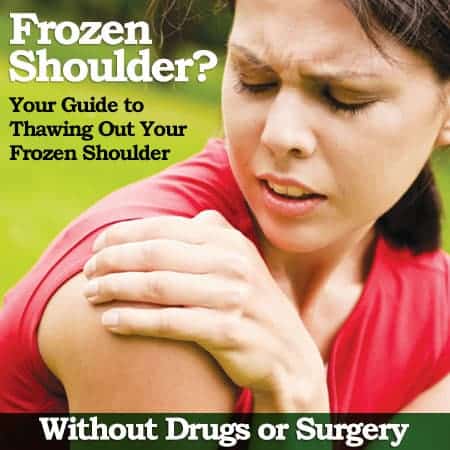
Frozen shoulder, also known as adhesive capsulitis, is a condition characterized by stiffness and restricted movement in the shoulder joint. In its early stages, individuals may experience limited mobility, which can gradually progress if left unaddressed.
Non-invasive strategies focusing on joint flexibility, muscular balance, and underlying structural concerns may help comfort shoulder mobility. The following sections outline common causes of shoulder pain and key contributors to frozen shoulder.
What Contributes to Frozen Shoulder and Shoulder Stiffness?
Frozen shoulder occurs when the soft tissues surrounding the shoulder joint undergo structural changes that limit movement. Over time, the joint capsule may thicken, restricting flexibility.
Common Contributing Factors:
- Repetitive Shoulder Strain: Repeated shoulder movements may place stress on muscles and tendons.
- Prolonged Immobility: Reduced shoulder use following injury or surgery may contribute to joint stiffness.
- Systemic Influences: Some metabolic or circulatory conditions have been linked to shoulder mobility adaptations.
Common Structural Influences on Shoulder Mobility
Shoulder mobility concerns may stem from musculoskeletal adaptations, including:
- Rotator Cuff Sensitivity: Structural changes affecting rotator cuff tendons may influence movement.
- Shoulder Impingement: Compression of soft tissues within the joint may contribute to restricted mobility.
- Thoracic Outlet Influence: Sensitivity in the upper chest region may extend to the shoulder.
- Spinal Function: Changes in spinal mobility or nerve pathways may influence shoulder function.
Structured Strategies for Shoulder Mobility Comfort
A well-rounded approach that considers joint mechanics, soft tissue flexibility, and postural influences may help restore shoulder mobility.
Common Non-Invasive Strategies:
- Manual Techniques: Gentle methods to comfort soft tissue flexibility and joint mobility.
- Technology-Assisted Approaches: Rehabilitation technology helping tissue recovery and circulation.
- Postural and Lifestyle Adjustments: Ergonomic modifications and structured movement plans to maintain long-term mobility.
Early intervention is essential for preventing progressive stiffness and maintaining optimal shoulder function.
Understanding the Differences Between Adhesive Capsulitis and Frozen Shoulder Syndrome (FSS)
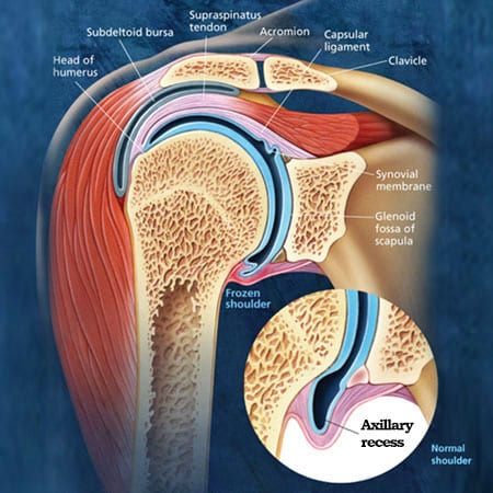
Although adhesive capsulitis and frozen shoulder syndrome (FSS) share similar characteristics, they differ in their underlying causes.
Key Differences:
- Adhesive Capsulitis: Primarily involves structural changes within the joint capsule.
- Frozen Shoulder Syndrome (FSS): Typically results from soft tissue influences, such as muscle or tendon adaptations.
Understanding these distinctions ensures that shoulder mobility strategies are directed toward the underlying factors affecting movement.
What Contributes to Adhesive Capsulitis?
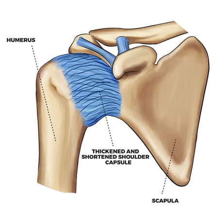
Adhesive capsulitis is commonly linked to joint capsule inflammation and structural thickening. While the exact cause remains debated, several factors may contribute.
Common Contributors to Adhesive Capsulitis:
- Systemic Influences: Some research suggests a connection between joint inflammation and immune function.
- Capsular Adaptations: Structural changes in the shoulder capsule may reduce flexibility.
- Soft Tissue Influence: Muscular or ligamentous adaptations near the shoulder may contribute to movement restrictions.
Breaking Down the Terminology
To better understand adhesive capsulitis, analyzing its components can provide clarity:
- Capsulitis: Refers to joint capsule inflammation, which may influence mobility.
- Adhesive: Describes the sticking or binding of joint structures, contributing to restricted movement.
In primary adhesive capsulitis, changes occur within the joint capsule. In secondary adhesive capsulitis, movement restrictions may result from surrounding tissue adaptations, such as rotator cuff changes or bursitis.
A structured, non-invasive approach that focuses on mobility, postural alignment, and joint function may help assist shouldermovement and long-term flexibility.
Distinguishing Between Primary and Secondary Adhesive Capsulitis
1. Primary Adhesive Capsulitis:
- Develops within the shoulder joint capsule, leading to progressive stiffness.
- Associated with joint-specific changes and structural degeneration.
- May have links to inflammatory or systemic factors affecting joint mobility.
2. Secondary Adhesive Capsulitis:
- Results from soft tissue injuries or inflammation, including muscles, tendons, or bursae.
- Commonly associated with rotator cuff injuries or shoulder bursitis.
Understanding whether shoulder stiffness is related to joint capsule changes or surrounding soft tissue influences helps determine the most efficient non-invasive management strategies.
The Influence of Ligaments in Shoulder Mobility
The shoulder joint capsule consists of soft tissues and ligaments that comfort joint stability and flexibility. The capsule contains synovial fluid, which helps nourish cartilage and connective tissues within the joint.
Effects of Ligament Changes on Shoulder Function:
- Structural Adaptations: Repetitive strain or trauma may contribute to ligament thickening (hypertrophy).
- Adhesion Formation: Thickened ligaments may develop fibrotic adhesions, reducing joint flexibility.
- Reduced Synovial Fluid Production: Changes in the capsule may influence joint lubrication and movement.
Potential Outcomes of Structural Ligament Changes:
- Restricted shoulder movement.
- Progressive joint stiffness.
- Challenges in daily activities such as lifting, brushing hair, or reaching overhead.
Recognizing Common Symptoms of Adhesive Capsulitis
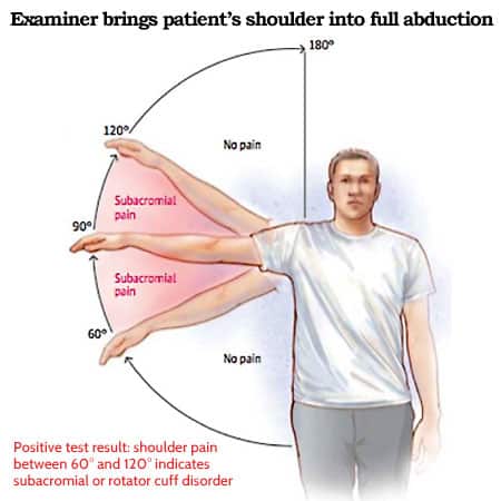
Frozen shoulder is often identified by a gradual reduction in joint mobility and increasing stiffness.
Common Symptoms Include:
- Limited Motion:
- Difficulty with overhead movements or across-the-body actions.
- Reduced rotational mobility, especially in chronic cases.
- Pain:
- Sensitivity during movement or at rest.
- Sensations that may become more noticeable at night.
- Progressive Stiffness:
- Shoulder movement becomes increasingly restricted over time.
- Prolonged immobility may lead to near-complete loss of movement.
- Potential Contributing Factors:
- Spinal influences such as neck or upper back mobility adaptations.
- Systemic influences, including metabolic or circulatory conditions.
- Post-surgical adaptations following shoulder procedures.
How the Shoulder Joint System Assists Mobility
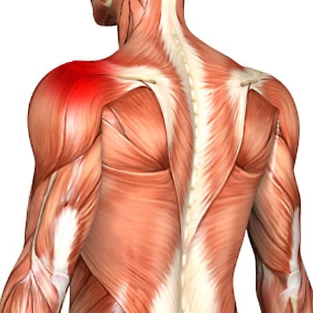
The shoulder is one of the most mobile regions in the body, allowing a wide range of movement due to its structural design. It consists of multiple joints that function together.
Four Main Shoulder Joints:
- Glenohumeral Joint: Primary ball-and-socket articulation between the humerus and scapula. Gelnohumeral joints responsible for the majority of shoulder movement.
- Acromioclavicular (AC) Joint: Connects the clavicle and acromion, playing a role in lifting and overhead motions.
- Sternoclavicular Joint: Links the sternum and clavicle, providing stability. It facilitates shoulder elevation and retraction.
- Scapulothoracic Joint: A functional articulation where the scapula glides over the ribcage. It is sssential for coordinated shoulder movement.
Types of Adhesive Capsulitis and Their Distinctions
Frozen shoulder is classified into two primary categories based on its underlying cause.
1. Primary Adhesive Capsulitis:
- May be linked to systemic conditions such as metabolic, circulatory, or neurological factors.
- Reported prevalence in individuals with diabetes is approximately 20%.
- Commonly presents as restricted external rotation in early stages.
2. Secondary Adhesive Capsulitis (Frozen Shoulder Syndrome - FSS):
- Develops due to localized shoulder injuries, such as rotator cuff strain or bursitis.
- Frequently results from overuse, trauma, or degenerative changes.
- Early symptoms often include reduced internal rotation, differentiating it from primary adhesive capsulitis.
A structured, non-invasive approach focusing on joint mobility, soft tissue flexibility, and postural comfort can help improve shoulder function and restore movement.
Understanding the Causes of Primary Adhesive Capsulitis
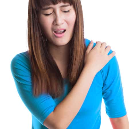
Primary adhesive capsulitis is often linked to systemic health conditions that may influence shoulder joint function. These conditions can contribute to inflammatory responses or capsular adaptations, leading to progressive stiffness and mobility limitations.
Common Factors Associated with Primary Adhesive Capsulitis:
- Autoimmune Conditions: Some immune-related conditions may contribute to joint inflammation.
- Metabolic Influences: Individuals with diabetes have an increased likelihood of developing shoulder mobility adaptations.
- Circulatory Health: Cardiovascular factors may influence joint capsule flexibility.
- Neurological Conditions: Stroke, Parkinson’s disease, and other nerve-related conditions may affect shoulder movement.
- Endocrine Function: Thyroid imbalances (hypothyroidism or hyperthyroidism) may play a role in joint mobility adaptations.
- Respiratory Factors: Some chronic respiratory conditions may influence shoulder movement patterns.
- Post-Surgical Factors: Procedures involving the shoulder or upper spine may contribute to capsular stiffness.
Identifying systemic influences is essential for structuring targeted, non-invasive strategies that comfort shoulder mobility and function.
Understanding the Causes of Secondary Adhesive Capsulitis (Frozen Shoulder Syndrome - FSS)
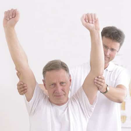
Secondary adhesive capsulitis, also referred to as true frozen shoulder syndrome (FSS), develops due to structural adaptations in the shoulder’s soft tissues. This condition is often associated with rotator cuff changes, joint irritation, or degenerative influences.
Common Contributors to Secondary Adhesive Capsulitis:
- Rotator Cuff Structural Changes:
- The supraspinatus tendon, which attaches to the humerus, is prone to repetitive strain.
- Structural adaptations may develop near the tendon’s attachment point, influencing mobility.
- Bursitis:
- Inflammation of the bursa (fluid-filled sac) within the shoulder may contribute to reduced movement.
- Tendon Sensitivity:
- Repetitive strain or tendon irritation may influence shoulder flexibility.
- Bone Spurs:
- Structural changes near the acromioclavicular (AC) joint may influence rotator cuff movement.
The Role of Rotator Cuff Muscles in Shoulder Function
The rotator cuff consists of four primary muscles that help shoulder movement and joint stability:
- Supraspinatus: Assists in lifting the arm.
- Infraspinatus: Helps external rotation of the shoulder.
- Teres Minor: Assists with external rotation.
- Subscapularis: Facilitates internal rotation of the shoulder.
Among these, the supraspinatus muscle and tendon are most frequently affected by structural changes. Adaptations near the tendon attachment at the humerus may influence shoulder mobility.
Distinguishing Between Primary and Secondary Adhesive Capsulitis

Although primary adhesive capsulitis and secondary adhesive capsulitis (FSS) share similar mobility limitations, their causes and clinical patterns differ.
Primary Adhesive Capsulitis:
- Linked to systemic factors such as metabolic, circulatory, or neurological conditions.
- Often involves difficulty with external rotation due to capsular changes.
- Both active and passive movement are restricted.
Secondary Adhesive Capsulitis (FSS):
- Results from localized musculoskeletal changes such as rotator cuff strain or bursitis.
- Individuals commonly experience difficulty with internal rotation and passive abduction.
- A defining feature is reduced passive mobility, even when assisted movement is attempted.
By identifying whether shoulder limitations are related to systemic factors or musculoskeletal adaptations, a structured, non-invasive approach can be developed to manage recovery and improve mobility.
MRI as a Preferred Imaging Method for Shoulder Assessment
Magnetic Resonance Imaging (MRI) is a highly efficient imaging method for distinguishing primary adhesive capsulitis from secondary adhesive capsulitis. It provides detailed visualization of the rotator cuff tendons, bursa, joint capsule, and surrounding structures, helping to identify contributing factors affecting shoulder mobility.
By determining whether the shoulder capsule (primary adhesive capsulitis) or surrounding soft tissues (secondary adhesive capsulitis) are involved, structured, non-invasive care strategies can be implemented promptly to comfort mobility and recovery.
MRI: The Recommended Imaging Method for Shoulder Conditions
While X-rays and ultrasound can provide insights into joint and soft tissue structure, MRI remains the preferred method for evaluating shoulder mobility concerns.
Why MRI is Preferred for Shoulder Assessment:
- Soft Tissue Imaging: Detects muscle, tendon, and ligament structures in detail.
- Joint Capsule and Cartilage Assessment: Provides clear visualization of joint surfaces and surrounding soft tissues.
- Structural Analysis: Identifies bone spurs, capsule changes, and early signs of degenerative influences.
For individuals experiencing persistent shoulder mobility limitations, consulting a healthcare professional to determine whether an MRI assessment is necessary may help a well-structured care plan.
At-Home Strategies for Assisting Shoulder Mobility
Managing shoulder mobility concerns at home involves protecting the joint, maintaining flexibility, and reducing stress on soft tissues.
Recommended Home Strategies:
- Minimize Excessive Strain: Avoiding repetitive overhead movements and sudden shoulder strain may help manage recovery. Some individuals may find shoulder slings beneficial in minimizing excessive movement during certain activities.
- Cold Applications for Localized Irritation: Applying a cloth-wrapped ice pack for 15 minutes every 2-3 hours may help manage localized irritation. Ice should never be applied directly to the skin, and icing sessions should be limited to under 20 minutes with at least a 1-hour gap between applications.
- Gentle Mobility Exercises: Performing controlled, pain-free arm movements may help maintain joint flexibility. Movements should be gentle and within a comfortable range, followed by cold application if needed.
- Professional Assessment: While home strategies may provide temporary alleviation, a professional evaluation is essential for structured recovery. Addressing shoulder mobility concerns early may help prevent further restrictions in movement.
Non-Invasive Strategies for Shoulder Mobility Care
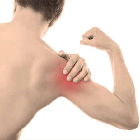
Both primary and secondary adhesive capsulitis benefit from structured, non-invasive approaches that focus on joint function, soft tissue mobility, and muscular stability.
Common Strategies for Shoulder Care:
- Comprehensive, Non-Invasive Approaches: A structured care plan may integrate chiropractic and physiotherapy-based approaches to promote joint mobility and soft tissue function.
- Multi-Modal Care Strategies: Manual techniques for targeted mobility, care. CSC offers rehabilitation technology-assisted approaches, including:
- Shockwave-assisted rehabilitation
- Ultrasound-based mobility helps
- Neuromuscular stimulation techniques
- Soft tissue mobility approaches
- Targeted movement rehabilitation
- Strength and Flexibility Care: Shoulder mobility limitations may lead to soft tissue changes over time. A structured strengthening and flexibility-focused approach can help maintain joint stability and mobility.
The Importance of Professional Assessment
Seeking structured, professional evaluation is essential for conditions such as adhesive capsulitis to ensure an accurate assessment and a targeted care strategy.
A structured, non-invasive approach can comfort shoulder mobility, minimize pain, and reduce the need for invasive interventions such as injections or surgical procedures.
Where to Be Seen in Kuala Lumpur
Assessment and care are available at both of our Kuala Lumpur centers. The main one is described on the page for our Kuala Lumpur center in Bukit Damansara.
A second center serves the northwest of the city. You can read about our Bandar Sri Damansara center.
For travel, parking and opening hours, see contact details for the Kuala Lumpur headquarters or contact details for Bandar Sri Damansara.
The Three Phases, and Why the Treatment Changes With Them
Frozen shoulder is one of the few conditions where the right treatment in one phase is the wrong treatment in another. Getting the phase right matters more than the choice of technique.
- Freezing, roughly the first two to nine months. Pain dominates, including at night, and range is falling. Aggressive stretching here tends to make things worse. The work is pain control and movement within comfort.
- Frozen, roughly four to twelve months. Pain settles, stiffness dominates. This is where stretching and loading earn their place, because the tissue is no longer as reactive.
- Thawing, up to two years or more. Range returns gradually. The job is to keep loading so that strength returns with the movement.
Those timelines are approximate and overlap, and the total course is long. Anyone offering to resolve a frozen shoulder in a few weeks is describing something other than frozen shoulder.
Two Things Worth Checking That Are Often Missed
- Blood sugar. Frozen shoulder is markedly more common in people with diabetes, and it tends to run a longer course. If you have not had blood sugar checked recently and you develop a frozen shoulder, it is worth asking your doctor.
- Thyroid function, which has a similar association and is a simple test.
Neither changes the shoulder program directly, but both change the expectations and the general health picture, and a clinic that never mentions them has not looked past the joint.
When Shoulder Stiffness Is Not Frozen Shoulder
- Stiffness after a fall or injury, where a fracture or dislocation needs ruling out first.
- Loss of power rather than loss of range, which points to the cuff rather than the capsule.
- Neck symptoms traveling into the arm with numbness, which points to the neck.
- A history of cancer with new shoulder pain, or unexplained weight loss and night pain.
Planning a visit? You can review the current chiropractic price in Kuala Lumpur and physiotherapy fees in Kuala Lumpur before you come in.
Peer-Reviewed References
- Diercks RL, Stevens M. Gentle thawing of the frozen shoulder: a prospective study of supervised neglect versus intensive physical therapy in seventy-seven patients with frozen shoulder syndrome followed up for two years. J Shoulder Elbow Surg. 2004;13(5):499-502.
- Mertens MG, Meert L, Struyf F, et al. Exercise therapy is effective for improvement in range of motion, function, and pain in patients with frozen shoulder: a systematic review and meta-analysis. Arch Phys Med Rehabil. 2022;103(5):1011-1021.
- Nakandala P, Nanayakkara I, Wadugodapitiya S, Gawarammana I. The efficacy of physiotherapy interventions in the treatment of adhesive capsulitis: a systematic review. J Back Musculoskelet Rehabil. 2021;34(3):409-418.
The Author of Frozen Shoulder Care: Non-Invasive Options in Malaysia
Frozen Shoulder Care: Non-Invasive Options in Malaysia was written by Yama Zafer, D.C., who has 30+ years in chiropractic and physiotherapy and founded Chiropractic Specialty Center in Kuala Lumpur in 2006.
Last Updated: Frozen Shoulder Care in Kuala Lumpur
Frozen Shoulder Care in Kuala Lumpur was last reviewed and updated on October 3, 2026.
Hi Yama, thank you for your effort in posting the articles on your blog.
On 25/10/23 and 28/10/23, I played badminton. On 29/10/23, I noticed my right collarbone popped out more than my left collarbone. I went to the doctor, and he diagnosed it as stenoclavicular anterior subluxation or dislocation. Most of the pain and swelling are gone, but my shoulder feels stiff sometimes, like some blood flow is incorrect. The doctor didn't advise surgery due to the risks, and he told me to "live with it". But it has been draining me mentally as I am afraid to exercise, play badminton again or even reach back for my wallet with my right hand because I would stress the joint. So my questions are: -
1. Are there any chiropractic doctors in your clinics who have successfully brought the raised stenoclavicular joint back down to the same level as the opposite joint?
2. May I know their name and which branch?
I want to fix the joint, ensure both clavicles are optimistically placed, and then take glucosamine and chondroitin supplements to help repair it. I will also go on a one-year upper body exercise regime to rebuild the muscle and tissue I have lost from ageing. I just turned 50. Are there any chiropractors in your clinics with proven experience in successfully restoring anterior stenoclavicular subluxation?
Thanks!
The reassuring part first: anterior is the version you want. The sternoclavicular joint can displace forward or backward, and those two are entirely different in seriousness. Backward displacement is a surgical emergency because the structures immediately behind that joint include the windpipe, the esophagus, and major blood vessels. Forward displacement, which is what you have, does not threaten any of those, and it is much the more common of the two.
The signs that would indicate the other kind, so you can dismiss them: difficulty breathing or swallowing, a change in your voice, a choking sensation, or swelling or color change in the arm on that side. If any of those ever develop, go to an emergency department the same day. Nothing in your description suggests it.
On what happened, two things are worth noting. The collarbone became prominent without a specific injury, after two days of badminton rather than a fall or collision. That pattern, displacement without significant trauma, often reflects laxity in the joint's ligaments rather than an injury that tore something. It is worth screening yourself for general hypermobility, since it is commonly associated: with knees straight can you put your palms flat on the floor, do your elbows or knees bend backward past straight, can you bend your little finger back beyond a right angle, can your thumb touch your forearm. Several yes answers make laxity the likely explanation, and that changes what helps.
On management, this joint is usually managed without surgery, and deliberately so. Attempts to hold a forward-displaced sternoclavicular joint back in position often fail to hold, and surgical stabilization of this joint carries a higher complication rate than most shoulder procedures, so it is generally reserved for cases with persistent significant pain and functional loss. The usual course for the forward version is that pain settles over weeks to months while some visible prominence remains. The prominence is a cosmetic matter rather than a functional one, and it is worth knowing that in advance so it does not read as failure.
What actually helps: avoiding the positions that provoke it, which for this joint means end-range reaching across the body and sustained overhead work, in the early phase. Then progressive strengthening of the muscles that control the shoulder blade and the shoulder itself, since the collarbone is stabilized indirectly by them. Serratus anterior, the lower trapezius, and the rotator cuff are the specific targets. Return to badminton gradually, with overhead smashing added last, and expect that to be weeks rather than days.
On imaging, if there is any question about the direction of displacement, CT is the study that shows this joint clearly. Plain X-rays are unreliable here, and an anterior versus posterior distinction should not rest on them.
If pain persists beyond a few months despite proper strengthening, or if the joint becomes unstable enough to interfere with daily activity, an orthopedic opinion from someone who deals with this joint specifically is the next step.
Any hope for a frozen shoulder plain?
Yes, and that is an accurate answer rather than an encouraging one: frozen shoulder resolves in the large majority of people. The difficulty is that it resolves slowly, typically over one to three years, and knowing which phase you are in changes what you should be doing.
There are three phases. The first is painful and progressively stiffening, with pain often worst at night and disturbing sleep. The second is stiff but considerably less painful, where movement is limited but the ache has settled. The third is gradual return of movement. Treating the first phase like the second is the most common error, because aggressive stretching during the painful phase tends to inflame it and prolong things.
First, confirm it is actually a frozen shoulder. The defining feature is loss of passive external rotation: with the elbow held at your side and someone else rotating your forearm outward while you relax completely, the movement is blocked. If that movement is full when someone else moves your arm for you, this is more likely a rotator cuff problem, which is common, feels similar, and is managed quite differently.
Second, and this is the most useful practical point: ask your doctor to check your blood sugar and your thyroid function if that has not been done. Frozen shoulder is markedly more common in people with diabetes, including undiagnosed diabetes, and in thyroid disorders, and where diabetes is present the condition tends to be more stubborn and to last longer. Finding an undiagnosed metabolic condition matters well beyond the shoulder.
On what helps by phase. During the painful phase the priorities are pain control, sleeping arrangements that keep the arm supported, and gentle movement within a comfortable range rather than pushing into pain. Corticosteroid injection into the joint has the best supporting evidence for this phase, both for pain and for speeding the overall course, and it is worth discussing early rather than after months of struggle. Once the pain has settled and stiffness dominates, progressive stretching and joint mobilization become the main work, and consistent daily effort over months is what restores range.
For shoulders that remain severely restricted after a genuine course of that, further options exist, including distension of the joint with fluid under imaging, and surgical release. Those are decisions for an orthopedic specialist.
One honest note: a proportion of people are left with some permanent restriction at the extremes of movement, most often reaching behind the back. That is usually of little functional consequence, but it is better to hear it now than to interpret it later as failure.