


MRI Scan: What It Shows, Safety and Preparation
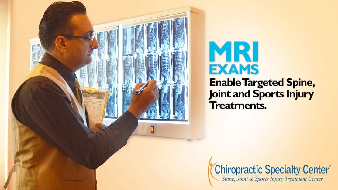
An MRI scan uses a strong magnetic field and radio waves to show soft tissues that ordinary X-rays cannot display well, including spinal discs, nerve roots, cartilage, ligaments and muscle. What an MRI cannot show is pain itself, which is why a scan finding must be interpreted alongside symptoms and examination findings.
A scan is therefore most useful when it answers a question the examination has already raised. Where symptoms and examination findings point to one level, imaging either confirms or contradicts that, and the plan changes accordingly. Where a scan is taken first and its findings are read backward into the symptoms, treatment tends to be aimed at something that was never the problem. Weakness, spreading numbness, or a symptom that will not settle are the situations in which imaging earns its place.
Key Takeaways: Top 3 MRI Insights
- MRI uses no ionizing radiation and shows soft tissues such as discs, nerves, ligaments and cartilage in detail.
- Safety screening for metal, implants and devices is essential, and preparation affects both safety and image quality.
- MRI findings such as disc bulges are common in people without pain, so the report is read together with the examination.
Understanding MRI Exams
Chiropractic Specialty Center does not perform MRI scans. When the examination raises a question that imaging can answer, the clinical team may recommend an MRI, which is carried out at a hospital or radiology center. MRI uses magnetic fields and radio waves to capture detailed images of soft tissue, which X-rays, CT scans and ultrasound show less clearly.
MRI Review and Spine Assessment at Two KL Centers
If you already have an MRI report, or are wondering whether you need one, the report and your symptoms can be reviewed together at either center of Chiropractic Specialty Center. Bring the report, and the images if you have them. Travel details are on the Bukit Damansara contact page and the Bandar Sri Damansara contact page.
- Bukit Damansara (KL, main center): Call +603 2093 1000 or WhatsApp +60 17 269 1873 to see the Bukit Damansara / KL center.
- Bandar Sri Damansara (KL): Call +603 6262 5777 or WhatsApp +60 12 455 6939 to see the Bandar Sri Damansara center.
Why Might You Need an MRI Exam?
The right care depends on understanding what is causing the symptoms. For most back and neck pain, the examination is enough to begin care, and imaging is not needed at the start. An MRI or X-ray is recommended when the findings raise a specific question, such as nerve involvement, a suspected fracture or symptoms that are not settling as expected.
MRI is especially useful for soft tissues such as ligaments, cartilage, spinal discs, muscles, bursae and tendons. It is commonly used to evaluate herniated discs, a disc bulge, frozen shoulder, knee pain, hip pain, shoulder injuries and Tarlov cysts, and brain MRI is sometimes used in headache investigations to rule out other causes. Care for the spine is then planned around the findings that match the examination.
MRI provides detailed images of the targeted region in several planes (coronal, sagittal and axial), using a strong magnetic field and computer processing.
Are There Different Types of MRI Scanners?
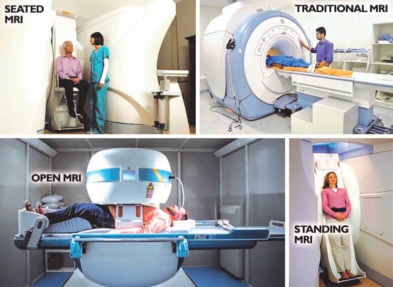
Most MRI scanners are used lying down. Upright or weight-bearing scanners, which image the spine while sitting or standing, exist but are uncommon. Among standard lying-down scanners, there are closed scanners, wide-bore scanners with a larger tunnel, and open scanners.
Open scanners suit some people who are larger or anxious in enclosed spaces, but most operate at a lower field strength, so image detail may be lower and scans may take longer. A wide-bore scanner is often a good middle ground. You will usually lie on your back, and the radiographer will help you get comfortable before the table moves into the scanner.
What to Do When Preparing for an MRI
Because the scanner uses a powerful magnet, all metal objects must be removed before the scan, including watches, jewelry, hearing aids, removable dentures and hairpins. Tell the radiology team in advance about any implanted device or metal in your body, such as a pacemaker, defibrillator, cochlear implant, stents, heart valves, joint replacements, surgical clips or dental braces. Many modern implants are safe in MRI under set conditions, but each one must be checked.
If you feel anxious in confined spaces, mention it when the scan is booked. Going in feet first, an eye mask, music through headphones or, if needed, a mild sedative prescribed by a doctor can make the scan manageable.
What to Expect Before and During the MRI Exam
You will usually complete a safety questionnaire and sign a consent form. Inside the scanner you will hear loud knocking and buzzing sounds as images are taken, so earplugs or headphones are provided.
The radiographer watches through a window and can speak to you through an intercom, and you hold a call button throughout. If contrast is needed, it is given through a small needle in the arm. Most scans take around 20 to 60 minutes, depending on the area and the number of sequences.
What Does It Feel Like to Take an MRI?
An MRI is painless. You lie still on a firm table, which some people find tiring, and the area being scanned may feel slightly warm. Tell the radiographer straight away if you feel dizzy, nauseated, short of breath or feel any burning sensation during the scan.
Are Radiation or Other Harmful Effects Associated with MRI Exams?
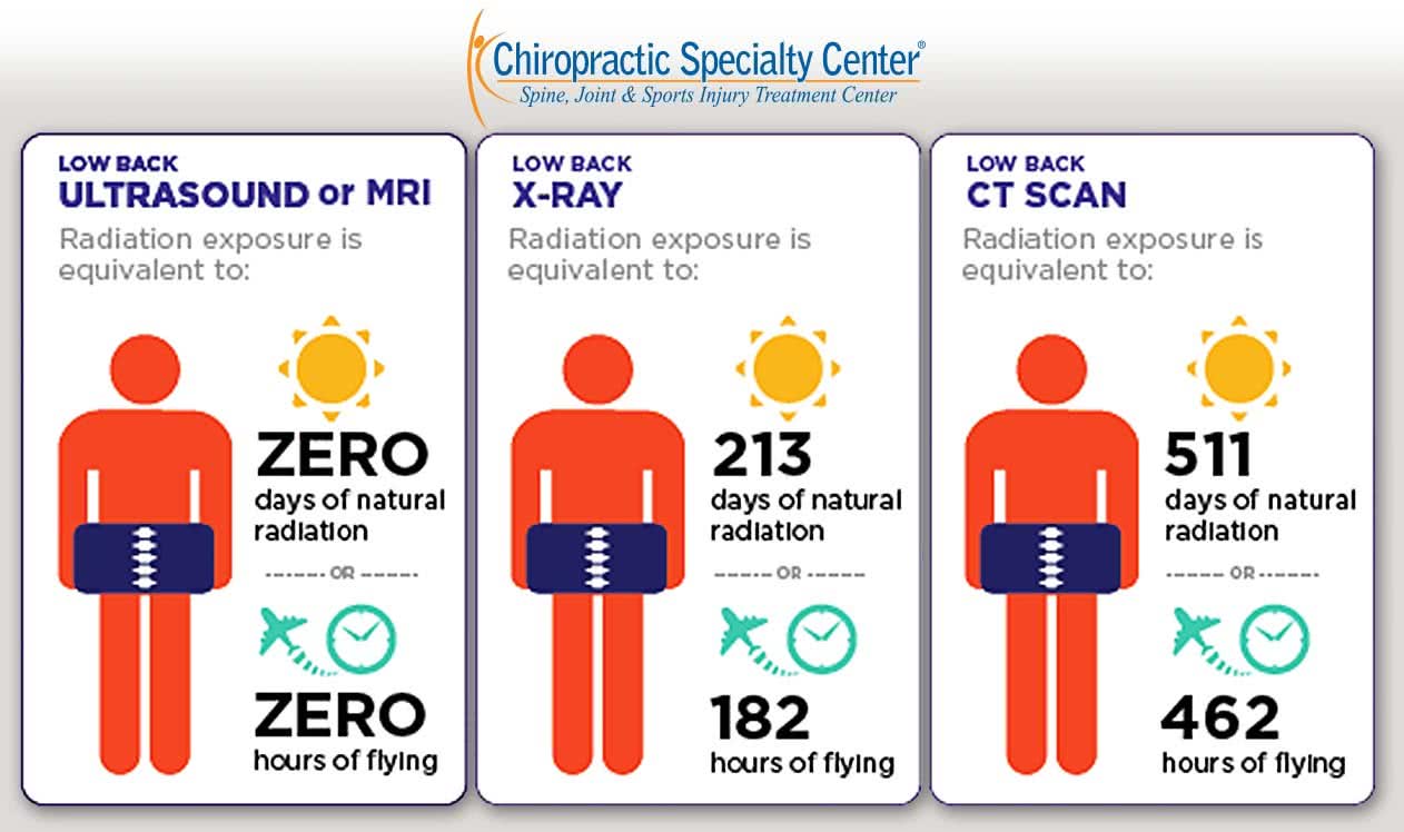
MRI does not use ionizing radiation, which is one reason it can be repeated where CT would not be appropriate. It is considered very safe when screening and safety protocols are followed. The main risks come from metal: the magnetic field can move or heat certain implants and metal objects.
People who have worked with metal, such as welders and metalworkers, may have tiny metal fragments in the eyes. An X-ray of the eyes may be done before the MRI to check. Always mention your work history and any previous injuries involving metal.
MRI is the preferred test for many soft-tissue questions, while X-ray and CT are better for others, such as alignment under load or fine bone detail. Each answers a different question.
Should You Be Concerned About the Magnetic Fields?
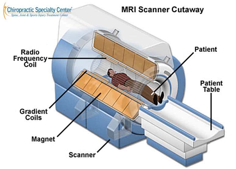
The magnetic fields used in clinical MRI have no known harmful effects on the body when safety screening is followed. Metal fragments in the eyes are a reason not to have an MRI until they are checked. Some tattoo pigments contain iron and can occasionally cause warmth or irritation in the skin during the scan.
Allergic reactions to MRI contrast are uncommon and usually mild. If you are breastfeeding, current radiology guidance generally allows breastfeeding to continue normally after contrast; if you prefer, you can express milk beforehand, and your radiology team can advise you.
What an MRI Report Can and Cannot Tell You
Disc bulges, disc degeneration and other age-related changes appear on MRI in many people who have no pain at all. In one well-known study, about half of adults without back pain had a disc bulge on at least one level. A large review found that disc degeneration was reported in about a third of pain-free 20-year-olds and in most pain-free 80-year-olds.
That is why the report is read together with the examination. The level responsible for symptoms is the one whose nerve pattern matches where the symptoms travel, which reflex is affected and which muscles test weak. Imaging is most useful when it answers a specific question, and guidelines advise against routine early imaging for uncomplicated low back pain.
The Author of MRI Scan: What It Shows, Safety and Preparation
MRI Scan: What It Shows, Safety and Preparation was written by Yama Zafer, D.C., a graduate of Cleveland University - Kansas City with 30+ years in chiropractic and physiotherapy.
Peer-Reviewed References
- Jensen MC, Brant-Zawadzki MN, Obuchowski N, et al. Magnetic resonance imaging of the lumbar spine in people without back pain. N Engl J Med. 1994;331(2):69-73.
- Brinjikji W, Luetmer PH, Comstock B, et al. Systematic literature review of imaging features of spinal degeneration in asymptomatic populations. AJNR Am J Neuroradiol. 2015;36(4):811-816.
- Brinjikji W, Diehn FE, Jarvik JG, et al. MRI findings of disc degeneration are more prevalent in adults with low back pain than in asymptomatic controls: a systematic review and meta-analysis. AJNR Am J Neuroradiol. 2015;36(12):2394-2399.
- Expert Panel on Neurological Imaging; Hutchins TA, Peckham M, Shah LM, et al. ACR Appropriateness Criteria low back pain: 2021 update. J Am Coll Radiol. 2021;18(11S):S361-S379.
- Jenkins HJ, Downie AS, Maher CG, et al. Imaging for low back pain: is clinical use consistent with guidelines? A systematic review and meta-analysis. Spine J. 2018;18(12):2266-2277.
- Urrutia J, Zamora T, Prada C. The prevalence of degenerative or incidental findings in the lumbar spine of pediatric patients: a study using magnetic resonance imaging as a screening tool. Eur Spine J. 2016;25(2):596-601.
- Knezevic NN, Candido KD, Vlaeyen JWS, et al. Low back pain. Lancet. 2021;398(10294):78-92.
These references cover MRI findings in people with and without back pain, imaging guidelines and the management of low back pain. None of them evaluates a care program at Chiropractic Specialty Center. Each one can be checked through its free PubMed record.
Last Updated: MRI Scan: What It Shows, Safety and Preparation
MRI Scan: What It Shows, Safety and Preparation was last reviewed and updated on September 25, 2026.
Planning a visit? You can review the current chiropractic price in Kuala Lumpur and physiotherapy fees in Kuala Lumpur before you come in.
Dear Sir.
I need to do a MRI of the Brain.
Can your open MRI do this procedure.
Kindly advise.
Thank you.
No. No MRI services of any kind are provided here, open or otherwise. This is not an imaging center, and scans are arranged through hospitals and radiology centers.
There are two things worth knowing before you arrange a brain scan elsewhere.
The first is that an open scanner is a poor choice for brain imaging specifically. Most open scanners operate at lower field strength, and brain imaging is where field strength matters most, because the detail required to assess brain tissue is considerably finer than for a knee or a lumbar spine. A brain study on a low-field open scanner can come back non-diagnostic, meaning the scan has to be repeated properly and you have paid twice. If the reason you want an open scanner is anything other than body size, ask for a standard or wide-bore 1.5 tesla or 3 tesla machine instead.
The second follows from that, if enclosed spaces are the concern. For a brain scan you cannot be positioned feet first, because your head has to be at the center of the scanner, so the usual workaround is not available. What does help: an eye mask, or keeping your eyes closed from before you enter and not opening them; a mirror attachment on the head coil, which many centers have and which lets you see out of the scanner; headphones with music; and asking the radiographer to tell you the length of each sequence and to speak to you between them. You will have a call button in your hand throughout and can stop the scan. If that is not enough, ask your doctor about a mild sedative taken beforehand, which is routine to arrange and means someone else drives you home.
You will need a referral in any case. Ask whoever refers you to state the clinical question rather than simply requesting a brain MRI, and to specify whether contrast is required, since that changes both the preparation and the cost.
Hello
Can I know which hospital in KL has an open MRI? Also, can I know how much it will cost me? Thank you for your help.
Naming a specific hospital would not be reliable, since scanners are replaced and upgraded and any list dates quickly. What holds is how to find one, and there is a distinction worth knowing first.
Open and wide-bore are not the same. An open scanner has no tunnel, but most operate at lower field strength, meaning longer scans and less detail. A wide-bore scanner is still a tunnel but a noticeably larger one, around seventy centimeters rather than sixty, while keeping full image quality. For spine imaging a wide-bore scanner is usually the better result if you can tolerate it. When you telephone a radiology department, ask three things: the bore diameter in centimeters, the table weight limit in kilograms, and whether the scanner is open, standard, or wide-bore. Those three answers settle it in a few minutes per hospital.
If the reason you want an open scanner is that enclosed spaces are difficult, several things make a standard scan manageable and are worth trying before accepting lower image quality.
Ask to go in feet first. For a lumbar spine study this is usually possible, and it means your head is either outside the bore or only just inside it, which removes most of the difficulty for most people.
Ask for an eye mask, or keep your eyes closed from before you enter the scanner and do not open them. A large part of the distress comes from seeing how close the surface is.
Ask about a mirror attachment on the head coil, which lets you see out of the scanner, and ask for headphones with music, which help with both the noise and the sense of enclosure.
Ask the radiographer to talk to you between sequences and to tell you the length of each one. Knowing that a sequence lasts three minutes is very different from lying in silence with no idea how long remains. You will also have a call button in your hand at all times, and you can stop the scan.
And if none of that is enough, ask your doctor about a mild sedative taken beforehand. This is common, straightforward to arrange, and you will need someone to drive you home.
On cost, it varies by facility, by how many regions are scanned, and by whether contrast is used. Two regions costs roughly double one, so ask whether both are genuinely needed now or whether one should come first. Government hospitals charge substantially less than private ones but usually have longer waits. Ask each department for a written quote for the specific region requested, and check with your insurer whether an MRI ordered for an investigated condition is claimable.
One last point: a referral that states the clinical question, rather than simply requesting a scan, produces a more useful report. Ask whoever refers you to write down what they are looking for.
How much do MRIs cost? I need to do for my long st of adding back pain resulting from a fall.
MRI is not performed at our center, so any figure quoted here would be secondhand. Cost depends on the facility, on how many regions are scanned, and on whether contrast is used, and government hospitals charge considerably less than private ones though usually with longer waits. Call the radiology department directly and ask for a written quote for the specific region requested.
The more important question is whether you need one, and in your case there is a clear reason to be assessed rather than to arrange the scan yourself.
Back pain that follows a fall belongs in a different category from ordinary back pain. Trauma is one of the specific situations where imaging is indicated rather than optional, because a compression fracture in a vertebra can produce exactly the picture you describe: pain that began with an event and has not settled. That is truer with increasing age, in anyone with thinning bones, and in anyone who has taken steroid medication for a long period. A fracture of this kind is frequently missed for months because the pain is assumed to be muscular.
The usual sequence is a plain X-ray first, which shows most compression fractures and costs a fraction of an MRI. MRI is added when the X-ray is normal but suspicion remains, when there are symptoms in a leg, or when a fracture needs to be dated as recent or old, which an X-ray cannot do and which changes management.
You will need a referral in any case, and a referral that states the clinical question rather than simply requesting a scan produces a more useful report. So the practical order is an assessment first, then the imaging it indicates, rather than an MRI ordered blind.
Seek medical attention promptly rather than arranging a scan at leisure if you have weakness in a leg, numbness in the saddle area, any change in bladder or bowel control, or pain that wakes you at night, and go the same day if any of the last two apply.
Hello
Can I know which hospital in kl that provides open mri? I want to do mri but I can’t because of obesity. Also, can I know how much it will cost me? Thank you for your help.
Naming a specific hospital would not be reliable, because scanners are replaced and upgraded and any list goes out of date quickly. What does not go out of date is how to find one, and there is a distinction that will save you wasted trips.
Open and wide-bore are not the same thing. An open scanner has no tunnel at all, but most of these operate at a lower field strength, which means longer scan times and less detail. A wide-bore scanner is still a tunnel but a noticeably larger one, typically around seventy centimeters against the sixty of a standard machine, while keeping full field strength and image quality. For a knee or a spine, a wide-bore scanner that fits you is generally the better result than an open one, so ask about both rather than only for open MRI.
There are also two separate limits, and being turned away usually comes down to one of them. The bore diameter, which is about width and shape, and the table weight limit, which is a fixed engineering figure. A person can be within one and outside the other. When you telephone a radiology department, ask three specific questions: what is the bore diameter in centimeters, what is the table weight limit in kilograms, and is the scanner open, standard, or wide-bore. Written down before you call, those three answers settle it in a few minutes per hospital and remove the guesswork.
For the knee specifically, ask whether they have a dedicated extremity scanner. These take only the limb into a small opening, so body size is not a factor at all. Not every center has one, but where it exists it solves the knee half of your problem immediately.
On cost, it varies by facility, by how many regions are scanned, and by whether contrast is used. Two regions is roughly double one, so ask whether both the knee and the back are genuinely needed at the same time or whether one should come first. Government hospitals charge substantially less than private ones but usually have longer waiting times. Ask each department for a written quote for the specific region requested, and if you have insurance, ask whether an MRI ordered for an investigated condition is claimable under your policy.
One more practical point. A referral letter that states the clinical question, rather than simply requesting a scan, gets you a more useful report. Ask whoever refers you to write what they are looking for.
Hi there
Yes, I know MRI needs a referral. Actually, I need MRI scans for my knee and back pain, but unfortunately.
MRI machine at Hospital Pantai Ampang cannot fit my body size. So they only do Xray. I will call the number given for further discussion. Tq for your help
Since Pantai Ampang could only do X-rays, two things are worth knowing before you call.
For the knee, ask specifically whether a center has a dedicated extremity MRI scanner. Only the limb goes into a small opening, so overall body size is not a constraint at all. That removes the problem entirely for the knee half of your question, and it is worth asking about first because it is the easiest to arrange.
For the back, the practical route is a wide-bore scanner rather than an open one. Ask for the bore diameter in centimeters and the table weight limit in kilograms as two separate questions, because those are two separate limits and being turned away usually comes down to one or the other. Written down before you call, those two figures settle it quickly.
The X-rays you already have are not wasted, and this is worth saying because people often assume they are. For a knee, X-rays show joint space narrowing, alignment, and arthritic change, which is the majority of what is needed in many cases. For the back, they show alignment, disc height, spur formation, and whether there is a slip between vertebrae, and flexion and extension views can show whether a segment is moving abnormally, which an MRI taken lying still cannot. Bring them to any assessment.
What imaging does not do is identify which finding is causing your pain. That comes from examination. So while you are arranging the scans, an assessment of the knee and the back can proceed on what you already have, and often narrows down what the MRI actually needs to answer, which makes the referral more useful when you get it.
Magnetic Resonance Image is so much advantageous. It is one of the best innovations in the entire history of the medical industry. Additionally, its radio waves and magnetic scan magnetic control are not at all hazardous to a patient’s body cells. Moreover, its scanned outcome gives you a more deep and flawless image as every patient wants.
Both points are fair, and the absence of ionizing radiation is the main reason MRI can be used freely where repeated CT would not be appropriate. Its real strength in spine care is soft tissue: discs, nerve roots, the spinal cord, ligaments, and marrow are visible in a way no X-ray can show.
The limitation is worth stating alongside the advantage, because it causes more difficulty in practice than the technology itself. MRI shows structure, not pain. Disc bulges, degenerative change, and narrowing appear in large numbers of people who have no symptoms at all, and the proportion rises steadily with age. So a scan will frequently report several findings, of which none, one, or more than one may be responsible for what a person feels.
The consequence is that the report cannot be read in isolation. The level responsible for symptoms is the one whose nerve pattern matches the clinical examination: where the symptoms travel, which reflex is affected, which specific muscles test weak. Where imaging and examination agree, confidence is high. Where they disagree, the examination is generally the better guide, and treating whichever level looked worst on the film is a common and avoidable mistake.
There is also a documented downside to imaging early in uncomplicated back pain: people shown a report full of degenerative findings tend to do worse, not better, partly because the wording sounds more alarming than the condition warrants. That is why imaging is most useful when there is a specific question to answer, rather than as a routine first step.
X-ray, CT, and MRI each answer different questions. Bone detail and alignment under load favor X-ray, fine bone and spur anatomy favor CT, and soft tissue favors MRI. The best of them is the one that answers the question the examination has raised.